Malate dehydrogenase 2

Last updated
MDH2
Protein MDH2 PDB 1mld.png
Available structures
PDB Ortholog search: PDBe RCSB
Identifiers
Aliases MDH2 , M-MDH, MDH, MGC:3559, MOR1, Malate dehydrogenase 2, EIEE51, DEE51
External IDs OMIM: 154100 MGI: 97050 HomoloGene: 55938 GeneCards: MDH2
Orthologs
SpeciesHumanMouse
Entrez
Ensembl
UniProt
RefSeq (mRNA)

NM_001282403
NM_001282404
NM_005918

NM_008617

RefSeq (protein)

NP_001269332
NP_001269333
NP_005909

NP_032643

Location (UCSC) Chr 7: 76.05 – 76.07 Mb Chr 5: 135.81 – 135.82 Mb
PubMed search [3] [4]
Wikidata
View/Edit Human View/Edit Mouse

Malate dehydrogenase, mitochondrial also known as malate dehydrogenase 2 is an enzyme that in humans is encoded by the MDH2 gene. [5]

Contents

Malate dehydrogenase catalyzes the reversible oxidation of malate to oxaloacetate, utilizing the NAD/NADH cofactor system in the citric acid cycle. The protein encoded by this gene is localized to the mitochondria and may play pivotal roles in the malate-aspartate shuttle that operates in the metabolic coordination between cytosol and mitochondria. [6]

Structure

The protein encoded by MDH2 exists as a dimer, which indicate the important connection between protein stability and enzymatic activity. Each subunit contains two structurally and functionally distinct domains. The first is the NAD-binding domain, which exists in the amino-terminal half of each molecule, and contains a parallel-sheet structure, otherwise known as a Rosman fold motif. The core dinucleotide binding structure is composed of four beta-sheets and one alpha-helix. The other domain is a carboxy-terminal domain that contains the substrate binding site and amino acids that are necessary for catalysis. The active site of these enzymes is in a cleft between two domains. [7] Crystallography reveals the dimer interface, which consists mainly of interacting alpha-helices that form a compact interaction. The active sites in these dimeric proteins are well separated from each other. [8]

Function

Because malate dehydrogenase is closely tied to the citric acid cycle, regulation is highly dependent on TCA products. [9] Citrate also affects MDH activity by very complex manner. It inhibits the reduction of oxaloacetate under all conditions. Citrate also inhibits malate oxidation, but only at low malate or NAD concentrations. When both malate and NAD concentrations are high (10 mmol/L and 5 mmol/L, respectively), citrate can actually augment MDH2 activity. [10] All three effectors (malate, oxaloacetate and citrate) bind to the same putative allosteric site. [11] Recent studies of mitochondrial malate dehydrogenase are focused into the nature of the inactivation processes. The oligomeric structure of MDH2 has a variety of biological implications. Some researches have suggested that the dimeric structure is critical for enzymatic activity. It was first proposed that the reciprocating compulsory ordered mechanism where each subunit alternates as the active and the helper subunit, but both are needed for activity. This mechanism predicts an inactive monomer, and was corroborated by studies that showed a dramatic reduction of enzymatic activity. [12] Studies with mitochondrial MDH2 have shown that this enzyme is allosterically regulated as a complex as well. Binding experiments indicate that mitochondrial aspartate aminotransferase can associate with the alpha-ketoglutarate dehydrogenase complex and that mitochondrial malate dehydrogenase can associate with this binary complex to form a ternary complex. Formation of this ternary complex enables low levels of the alpha-ketoglutarate dehydrogenase complex, in the presence of the aminotransferase, to reverse inhibition of malate oxidation by glutamate. Thus, glutamate can react with the aminotransferase in this complex without glutamate inhibiting production of oxaloacetate by the malate dehydrogenase in the complex. The conversion of glutamate to alpha-ketoglutarate could also be facilitated because in the trienzyme complex, oxaloacetate might be directly transferred from malate dehydrogenase to the aminotransferase. In addition, association of malate dehydrogenase with these other two enzymes enhances malate dehydrogenase activity due to a marked decrease in the Km of malate. The potential ability of the aminotransferase to transfer directly alpha-ketoglutarate to the alpha-ketoglutarate dehydrogenase complex in this multienzyme system plus the ability of succinyl-CoA, a product of this transfer, to inhibit citrate synthase could play a role in preventing alpha-ketoglutarate and citrate from accumulating in high levels. This would maintain the catalytic activity of the multienzyme system because alpha-ketoglutarate and citrate allosterically inhibit malate dehydrogenase and dissociate this enzyme from the multienzyme system. [13]

Clinical Significance

Mutations in the MDH2 gene have been associated with several cancers, including uterine cancer, prostate cancer, pheochromocytoma and other paragangliomas. [14] [15] In particular, MDH2 has been found to be overexpressed in doxorubicin-resistant uterine cancer cells and may contribute to drug resistance. Since MDH2 plays a major role in malate-aspartate shuttling in ATP production, its overexpression likely supplies additional energy for P-glycoprotein to pump chemotherapeutic drugs out of the cells. Likewise, MDH2 contributes to docetaxel resistance in prostate cancer cells via the JNK pathway, and its knockdown reduced ATP levels as well as increased drug sensitivity. Thus, MDH2 may be an effective therapeutic target to enhance drug treatments for cancer. [14]

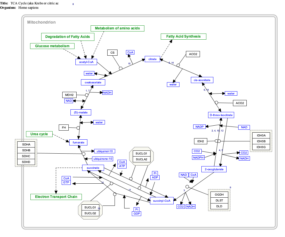
Interactive pathway map

Click on genes, proteins and metabolites below to link to respective articles. [§ 1]
[[File:
TCACycle WP78.png Go to articleGo to articleGo to articleGo to articleGo to articleGo to articleGo to articleGo to articleGo to articleGo to articleGo to articleGo to articleGo to articleGo to articleGo to articleGo to articleGo to articleGo to articleGo to articleGo to articleGo to articleGo to articleGo to articleGo to articleGo to articleGo to articleGo to articleGo to articleGo to articleGo to articleGo to articleGo to articleGo to articleGo to articleGo to articleGo to articleGo to articleGo to articleGo to articleGo to articleGo to articleGo to HMDBGo to articleGo to articleGo to articleGo to articleGo to articleGo to articleGo to articleGo to articleGo to articleGo to article
[[
]]
[[
]]
[[
]]
[[
]]
[[
]]
[[
]]
[[
]]
[[
]]
[[
]]
[[
]]
[[
]]
[[
]]
[[
]]
[[
]]
[[
]]
[[
]]
[[
]]
[[
]]
[[
]]
[[
]]
[[
]]
[[
]]
[[
]]
[[
]]
[[
]]
[[
]]
[[
]]
[[
]]
[[
]]
[[
]]
[[
]]
[[
]]
[[
]]
[[
]]
[[
]]
[[
]]
[[
]]
[[
]]
[[
]]
[[
]]
[[
]]
[[
]]
[[
]]
[[
]]
[[
]]
[[
]]
[[
]]
[[
]]
[[
]]
[[
]]
[[
]]
[[
]]
[[
]]
[[
]]
[[
]]
[[
]]
[[
]]
[[
]]
[[
]]
[[
]]
[[
]]
[[
]]
[[
]]
[[
]]
TCACycle WP78.png Go to articleGo to articleGo to articleGo to articleGo to articleGo to articleGo to articleGo to articleGo to articleGo to articleGo to articleGo to articleGo to articleGo to articleGo to articleGo to articleGo to articleGo to articleGo to articleGo to articleGo to articleGo to articleGo to articleGo to articleGo to articleGo to articleGo to articleGo to articleGo to articleGo to articleGo to articleGo to articleGo to articleGo to articleGo to articleGo to articleGo to articleGo to articleGo to articleGo to articleGo to articleGo to HMDBGo to articleGo to articleGo to articleGo to articleGo to articleGo to articleGo to articleGo to articleGo to articleGo to article
|alt=TCACycle_WP78 edit]]
TCACycle_WP78 edit
  1. The interactive pathway map can be edited at WikiPathways: "TCACycle_WP78".
Click on genes, proteins and metabolites below to link to respective articles. [§ 1]
[[File:
WP534.png go to articlego to articlego to articlego to articlego to articlego to articlego to articlego to articlego to articlego to articlego to articlego to articlego to articlego to articlego to articlego to articlego to articlego to articlego to articlego to articlego to articlego to articlego to articlego to articlego to articlego to articlego to articlego to articlego to articlego to articlego to articlego to articlego to articlego to articlego to articlego to articlego to articlego to articlego to articlego to articlego to articlego to articlego to articlego to articlego to articlego to articlego to articlego to articlego to articlego to articlego to articlego to articlego to articlego to articlego to articlego to articlego to articlego to articlego to articlego to articlego to articlego to articlego to articlego to articlego to articlego to articlego to articlego to articlego to articlego to articlego to articlego to article
[[
]]
[[
]]
[[
]]
[[
]]
[[
]]
[[
]]
[[
]]
[[
]]
[[
]]
[[
]]
[[
]]
[[
]]
[[
]]
[[
]]
[[
]]
[[
]]
[[
]]
[[
]]
[[
]]
[[
]]
[[
]]
[[
]]
[[
]]
[[
]]
[[
]]
[[
]]
[[
]]
[[
]]
[[
]]
[[
]]
[[
]]
[[
]]
[[
]]
[[
]]
[[
]]
[[
]]
[[
]]
[[
]]
[[
]]
[[
]]
[[
]]
[[
]]
[[
]]
[[
]]
[[
]]
[[
]]
[[
]]
[[
]]
[[
]]
[[
]]
[[
]]
[[
]]
[[
]]
[[
]]
[[
]]
[[
]]
[[
]]
[[
]]
[[
]]
[[
]]
[[
]]
[[
]]
[[
]]
[[
]]
[[
]]
[[
]]
[[
]]
[[
]]
[[
]]
[[
]]
[[
]]
[[
]]
[[
]]
[[
]]
[[
]]
WP534.png go to articlego to articlego to articlego to articlego to articlego to articlego to articlego to articlego to articlego to articlego to articlego to articlego to articlego to articlego to articlego to articlego to articlego to articlego to articlego to articlego to articlego to articlego to articlego to articlego to articlego to articlego to articlego to articlego to articlego to articlego to articlego to articlego to articlego to articlego to articlego to articlego to articlego to articlego to articlego to articlego to articlego to articlego to articlego to articlego to articlego to articlego to articlego to articlego to articlego to articlego to articlego to articlego to articlego to articlego to articlego to articlego to articlego to articlego to articlego to articlego to articlego to articlego to articlego to articlego to articlego to articlego to articlego to articlego to articlego to articlego to articlego to article
|alt=Glycolysis and Gluconeogenesis edit]]
Glycolysis and Gluconeogenesis edit
  1. The interactive pathway map can be edited at WikiPathways: "GlycolysisGluconeogenesis_WP534".

See also

Related Research Articles

<span class="mw-page-title-main">Citric acid cycle</span> Chemical reactions to release energy in cells

The citric acid cycle (CAC)—also known as the Krebs cycle, Szent-Györgyi-Krebs cycle or the TCA cycle (tricarboxylic acid cycle)—is a series of chemical reactions to release stored energy through the oxidation of acetyl-CoA derived from carbohydrates, fats, and proteins. The Krebs cycle is used by organisms that respire (as opposed to organisms that ferment) to generate energy, either by anaerobic respiration or aerobic respiration. In addition, the cycle provides precursors of certain amino acids, as well as the reducing agent NADH, that are used in numerous other reactions. Its central importance to many biochemical pathways suggests that it was one of the earliest components of metabolism. Even though it is branded as a 'cycle', it is not necessary for metabolites to follow only one specific route; at least three alternative segments of the citric acid cycle have been recognized.

<span class="mw-page-title-main">Pyruvate dehydrogenase complex</span>

Pyruvate dehydrogenase complex (PDC) is a complex of three enzymes that converts pyruvate into acetyl-CoA by a process called pyruvate decarboxylation. Acetyl-CoA may then be used in the citric acid cycle to carry out cellular respiration, and this complex links the glycolysis metabolic pathway to the citric acid cycle. Pyruvate decarboxylation is also known as the "pyruvate dehydrogenase reaction" because it also involves the oxidation of pyruvate.

<span class="mw-page-title-main">Glutamate dehydrogenase</span> Hexameric enzyme

Glutamate dehydrogenase is an enzyme observed in both prokaryotes and eukaryotic mitochondria. The aforementioned reaction also yields ammonia, which in eukaryotes is canonically processed as a substrate in the urea cycle. Typically, the α-ketoglutarate to glutamate reaction does not occur in mammals, as glutamate dehydrogenase equilibrium favours the production of ammonia and α-ketoglutarate. Glutamate dehydrogenase also has a very low affinity for ammonia, and therefore toxic levels of ammonia would have to be present in the body for the reverse reaction to proceed. However, in brain, the NAD+/NADH ratio in brain mitochondria encourages oxidative deamination. In bacteria, the ammonia is assimilated to amino acids via glutamate and aminotransferases. In plants, the enzyme can work in either direction depending on environment and stress. Transgenic plants expressing microbial GLDHs are improved in tolerance to herbicide, water deficit, and pathogen infections. They are more nutritionally valuable.

<span class="mw-page-title-main">Malate dehydrogenase</span> Class of enzymes

Malate dehydrogenase (EC 1.1.1.37) (MDH) is an enzyme that reversibly catalyzes the oxidation of malate to oxaloacetate using the reduction of NAD+ to NADH. This reaction is part of many metabolic pathways, including the citric acid cycle. Other malate dehydrogenases, which have other EC numbers and catalyze other reactions oxidizing malate, have qualified names like malate dehydrogenase (NADP+).

<span class="mw-page-title-main">Isocitrate dehydrogenase</span> Class of enzymes

Isocitrate dehydrogenase (IDH) (EC 1.1.1.42) and (EC 1.1.1.41) is an enzyme that catalyzes the oxidative decarboxylation of isocitrate, producing alpha-ketoglutarate (α-ketoglutarate) and CO2. This is a two-step process, which involves oxidation of isocitrate (a secondary alcohol) to oxalosuccinate (a ketone), followed by the decarboxylation of the carboxyl group beta to the ketone, forming alpha-ketoglutarate. In humans, IDH exists in three isoforms: IDH3 catalyzes the third step of the citric acid cycle while converting NAD+ to NADH in the mitochondria. The isoforms IDH1 and IDH2 catalyze the same reaction outside the context of the citric acid cycle and use NADP+ as a cofactor instead of NAD+. They localize to the cytosol as well as the mitochondrion and peroxisome.

<span class="mw-page-title-main">Mitochondrial matrix</span> Space within the inner membrane of the mitochondrion

In the mitochondrion, the matrix is the space within the inner membrane. The word "matrix" stems from the fact that this space is viscous, compared to the relatively aqueous cytoplasm. The mitochondrial matrix contains the mitochondrial DNA, ribosomes, soluble enzymes, small organic molecules, nucleotide cofactors, and inorganic ions.[1] The enzymes in the matrix facilitate reactions responsible for the production of ATP, such as the citric acid cycle, oxidative phosphorylation, oxidation of pyruvate, and the beta oxidation of fatty acids.

The oxoglutarate dehydrogenase complex (OGDC) or α-ketoglutarate dehydrogenase complex is an enzyme complex, most commonly known for its role in the citric acid cycle.

<span class="mw-page-title-main">Pyruvate carboxylase</span>

Pyruvate carboxylase (PC) encoded by the gene PC is an enzyme of the ligase class that catalyzes the physiologically irreversible carboxylation of pyruvate to form oxaloacetate (OAA).

<span class="mw-page-title-main">Glutamate dehydrogenase 1</span> Enzyme

GLUD1 is a mitochondrial matrix enzyme, one of the family of glutamate dehydrogenases that are ubiquitous in life, with a key role in nitrogen and glutamate (Glu) metabolism and energy homeostasis. This dehydrogenase is expressed at high levels in liver, brain, pancreas and kidney, but not in muscle. In the pancreatic cells, GLUD1 is thought to be involved in insulin secretion mechanisms. In nervous tissue, where glutamate is present in concentrations higher than in the other tissues, GLUD1 appears to function in both the synthesis and the catabolism of glutamate and perhaps in ammonia detoxification.

The branched-chain α-ketoacid dehydrogenase complex is a multi-subunit complex of enzymes that is found on the mitochondrial inner membrane. This enzyme complex catalyzes the oxidative decarboxylation of branched, short-chain alpha-ketoacids. BCKDC is a member of the mitochondrial α-ketoacid dehydrogenase complex family comprising pyruvate dehydrogenase and alpha-ketoglutarate dehydrogenase, key enzymes that function in the Krebs cycle.

<span class="mw-page-title-main">Malate-aspartate shuttle</span> Biochemical system for transporting electrons produced during glycolysis

The malate-aspartate shuttle is a biochemical system for translocating electrons produced during glycolysis across the semipermeable inner membrane of the mitochondrion for oxidative phosphorylation in eukaryotes. These electrons enter the electron transport chain of the mitochondria via reduction equivalents to generate ATP. The shuttle system is required because the mitochondrial inner membrane is impermeable to NADH, the primary reducing equivalent of the electron transport chain. To circumvent this, malate carries the reducing equivalents across the membrane.

<span class="mw-page-title-main">Amino acid synthesis</span> The set of biochemical processes by which amino acids are produced

Amino acid synthesis is the set of biochemical processes by which the amino acids are produced. The substrates for these processes are various compounds in the organism's diet or growth media. Not all organisms are able to synthesize all amino acids. For example, humans can synthesize 11 of the 20 standard amino acids. These 11 are called the non-essential amino acids).

The mitochondrial shuttles are biochemical transport systems used to transport reducing agents across the inner mitochondrial membrane. NADH as well as NAD+ cannot cross the membrane, but it can reduce another molecule like FAD and [QH2] that can cross the membrane, so that its electrons can reach the electron transport chain.

<span class="mw-page-title-main">GOT2</span> Mitochondrial enzyme involved in amino acid metabolism

Aspartate aminotransferase, mitochondrial is an enzyme that in humans is encoded by the GOT2 gene. Glutamic-oxaloacetic transaminase is a pyridoxal phosphate-dependent enzyme which exists in cytoplasmic and inner-membrane mitochondrial forms, GOT1 and GOT2, respectively. GOT plays a role in amino acid metabolism and the urea and Kreb's cycle. Also, GOT2 is a major participant in the malate-aspartate shuttle, which is a passage from the cytosol to the mitochondria. The two enzymes are homodimeric and show close homology. GOT2 has been seen to have a role in cell proliferation, especially in terms of tumor growth.

<span class="mw-page-title-main">IDH3A</span> Protein-coding gene in the species Homo sapiens

Isocitrate dehydrogenase [NAD] subunit alpha, mitochondrial (IDH3α) is an enzyme that in humans is encoded by the IDH3A gene.

<span class="mw-page-title-main">IDH3G</span> Protein-coding gene in the species Homo sapiens

Isocitrate dehydrogenase [NAD] subunit gamma, mitochondrial is an enzyme that in humans is encoded by the IDH3G gene.

Glutaminolysis (glutamine + -lysis) is a series of biochemical reactions by which the amino acid glutamine is lysed to glutamate, aspartate, CO2, pyruvate, lactate, alanine and citrate.

<span class="mw-page-title-main">IDH3B</span> Protein-coding gene in the species Homo sapiens

Isocitrate dehydrogenase [NAD] subunit beta, mitochondrial is an enzyme that in humans is encoded by the IDH3B gene.

<span class="mw-page-title-main">MDH1</span> Protein-coding gene in the species Homo sapiens

Malate dehydrogenase, cytoplasmic also known as malate dehydrogenase 1 is an enzyme that in humans is encoded by the MDH1 gene.

The citrate-malate shuttle is a series of chemical reactions – commonly referred to as a biochemical cycle or system – that transports acetyl-CoA in the mitochondrial matrix across the inner and outer mitochondrial membrane for fatty acid synthesis. Mitochondria, also known as the powerhouse of a cell, is enclosed in a double membrane. As the inner mitochondrial membrane is impermeable to acetyl-CoA, the shuttle system is essential to fatty acid synthesis in the cytosol. It plays an important role in the generation of lipids in the liver.

References

  1. 1 2 3 GRCh38: Ensembl release 89: ENSG00000146701 - Ensembl, May 2017
  2. 1 2 3 GRCm38: Ensembl release 89: ENSMUSG00000019179 - Ensembl, May 2017
  3. "Human PubMed Reference:". National Center for Biotechnology Information, U.S. National Library of Medicine.
  4. "Mouse PubMed Reference:". National Center for Biotechnology Information, U.S. National Library of Medicine.
  5. Habets GG, van der Kammen RA, Willemsen V, Balemans M, Wiegant J, Collard JG (1992). "Sublocalization of an invasion-inducing locus and other genes on human chromosome 7". Cytogenetics and Cell Genetics. 60 (3–4): 200–5. doi:10.1159/000133336. PMID   1505215.
  6. "Entrez Gene: MDH2 malate dehydrogenase 2, NAD (mitochondrial)".
  7. Hall MD, Levitt DG, Banaszak LJ (Aug 1992). "Crystal structure of Escherichia coli malate dehydrogenase. A complex of the apoenzyme and citrate at 1.87 A resolution". Journal of Molecular Biology. 226 (3): 867–82. doi:10.1016/0022-2836(92)90637-Y. PMID   1507230.
  8. Breiter DR, Resnik E, Banaszak LJ (Nov 1994). "Engineering the quaternary structure of an enzyme: construction and analysis of a monomeric form of malate dehydrogenase from Escherichia coli". Protein Science. 3 (11): 2023–32. doi:10.1002/pro.5560031115. PMC   2142640 . PMID   7703849.
  9. Mullinax TR, Mock JN, McEvily AJ, Harrison JH (Nov 1982). "Regulation of mitochondrial malate dehydrogenase. Evidence for an allosteric citrate-binding site". The Journal of Biological Chemistry. 257 (22): 13233–9. doi: 10.1016/S0021-9258(18)33435-5 . PMID   7142142.
  10. Gelpí JL, Dordal A, Montserrat J, Mazo A, Cortés A (Apr 1992). "Kinetic studies of the regulation of mitochondrial malate dehydrogenase by citrate". The Biochemical Journal. 283 (1): 289–97. doi:10.1042/bj2830289. PMC   1131027 . PMID   1567375.
  11. Mullinax TR, Mock JN, McEvily AJ, Harrison JH (Nov 1982). "Regulation of mitochondrial malate dehydrogenase. Evidence for an allosteric citrate-binding site". The Journal of Biological Chemistry. 257 (22): 13233–9. doi: 10.1016/S0021-9258(18)33435-5 . PMID   7142142.
  12. Harada K, Wolfe RG (Aug 1968). "Malic dehydrogenase. VII. The catalytic mechanism and possible role of identical protein subunits". The Journal of Biological Chemistry. 243 (15): 4131–7. doi: 10.1016/S0021-9258(18)93289-8 . PMID   4299102.
  13. Fahien LA, Kmiotek EH, MacDonald MJ, Fibich B, Mandic M (Aug 1988). "Regulation of malate dehydrogenase activity by glutamate, citrate, alpha-ketoglutarate, and multienzyme interaction". The Journal of Biological Chemistry. 263 (22): 10687–97. doi: 10.1016/S0021-9258(18)38026-8 . PMID   2899080.
  14. 1 2 Lo, YW; Lin, ST; Chang, SJ; Chan, CH; Lyu, KW; Chang, JF; May, EW; Lin, DY; Chou, HC; Chan, HL (April 2015). "Mitochondrial proteomics with siRNA knockdown to reveal ACAT1 and MDH2 in the development of doxorubicin-resistant uterine cancer". Journal of Cellular and Molecular Medicine. 19 (4): 744–59. doi:10.1111/jcmm.12388. PMC   4395189 . PMID   25639359.
  15. Cascón, A; Comino-Méndez, I; Currás-Freixes, M; de Cubas, AA; Contreras, L; Richter, S; Peitzsch, M; Mancikova, V; Inglada-Pérez, L; Pérez-Barrios, A; Calatayud, M; Azriel, S; Villar-Vicente, R; Aller, J; Setién, F; Moran, S; Garcia, JF; Río-Machín, A; Letón, R; Gómez-Graña, Á; Apellániz-Ruiz, M; Roncador, G; Esteller, M; Rodríguez-Antona, C; Satrústegui, J; Eisenhofer, G; Urioste, M; Robledo, M (11 March 2015). "Whole-exome sequencing identifies MDH2 as a new familial paraganglioma gene". Journal of the National Cancer Institute. 107 (5). doi: 10.1093/jnci/djv053 . PMID   25766404.