Related Research Articles

Magnetic resonance imaging (MRI) is a medical imaging technique used in radiology to form pictures of the anatomy and the physiological processes inside the body. MRI scanners use strong magnetic fields, magnetic field gradients, and radio waves to generate images of the organs in the body. MRI does not involve X-rays or the use of ionizing radiation, which distinguishes it from computed tomography (CT) and positron emission tomography (PET) scans. MRI is a medical application of nuclear magnetic resonance (NMR) which can also be used for imaging in other NMR applications, such as NMR spectroscopy.

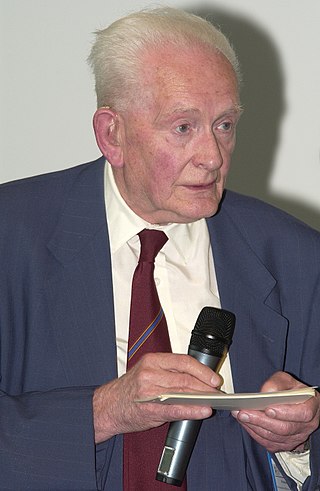
Sir Peter Mansfield was an English physicist who was awarded the 2003 Nobel Prize in Physiology or Medicine, shared with Paul Lauterbur, for discoveries concerning Magnetic Resonance Imaging (MRI). Mansfield was a professor at the University of Nottingham.

Neuroimaging is the use of quantitative (computational) techniques to study the structure and function of the central nervous system, developed as an objective way of scientifically studying the healthy human brain in a non-invasive manner. Increasingly it is also being used for quantitative research studies of brain disease and psychiatric illness. Neuroimaging is highly multidisciplinary involving neuroscience, computer science, psychology and statistics, and is not a medical specialty. Neuroimaging is sometimes confused with neuroradiology.

Magnetic resonance microscopy is magnetic resonance imaging (MRI) at a microscopic level down to the scale of microns. The first definition of MRM was MRI having voxel resolutions of better than 100 μm.
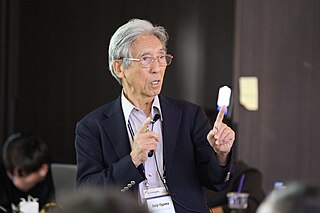
Seiji Ogawa is a Japanese biophysicist and neuroscientist known for discovering the technique that underlies Functional Magnetic Resonance Imaging (fMRI). He is regarded as the father of modern functional brain imaging. He determined that the changes in blood oxygen levels cause its magnetic resonance imaging properties to change, allowing a map of blood, and hence, functional, activity in the brain to be created. This map reflected which neurons of the brain responded with electrochemical signals to mental processes. He was the first scientist who demonstrated that the functional brain imaging is dependent on the oxygenation status of the blood, the BOLD effect. The technique was therefore called blood oxygenation level-dependent or BOLD contrast. Functional MRI (fMRI) has been used to map the visual, auditory, and sensory regions and moving toward higher brain functions such as cognitive functions in the brain.
John Rowland Mallard OBE FRSE FREng was an English physicist and professor of Medical Physics at the University of Aberdeen from 1965 until his retirement in 1992. He was known for setting up and leading the team that developed the first magnetic resonance imaging (MRI) full body scanner and, in particular, positron emission tomography (PET). He was born in Kingsthorpe, Northampton, England.
During nuclear magnetic resonance observations, spin–lattice relaxation is the mechanism by which the longitudinal component of the total nuclear magnetic moment vector (parallel to the constant magnetic field) exponentially relaxes from a higher energy, non-equilibrium state to thermodynamic equilibrium with its surroundings (the "lattice"). It is characterized by the spin–lattice relaxation time, a time constant known as T1.
MRI contrast agents are contrast agents used to improve the visibility of internal body structures in magnetic resonance imaging (MRI). The most commonly used compounds for contrast enhancement are gadolinium-based contrast agents (GBCAs). Such MRI contrast agents shorten the relaxation times of nuclei within body tissues following oral or intravenous administration.

Cardiac magnetic resonance imaging, also known as cardiovascular MRI, is a magnetic resonance imaging (MRI) technology used for non-invasive assessment of the function and structure of the cardiovascular system. Conditions in which it is performed include congenital heart disease, cardiomyopathies and valvular heart disease, diseases of the aorta such as dissection, aneurysm and coarctation, coronary heart disease. It can also be used to look at pulmonary veins.
The Libin Cardiovascular Institute is an entity of Alberta Health Services and the University of Calgary. It connects all cardiovascular research, education and patient care in Southern Alberta, serving a population of about two million. Its more than 1,500 members include physicians, clinicians and other health professionals, researchers and trainees.

Magnetic resonance imaging (MRI) is a medical imaging technique mostly used in radiology and nuclear medicine in order to investigate the anatomy and physiology of the body, and to detect pathologies including tumors, inflammation, neurological conditions such as stroke, disorders of muscles and joints, and abnormalities in the heart and blood vessels among other things. Contrast agents may be injected intravenously or into a joint to enhance the image and facilitate diagnosis. Unlike CT and X-ray, MRI uses no ionizing radiation and is, therefore, a safe procedure suitable for diagnosis in children and repeated runs. Patients with specific non-ferromagnetic metal implants, cochlear implants, and cardiac pacemakers nowadays may also have an MRI in spite of effects of the strong magnetic fields. This does not apply on older devices, and details for medical professionals are provided by the device's manufacturer.
Robert Turner is a British neuroscientist, physicist, and social anthropologist. He has been a director and professor at the Max Planck Institute for Human Cognitive and Brain Sciences in Leipzig, Germany, and is an internationally recognized expert in brain physics and magnetic resonance imaging (MRI). Coils inside every MRI scanner owe their shape to his ideas.

Jürgen Klaus Hennig is a German chemist and medical physicist. Internationally he is considered to be one of the pioneers of Magnetic Resonance Imaging for clinical diagnostics. He is the Scientific Director of the Department of Diagnostic Radiology and Chairman of the Magnetic Resonance Development and Application Center (MRDAC) at the University Medical Center Freiburg. In the year 2003 he was awarded the Max Planck Research Award in the category of Biosciences and Medicine.

Val Murray Runge is an American and Swiss professor of radiology and the editor-in-chief of Investigative Radiology. Runge was one of the early researchers to investigate the use of gadolinium-based contrast agents for magnetic resonance imaging (MRI), giving the first presentation in this field, followed two years later by the first presentation of efficacy. His research also pioneered many early innovations in MRI, including the use of tilted planes and respiratory gating. His publication on multiple sclerosis in 1984 represented the third and largest clinical series investigating the role of MRI in this disease, and the first to show characteristic abnormalities on MRI in patients whose CT was negative.
Hyperpolarized carbon-13 MRI is a functional medical imaging technique for probing perfusion and metabolism using injected substrates.
Synthetic MRI is a simulation method in Magnetic Resonance Imaging (MRI), for generating contrast weighted images based on measurement of tissue properties. The synthetic (simulated) images are generated after an MR study, from parametric maps of tissue properties. It is thereby possible to generate several contrast weightings from the same acquisition. This is different from conventional MRI, where the signal acquired from the tissue is used to generate an image directly, often generating only one contrast weighting per acquisition. The synthetic images are similar in appearance to those normally acquired with an MRI scanner.

The history of magnetic resonance imaging (MRI) includes the work of many researchers who contributed to the discovery of nuclear magnetic resonance (NMR) and described the underlying physics of magnetic resonance imaging, starting early in the twentieth century. One researcher was American physicist Isidor Isaac Rabi who won the Nobel Prize in Physics in 1944 for his discovery of nuclear magnetic resonance, which is used in magnetic resonance imaging. MR imaging was invented by Paul C. Lauterbur who developed a mechanism to encode spatial information into an NMR signal using magnetic field gradients in September 1971; he published the theory behind it in March 1973.

An MRI pulse sequence in magnetic resonance imaging (MRI) is a particular setting of pulse sequences and pulsed field gradients, resulting in a particular image appearance.
Lindsay Cahill is a Canadian chemist who uses Magnetic Resonance Imaging to study metabolic abnormalities in pregnancy. She has published more than 70 articles on her research related to nuclear magnetic resonance in studying electrochemical materials and for imaging animal fetuses and placenta. She has published widely-used protocols for the imaging of mouse brains.
Brian Worthington was the first radiologist to be elected a Fellow of the Royal Society and is acknowledged as a pioneer in clinical magnetic resonance imaging. He was born in Oldham, England and was educated at Hulme Grammar School, training at Guy's Hospital after graduating in physiology and medicine. After graduation his career developed rapidly, particularly in the field of MRI research and he was subsequently admitted as a Fellow of the Royal College of Radiologists.
References
- 1 2 3 4 Children, The Hospital for Sick. "Henkelman to receive 2010 Killam Prize in health sciences". www.sickkids.ca. Retrieved 1 January 2020.
- 1 2 3 4 Bronskill, Michael (2 April 2001). "Mark Henkelman awarded Canada Research Chair". Canadian Medical Physics Newsletter. Retrieved 1 January 2020.
- ↑ "Cancer treatment promising, if you're a mouse" . Retrieved 1 January 2020.
- 1 2 3 "U of T faculty, alumni and other members of university community named to Order of Canada". University of Toronto News. Retrieved 1 January 2020.
- ↑ TAYLOR, SUZANNE. "THE MOUSE HOUSE | Maclean's | 22 OCT. 2007". Maclean's | The Complete Archive. Retrieved 1 January 2020.
- ↑ Damadian, Raymond V (January 1997). "5606970 Multiple patient scanning on a magnetic resonance imaging apparatus". Magnetic Resonance Imaging. 15 (5): XIII. doi:10.1016/s0730-725x(97)89749-4. ISSN 0730-725X.
- ↑ "Alumni Stories | University of Lethbridge". www.uleth.ca. Retrieved 1 January 2020.
- 1 2 Association, Canadian Medical (18 May 2010). "Briefly". CMAJ. 182 (8): E344–E346. doi: 10.1503/cmaj.109-3234 . ISSN 0820-3946. PMC 2871237 .
- ↑ "Mark Henkelman – Google Scholar Citations". scholar.google.com. Retrieved 1 January 2020.
- ↑ General, Office of the Secretary to the Governor (20 December 2019). "Governor General Announces 120 New Appointments to the Order of Canada". The Governor General of Canada. Retrieved 1 January 2020.
- ↑ "Five Scholars Awarded $100,000 Killam Prize". www.everythingzoomer.com. Retrieved 1 January 2020.
| International | |
|---|---|
| Academics | |