NDH-2

Last updated
Enzymatic reaction catalyzed by NDH-2. In yellow is represented the protein surface, sitting in the membrane (in gray) NDH-2.tif
Enzymatic reaction catalyzed by NDH-2. In yellow is represented the protein surface, sitting in the membrane (in gray)

NDH-2, also known as type II NADH:quinone oxidoreductase or alternative NADH dehydrogenase, is an enzyme (EC: 1.6.99.3) which catalyzes the electron transfer from NADH (electron donor) to a quinone (electron acceptor), being part of the electron transport chain. [1] NDH-2 are peripheral membrane protein, functioning as dimers in vivo, with approximately 45 KDa per subunit and a single FAD as their cofactor. [2]

Contents

NDH-2 are the only enzymes, with NADH dehydrogenase activity, expressed in the respiratory chain of some pathogenic organisms (e.g. Staphylococcus aureus ), and for that they have been proposed as new targets for rational drug design. [3]

Structure

NDH-2 structure colored by domains Estrutura da NDH-2 colorida por dominios.png
NDH-2 structure colored by domains

The structure/fold from these proteins may be divided into three domains: first dinucleotide binding domain (green in the figure), second dinucleotide binding domain (orange in the figure) and C-terminal domain (blue in the figure).

The first domain is responsible for the noncovalent binding of FAD, while the second dinucleotide binding domain binds NADH. Both these domain are structurally organized in Rossmann folds, with the characteristic GxGxxG motif present.

The third domain, C-terminal, is responsible for the protein-membrane interaction. Upon expression of a C-terminal truncated version of NDH-2, it was observed an intracellular delocalization from the membrane to the cytoplasm. [4] The third domain, together with part of the first domain, is also partially responsible for the binding of the electron acceptor (quinone).

There are currently crystallographic structures for NDH-2 from four different organisms:

Reaction

NAD+ to NADH NAD+toNADH.png
NAD+ to NADH

The enzymatic oxidoreduction reaction catalyzed by NDH-2 may be described as follows:

NADH + Q + H+ -----> NAD+ + QH2

(Q - quinone; QH2 - quinol)

In this case, the electron donor is NADH and the electron acceptor is the quinone. Depending on the organism, the reduced quinone changes between menaquinone, ubiquinone or plastoquinone. The mechanism of the reaction may be divided in two half-reactions: 1stHR and 2ndHR.

In the 1stHR, 2 electrons and 1 proton from NADH are transferred (simultaneously with an additional proton from the bulk) to the prosthetic group (FAD), giving rise to its protonated form FADH2. In this phase, an Enzyme-Substrate complex is established, characterized by the appearance of a "Charge-transfer complex". Na 2stHR, the quinone binds and the 2 electrons and one of the FAD protons are transferred for this second substrate (again, with an additional proton from the bulk), forming the product quinol.

It is now accepted that the overall mechanism occurs by a ternary complex (simultaneous binding of both substrates to the enzyme), [9] instead of the previously proposed ping-pong mechanism.

Phylogenetic distribution

The presence of NDH-2 in organisms which genome as already been fully sequenced was studied by Bioinformatics. [10] In this study, NDH-2 were identified in 83% of Eukaryotes, 60% of bacteria and 32% of Archaeas. It was also observed the absence of NDH-2 in phyla composed of anaerobic organisms.

Despite being considered absent (hence being considered as drug targets), in this same study, the presence of a gene coding for a NDH-2 homolog was observed in the human genome.

Related Research Articles

Oxidative phosphorylation The phosphorylation of ADP to ATP that accompanies the oxidation of a metabolite through the operation of the respiratory chain. Oxidation of compounds establishes a proton gradient across the membrane, providing the energy for ATP synthesis.

Oxidative phosphorylation or electron transport-linked phosphorylation or terminal oxidation is the metabolic pathway in which cells use enzymes to oxidize nutrients, thereby releasing chemical energy in order to produce adenosine triphosphate (ATP). In eukaryotes, this takes place inside mitochondria. Almost all aerobic organisms carry out oxidative phosphorylation. This pathway is so pervasive because it releases more energy than alternative fermentation processes such as anaerobic glycolysis.

Electron transport chain Cellular electron transfer

An electron transport chain (ETC) is a series of protein complexes and other molecules that transfer electrons from electron donors to electron acceptors via redox reactions (both reduction and oxidation occurring simultaneously) and couples this electron transfer with the transfer of protons (H+ ions) across a membrane. Many of the enzymes in the electron transport chain are membrane-bound.

Respiratory complex I

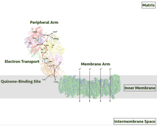
Respiratory complex I, EC 7.1.1.2 is the first large protein complex of the respiratory chains of many organisms from bacteria to humans. It catalyzes the transfer of electrons from NADH to coenzyme Q10 (CoQ10) and translocates protons across the inner mitochondrial membrane in eukaryotes or the plasma membrane of bacteria.

Coenzyme Q – cytochrome c reductase Class of enzymes

The coenzyme Q : cytochrome c – oxidoreductase, sometimes called the cytochrome bc1 complex, and at other times complex III, is the third complex in the electron transport chain, playing a critical role in biochemical generation of ATP. Complex III is a multisubunit transmembrane protein encoded by both the mitochondrial and the nuclear genomes. Complex III is present in the mitochondria of all animals and all aerobic eukaryotes and the inner membranes of most eubacteria. Mutations in Complex III cause exercise intolerance as well as multisystem disorders. The bc1 complex contains 11 subunits, 3 respiratory subunits, 2 core proteins and 6 low-molecular weight proteins.

Succinate dehydrogenase

Succinate dehydrogenase (SDH) or succinate-coenzyme Q reductase (SQR) or respiratory complex II is an enzyme complex, found in many bacterial cells and in the inner mitochondrial membrane of eukaryotes. It is the only enzyme that participates in both the citric acid cycle and the electron transport chain. Histochemical analysis showing high succinate dehydrogenase in muscle demonstrates high mitochondrial content and high oxidative potential.

Flavin adenine dinucleotide Redox-active coenzyme

In biochemistry, flavin adenine dinucleotide (FAD) is a redox-active coenzyme associated with various proteins, which is involved with several enzymatic reactions in metabolism. A flavoprotein is a protein that contains a flavin group, which may be in the form of FAD or flavin mononucleotide (FMN). Many flavoproteins are known: components of the succinate dehydrogenase complex, α-ketoglutarate dehydrogenase, and a component of the pyruvate dehydrogenase complex.

Glycerol-3-phosphate dehydrogenase Class of enzymes

Glycerol-3-phosphate dehydrogenase (GPDH) is an enzyme that catalyzes the reversible redox conversion of dihydroxyacetone phosphate to sn-glycerol 3-phosphate.

Electron-transferring-flavoprotein dehydrogenase

Electron-transferring-flavoprotein dehydrogenase is an enzyme that transfers electrons from electron-transferring flavoprotein in the mitochondrial matrix, to the ubiquinone pool in the inner mitochondrial membrane. It is part of the electron transport chain. The enzyme is found in both prokaryotes and eukaryotes and contains a flavin and FE-S cluster. In humans, it is encoded by the ETFDH gene. Deficiency in ETF dehydrogenase causes the human genetic disease multiple acyl-CoA dehydrogenase deficiency.

NADH dehydrogenase (quinone)

In enzymology, a NADH dehydrogenase (quinone) (EC 1.6.5.11) is an enzyme that catalyzes the chemical reaction

NAD(P)H dehydrogenase (quinone)

In enzymology, a NAD(P)H dehydrogenase (quinone) (EC 1.6.5.2) is an enzyme that catalyzes the chemical reaction

In enzymology, a NADPH dehydrogenase (quinone) (EC 1.6.5.10) is an enzyme that catalyzes the chemical reaction

NDUFB6 Protein-coding gene in the species Homo sapiens

NADH dehydrogenase [ubiquinone] 1 beta subcomplex subunit 6, also known as complex I-B17, is a protein that in humans is encoded by the NDUFB6 gene. NADH dehydrogenase (ubiquinone) 1 beta subcomplex subunit 6, is an accessory subunit of the NADH dehydrogenase (ubiquinone) complex, located in the mitochondrial inner membrane. It is also known as Complex I and is the largest of the five complexes of the electron transport chain.

FOXRED1

FAD-dependent oxidoreductase domain-containing protein 1 (FOXRED1), also known as H17, or FP634 is an enzyme that in humans is encoded by the FOXRED1 gene. FOXRED1 is an oxidoreductase and complex I-specific molecular chaperone involved in the assembly and stabilization of NADH dehydrogenase (ubiquinone) also known as complex I, which is located in the mitochondrial inner membrane and is the largest of the five complexes of the electron transport chain. Mutations in FOXRED1 have been associated with Leigh syndrome and infantile-onset mitochondrial encephalopathy.

NDUFB1

NADH dehydrogenase [ubiquinone] 1 beta subcomplex subunit 1 is an enzyme that in humans is encoded by the NDUFB1 gene. NADH dehydrogenase (ubiquinone) 1 beta subcomplex, 1, 7kDa is an accessory subunit of the NADH dehydrogenase (ubiquinone) complex, located in the mitochondrial inner membrane. It is also known as Complex I and is the largest of the five complexes of the electron transport chain.

NDUFB7 Protein-coding gene in the species Homo sapiens

NADH dehydrogenase [ubiquinone] 1 beta subcomplex subunit 7, also known as complex I-B18, is an enzyme that in humans is encoded by the NDUFB7 gene. NADH dehydrogenase (ubiquinone) 1 beta subcomplex subunit 7 is an accessory subunit of the NADH dehydrogenase (ubiquinone) complex, located in the mitochondrial inner membrane. It is also known as Complex I and is the largest of the five complexes of the electron transport chain.

NDUFB10

NADH dehydrogenase [ubiquinone] 1 beta subcomplex subunit 10 is an enzyme that in humans is encoded by the NDUFB10 gene. NADH dehydrogenase (ubiquinone) 1 beta subcomplex subunit 10 is an accessory subunit of the NADH dehydrogenase (ubiquinone) complex, located in the mitochondrial inner membrane. It is also known as Complex I and is the largest of the five complexes of the electron transport chain.

Dihydroorotate dehydrogenase (quinone)

Class 2 dihydroorotate dehydrogenases is an enzyme with systematic name (S)-dihydroorotate:quinone oxidoreductase. This enzyme catalyses the electron transfer from dihydroorotate to a quinone :

Fumarate reductase (quinol)

Fumarate reductase (quinol) is an enzyme with systematic name succinate:quinone oxidoreductase. This enzyme catalyzes the following chemical reaction:

The Monovalent Cation (K+ or Na+):Proton Antiporter-3 (CPA3) Family (TC# 2.A.63) is a member of the Na+ transporting Mrp superfamily. The CPA3 family consists of bacterial multicomponent K+:H+ and Na+:H+ antiporters. The best characterized systems are the PhaABCDEFG system of Sinorhizobium meliloti (TC# 2.A.63.1.1) that functions in pH adaptation and as a K+ efflux system, and the MnhABCDEFG system of Staphylococcus aureus (TC# 2.A.63.1.3) that functions as a Na+ efflux Na+:H+ antiporter.

The H+-translocating F420H2 Dehydrogenase (F420H2DH) Family(TC# 3.D.9) is a member of the Na+ transporting Mrp superfamily. A single F420H2 dehydrogenase (also referred to as F420H2:quinol oxidoreductase) from the methanogenic archaeon, Methanosarcina mazei Gö1, has been shown to be a redox driven proton pump. The F420H2DH of M. mazei has a molecular size of about 120 kDa and contains Fe-S clusters and FAD. A similar five-subunit enzyme has been isolated from Methanolobus tindarius. The sulfate-reducing Archaeoglobus fulgidus (and several other archaea) also have this enzyme.

References

  1. B.C. Marreiros, F. Calisto, P.J. Castro, A.M. Duarte, F. V. Sena, A.F. Silva, F.M. Sousa, M. Teixeira, P.N. Refojo, M.M. Pereira, Exploring membrane respiratory chains, Biochim. Biophys. Acta - Bioenerg. 1857 (2016) 1039–1067.
  2. Kerscher, S., Dröse, S., Zickermann, V., and Brandt, U. (2007) The Three Families of Respiratory NADH Dehydrogenases, in Bioenergetics (Schäfer, G., and Penefsky, H. S., Eds.), pp 185-222, Springer Berlin Heidelberg.
  3. F. V. Sena, A.P. Batista, T. Catarino, J.A. Brito, M. Archer, M. Viertler, T. Madl, E.J. Cabrita, M.M. Pereira, Type-II NADH: Quinone oxidoreductase from Staphylococcus aureus has two distinct binding sites and is rate limited by quinone reduction, Mol. Microbiol. 98 (2015) 272–288. doi:10.1111/mmi.13120.
  4. Y. Feng, W. Li, J. Li, J. Wang, J. Ge, D. Xu, Y. Liu, K. Wu, Q. Zeng, J.-W. Wu, C. Tian, B. Zhou, M. Yang, Structural insight into the type-II mitochondrial NADH dehydrogenases., Nature. 491 (2012) 478–82. doi:10.1038/nature11541
  5. Sousa, Filipe M.; Sena, Filipa V.; Batista, Ana P.; Athayde, Diogo; Brito, José A.; Archer, Margarida; Oliveira, A. Sofia F.; Soares, Cláudio M.; Catarino, Teresa (October 2017). "The key role of glutamate 172 in the mechanism of type II NADH:quinone oxidoreductase of Staphylococcus aureus". Biochimica et Biophysica Acta (BBA) - Bioenergetics. 1858 (10): 823–832. doi: 10.1016/j.bbabio.2017.08.002 . ISSN   0005-2728. PMID   28801048.
  6. Y. Feng, W. Li, J. Li, J. Wang, J. Ge, D. Xu, Y. Liu, K. Wu, Q. Zeng, J.-W. Wu, C. Tian, B. Zhou, M. Yang, Structural insight into the type-II mitochondrial NADH dehydrogenases., Nature. 491 (2012) 478–82. doi:10.1038/nature11541
  7. A. Heikal, Y. Nakatani, E. Dunn, M.R. Weimar, C.L. Day, E.N. Baker, J.S. Lott, L.A. Sazanov, G.M. Cook, Structure of the bacterial type II NADH dehydrogenase: a monotopic membrane protein with an essential role in energy generation, Mol Microbiol. 91 (2014) 950–964. doi:10.1111/mmi.12507.
  8. Yang, Yiqing; Yu, You; Li, Xiaolu; Li, Jing; Wu, Yue; Yu, Jie; Ge, Jingpeng; Huang, Zhenghui; Jiang, Lubin (2017-02-22). "Target Elucidation by Cocrystal Structures of NADH-Ubiquinone Oxidoreductase of Plasmodium falciparum (PfNDH2) with Small Molecule To Eliminate Drug-Resistant Malaria". Journal of Medicinal Chemistry. 60 (5): 1994–2005. doi:10.1021/acs.jmedchem.6b01733. ISSN   0022-2623. PMID   28195463.
  9. Sena, Filipa V.; Batista, Ana P.; Catarino, Teresa; Brito, José A.; Archer, Margarida; Viertler, Martin; Madl, Tobias; Cabrita, Eurico J.; Pereira, Manuela M. (2015-07-30). "Type-II NADH:quinone oxidoreductase from Staphylococcus aureushas two distinct binding sites and is rate limited by quinone reduction". Molecular Microbiology. 98 (2): 272–288. doi: 10.1111/mmi.13120 . ISSN   0950-382X. PMID   26172206.
  10. B.C. Marreiros, F. V Sena, F.M. Sousa, A.P. Batista, M.M. Pereira, Type II NADH:Quinone oxidoreductase family: Phylogenetic distribution, Structural diversity and Evolutionary divergences., Environ. Microbiol. 0 (2016). doi:10.1111/1462-2920.13352.