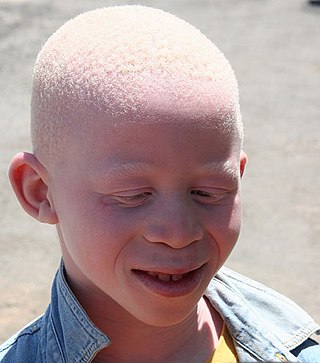
Albinism is a congenital condition characterized in humans by the partial or complete absence of pigment in the skin, hair and eyes. Albinism is associated with a number of vision defects, such as photophobia, nystagmus, and amblyopia. Lack of skin pigmentation makes for more susceptibility to sunburn and skin cancers. In rare cases such as Chédiak–Higashi syndrome, albinism may be associated with deficiencies in the transportation of melanin granules. This also affects essential granules present in immune cells leading to increased susceptibility to infection.

Human skin color ranges from the darkest brown to the lightest hues. Differences in skin color among individuals is caused by variation in pigmentation, which is the result of genetics, exposure to the sun, natural and sexual selection, or all of these. Differences across populations evolved through natural or sexual selection, because of social norms and differences in environment, as well as regulations of the biochemical effects of ultraviolet radiation penetrating the skin.

Melanin is a broad term for a group of natural pigments found in most organisms. Eumelanin is produced through a multistage chemical process known as melanogenesis, where the oxidation of the amino acid tyrosine is followed by polymerization. The melanin pigments are produced in a specialized group of cells known as melanocytes. Functionally, eumelanin serves as protection against UV radiation.

Melanocytes are melanin-producing neural crest-derived cells located in the bottom layer of the skin's epidermis, the middle layer of the eye, the inner ear, vaginal epithelium, meninges, bones, and heart. Melanin is a dark pigment primarily responsible for skin color. Once synthesized, melanin is contained in special organelles called melanosomes which can be transported to nearby keratinocytes to induce pigmentation. Thus darker skin tones have more melanosomes present than lighter skin tones. Functionally, melanin serves as protection against UV radiation. Melanocytes also have a role in the immune system.

Tiger eye or goat eye is a gene causing diluted eye color in horses. There are two variants, Tiger-eye 1 (TE1) and Tiger-eye 2 (TE2), which are both recessive. Horses displaying tiger eye typically have a yellow, orange, or amber iris. Tiger eye has only been found in Puerto Rican Paso Fino horses. Horses of related breeds were tested, and none were found to have either tiger eye allele. No obvious link between eye shade and coat color was seen, making this the first studied gene in horses to affect eye color but not coat color. Tiger eye does not appear to affect vision, and there were no signs of reduced pigment on the retina or retinal pigment epithelium.

Eye color is a polygenic phenotypic character determined by two factors: the pigmentation of the eye's iris and the frequency-dependence of the scattering of light by the turbid medium in the stroma of the iris.

The cream gene is responsible for a number of horse coat colors. Horses that have the cream gene in addition to a base coat color that is chestnut will become palomino if they are heterozygous, having one copy of the cream gene, or cremello, if they are homozygous. Similarly, horses with a bay base coat and the cream gene will be buckskin or perlino. A black base coat with the cream gene becomes the not-always-recognized smoky black or a smoky cream. Cream horses, even those with blue eyes, are not white horses. Dilution coloring is also not related to any of the white spotting patterns.

Equine coat color genetics determine a horse's coat color. Many colors are possible, but all variations are produced by changes in only a few genes. The "base" colors of the horse are determined by the Extension locus, which in recessive form (e) creates a solid chestnut or "red" coat. When dominant (E), a horse is black. The next gene that strongly affects coat color, Agouti, when present on a horse dominant for E, limits the black color to the points, creating a shade known as Bay that is so common and dominant in horses that it is informally grouped as a "base" coat color.

Tyrosinase is an oxidase that is the rate-limiting enzyme for controlling the production of melanin. The enzyme is mainly involved in two distinct reactions of melanin synthesis otherwise known as the Raper Mason pathway. Firstly, the hydroxylation of a monophenol and secondly, the conversion of an o-diphenol to the corresponding o-quinone. o-Quinone undergoes several reactions to eventually form melanin. Tyrosinase is a copper-containing enzyme present in plant and animal tissues that catalyzes the production of melanin and other pigments from tyrosine by oxidation. It is found inside melanosomes which are synthesized in the skin melanocytes. In humans, the tyrosinase enzyme is encoded by the TYR gene.

Heřmanský–Pudlák syndrome is an extremely rare autosomal recessive disorder which results in oculocutaneous albinism, bleeding problems due to a platelet abnormality, and storage of an abnormal fat-protein compound. It is considered to affect around 1 in 500,000 people worldwide, with a significantly higher occurrence in Puerto Ricans, with a prevalence of 1 in 1800. Many of the clinical research studies on the disease have been conducted in Puerto Rico.
Oculocutaneous albinism is a form of albinism involving the eyes, the skin, and the hair. Overall, an estimated 1 in 20,000 people worldwide are born with oculocutaneous albinism. OCA is caused by mutations in several genes that control the synthesis of melanin within the melanocytes. Seven types of oculocutaneous albinism have been described, all caused by a disruption of melanin synthesis and all autosomal recessive disorders. Oculocutaneous albinism is also found in non-human animals.

A white horse is born predominantly white and stays white throughout its life. A white horse has mostly pink skin under its hair coat, and may have brown, blue, or hazel eyes. "True white" horses, especially those that carry one of the dominant white (W) genes, are rare. Most horses that are commonly referred to as "white" are actually "gray" horses whose hair coats are completely white. Gray horses may be born of any color and their hairs gradually turn white as time goes by and take on a white appearance. Nearly all gray horses have dark skin, except under any white markings present at birth. Skin color is the most common method for an observer to distinguish between mature white and gray horses.

Tyrosinase-related protein 1, also known as TYRP1, is an intermembrane enzyme which in humans is encoded by the TYRP1 gene.

Membrane-associated transporter protein (MATP), also known as solute carrier family 45 member 2 (SLC45A2) or melanoma antigen AIM1, is a protein that in humans is encoded by the SLC45A2 gene.
HERC2 is a giant E3 ubiquitin protein ligase, implicated in DNA repair regulation, pigmentation and neurological disorders. It is encoded by a gene of the same name belonging to the HERC family, which typically encodes large protein products with C-terminal HECT domains and one or more RCC1-like (RLD) domains.
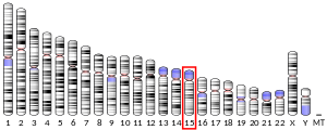
Ocular albinism is a form of albinism which, in contrast to oculocutaneous albinism, presents primarily in the eyes. There are multiple forms of ocular albinism, which are clinically similar.
Oculocutaneous albinism type I or type 1A is an autosomal recessive skin disease. This subtype of oculocutaneous albinism is caused when the gene for tyrosinase does not function properly.

Ocular albinism type 1(OA1) is the most common type of ocular albinism, with a prevalence rate of 1:50,000. It is an inheritable classical Mendelian type X-linked recessive disorder wherein the retinal pigment epithelium lacks pigment while hair and skin appear normal. Since it is usually an X-linked disorder, it occurs mostly in males, while females are carriers unless they are homozygous. About 60 missense and nonsense mutations, insertions, and deletions have been identified in Oa1. Mutations in OA1 have been linked to defective glycosylation and thus improper intracellular transportation.

Amelanism is a pigmentation abnormality characterized by the lack of pigments called melanins, commonly associated with a genetic loss of tyrosinase function. Amelanism can affect fish, amphibians, reptiles, birds, and mammals including humans. The appearance of an amelanistic animal depends on the remaining non-melanin pigments. The opposite of amelanism is melanism, a higher percentage of melanin.

Albinism is the congenital absence of melanin in an animal or plant resulting in white hair, feathers, scales and skin and pink or blue eyes. Individuals with the condition are referred to as albino.