ORAI1

Last updated
ORAI1
Available structures
PDB Ortholog search: PDBe RCSB
Identifiers
Aliases ORAI1 , CRACM1, IMD9, ORAT1, TAM2, TMEM142A, ORAI calcium release-activated calcium modulator 1
External IDs OMIM: 610277 MGI: 1925542 HomoloGene: 13117 GeneCards: ORAI1
Orthologs
SpeciesHumanMouse
Entrez
Ensembl
UniProt
RefSeq (mRNA)

NM_032790

NM_175423

RefSeq (protein)

NP_116179

NP_780632

Location (UCSC) Chr 12: 121.63 – 121.64 Mb Chr 5: 123.15 – 123.17 Mb
PubMed search [3] [4]
Wikidata
View/Edit Human View/Edit Mouse

Calcium release-activated calcium channel protein 1 is a calcium selective ion channel that in humans is encoded by the ORAI1 gene. [5] [6] [7] Orai channels play an important role in the activation of T-lymphocytes. The loss of function mutation of Orai1 causes severe combined immunodeficiency (SCID) in humans [5] The mammalian orai family has two additional homologs, Orai2 and Orai3. Orai proteins share no homology with any other ion channel family of any other known proteins. They have 4 transmembrane domains and form hexamers.

Contents

Structure and function

ORAI channels are activated upon the depletion of internal calcium stores, which is called the "store-operated" or the "capacitative" mechanism. [8] They are molecular constituents of the "calcium release activated calcium currents" (ICRAC). Upon activation of phospholipase C by various cell surface receptors, inositol trisphosphate is formed that releases calcium from the endoplasmic reticulum. The decreased calcium concentration in the endoplasmic reticulum is sensed by the STIM1 protein. STIM1 clusters upon the depletion of the calcium stores and forms "puncta", and relocates near the plasma membrane, where it activates ORAI1 via protein-protein interaction. [9] [10] [11] [12]

In 2012, a 3.35-angstrom (Å) crystal structure of the Drosophila Orai channel, which shares 73% sequence identity with human Orai1 within its transmembrane region, was published. [13] The structure, thought to show the closed state of the channel, revealed that a single channel is composed of six Orai subunits, with the transmembrane domains arranged in concentric rings around a central aqueous pore formed exclusively by the first transmembrane helix of each subunit. Transmembrane helices 2 and 3 surround TM1 and are hypothesized to shield it from the surrounding lipid bilayer and provide structural support. The fourth transmembrane helix forms the outermost layer.

Clinical relevance

ORAI1 mutations are associated with Immunodeficiency 9 and Tubular aggregate myopathy type 2 (TAM2). [14]

Ligands

Inhibitors

Related Research Articles

Calcium release-activated channels (CRAC) are specialized plasma membrane Ca2+ ion channels. When calcium ions (Ca2+) are depleted from the endoplasmic reticulum (a major store of Ca2+) of mammalian cells, the CRAC channel is activated to slowly replenish the level of calcium in the endoplasmic reticulum. The Ca2+ Release-activated Ca2+ (CRAC) Channel (CRAC-C) Family (TC# 1.A.52) is a member of the Cation Diffusion Facilitator (CDF) Superfamily. These proteins typically have between 4 and 6 transmembrane α-helical spanners (TMSs). The 4 TMS CRAC channels arose by loss of 2TMSs from 6TMS CDF carriers, an example of 'reverse' evolution'.

<span class="mw-page-title-main">Calcium signaling</span> Intracellular communication process

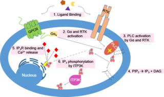
Calcium signaling is the use of calcium ions (Ca2+) to communicate and drive intracellular processes often as a step in signal transduction. Ca2+ is important for cellular signalling, for once it enters the cytosol of the cytoplasm it exerts allosteric regulatory effects on many enzymes and proteins. Ca2+ can act in signal transduction resulting from activation of ion channels or as a second messenger caused by indirect signal transduction pathways such as G protein-coupled receptors.

<span class="mw-page-title-main">Calnexin</span> Mammalian protein found in Homo sapiens

Calnexin (CNX) is a 67kDa integral protein (that appears variously as a 90kDa, 80kDa, or 75kDa band on western blotting depending on the source of the antibody) of the endoplasmic reticulum (ER). It consists of a large (50 kDa) N-terminal calcium-binding lumenal domain, a single transmembrane helix and a short (90 residues), acidic cytoplasmic tail.

Store-operated channels (SOCs) are ion channels located in the plasma membrane of cells. These channels are most studied in regard to their role in calcium entry into the cytoplasm from extracellular milieu. There are other SOC channels selective to other ions. Calcium SOCs are especially important for the cell because they are the major source of intracellular calcium; and calcium itself is involved in a wide array of vital cellular functions. SOCs are so called because they are activated by intracellular calcium stores depletion by both physiological or pharmacological processes.

<span class="mw-page-title-main">STIM1</span> Protein-coding gene in the species Homo sapiens

Stromal interaction molecule 1 is a protein that in humans is encoded by the STIM1 gene. STIM1 has a single transmembrane domain, and is localized to the endoplasmic reticulum, and to a lesser extent to the plasma membrane.

<span class="mw-page-title-main">ATP2A1</span> Protein-coding gene in the species Homo sapiens

Sarcoplasmic/endoplasmic reticulum calcium ATPase 1 (SERCA1) also known as Calcium pump 1, is an enzyme that in humans is encoded by the ATP2A1 gene.

<span class="mw-page-title-main">EIF2AK3</span> Human protein and coding gene

Eukaryotic translation initiation factor 2-alpha kinase 3, also known as protein kinase R (PKR)-like endoplasmic reticulum kinase (PERK), is an enzyme that in humans is encoded by the EIF2AK3 gene.

Ca<sub>v</sub>1.1 Mammalian protein found in Homo sapiens

Cav1.1 also known as the calcium channel, voltage-dependent, L type, alpha 1S subunit, (CACNA1S), is a protein which in humans is encoded by the CACNA1S gene. It is also known as CACNL1A3 and the dihydropyridine receptor.

<span class="mw-page-title-main">CLN6</span> Protein-coding gene in humans

Ceroid-lipofuscinosis neuronal protein 6 is a protein that in humans is encoded by the CLN6 gene.

<span class="mw-page-title-main">KDELR1</span> Protein-coding gene in the species Homo sapiens

KDEL (Lys-Asp-Glu-Leu) endoplasmic reticulum protein retention receptor 1, also known as KDELR1, is a protein which in humans is encoded by the KDELR1 gene.

<span class="mw-page-title-main">SEC61G</span> Mammalian protein found in Homo sapiens

Protein transport protein Sec61 subunit gamma is a protein that in humans is encoded by the SEC61G gene.

<span class="mw-page-title-main">STIM2</span> Protein-coding gene in the species Homo sapiens

Stromal interaction molecule 2 (STIM2) is a protein that in humans is encoded by the STIM2 gene.

<span class="mw-page-title-main">TMEM66</span> Protein-coding gene in the species Homo sapiens

SARAF is a protein that in humans is encoded by the SARAF gene, formerly known as TMEM66.

<span class="mw-page-title-main">ORAI2</span> Protein-coding gene in the species Homo sapiens

Protein orai-2 is a protein that in humans is encoded by the ORAI2 gene.

Membrane contact sites (MCS) are close appositions between two organelles. Ultrastructural studies typically reveal an intermembrane distance in the order of the size of a single protein, as small as 10 nm or wider, with no clear upper limit. These zones of apposition are highly conserved in evolution. These sites are thought to be important to facilitate signalling, and they promote the passage of small molecules, including ions, lipids and reactive oxygen species. MCS are important in the function of the endoplasmic reticulum (ER), since this is the major site of lipid synthesis within cells. The ER makes close contact with many organelles, including mitochondria, Golgi, endosomes, lysosomes, peroxisomes, chloroplasts and the plasma membrane. Both mitochondria and sorting endosomes undergo major rearrangements leading to fission where they contact the ER. Sites of close apposition can also form between most of these organelles most pairwise combinations. First mentions of these contact sites can be found in papers published in the late 1950s mainly visualized using electron microscopy (EM) techniques. Copeland and Dalton described them as “highly specialized tubular form of endoplasmic reticulum in association with the mitochondria and apparently in turn, with the vascular border of the cell”.

LiMETER stands for light-inducible membrane-tethered peripheral endoplasmic reticulum (ER). LiMETER is an optogenetics tool designed to reversibly label cortical ER or the apposition between plasma membrane (PM) and endoplasmic reticulum (ER) membranes.

The ryanodine-inositol 1,4,5-triphosphate receptor Ca2+ channel (RIR-CaC) family includes Ryanodine receptors and Inositol trisphosphate receptors. Members of this family are large proteins, some exceeding 5000 amino acyl residues in length. This family belongs to the Voltage-gated ion channel (VIC) superfamily. Ry receptors occur primarily in muscle cell sarcoplasmic reticular (SR) membranes, and IP3 receptors occur primarily in brain cell endoplasmic reticular (ER) membranes where they effect release of Ca2+ into the cytoplasm upon activation (opening) of the channel. They are redox sensors, possibly providing a partial explanation for how they control cytoplasmic Ca2+. Ry receptors have been identified in heart mitochondria where they provide the main pathway for Ca2+ entry. Sun et al. (2011) have demonstrated oxygen-coupled redox regulation of the skeletal muscle ryanodine receptor-Ca2+ release channel (RyR1;TC# 1.A.3.1.2) by NADPH oxidase 4.

Anjana Rao is a cellular and molecular biologist of Indian ethnicity, working in the US. She uses immune cells as well as other types of cells to understand intracellular signaling and gene expression. Her research focuses on how signaling pathways control gene expression.

Patrick G. Hogan is a cellular and molecular biologist who studies how cellular signaling leads to gene expression. He obtained his bachelor’s degree from Harvard University and a PhD in neurobiology from Harvard Medical School. In 2010, he moved to the La Jolla Institute for Immunology in San Diego as a Professor in the Division of Signaling and Gene Expression. He is a Founder and Member of the Scientific Advisory Board, CalciMedica Inc, La Jolla, CA.

References

  1. 1 2 3 GRCh38: Ensembl release 89: ENSG00000276045 - Ensembl, May 2017
  2. 1 2 3 GRCm38: Ensembl release 89: ENSMUSG00000049686 - Ensembl, May 2017
  3. "Human PubMed Reference:". National Center for Biotechnology Information, U.S. National Library of Medicine.
  4. "Mouse PubMed Reference:". National Center for Biotechnology Information, U.S. National Library of Medicine.
  5. 1 2 Feske S, Gwack Y, Prakriya M, Srikanth S, Puppel SH, Tanasa B, Hogan PG, Lewis RS, Daly M, Rao A (May 2006). "A mutation in Orai1 causes immune deficiency by abrogating CRAC channel function". Nature. 441 (7090): 179–85. Bibcode:2006Natur.441..179F. doi:10.1038/nature04702. PMID   16582901. S2CID   1605392.
  6. Vig M, Peinelt C, Beck A, Koomoa DL, Rabah D, Koblan-Huberson M, Kraft S, Turner H, Fleig A, Penner R, Kinet JP (May 2006). "CRACM1 is a plasma membrane protein essential for store-operated Ca2+ entry". Science. 312 (5777): 1220–3. Bibcode:2006Sci...312.1220V. doi:10.1126/science.1127883. PMC   5685805 . PMID   16645049.
  7. Zhang SL, Yeromin AV, Zhang XH, Yu Y, Safrina O, Penna A, Roos J, Stauderman KA, Cahalan MD (June 2006). "Genome-wide RNAi screen of Ca(2+) influx identifies genes that regulate Ca(2+) release-activated Ca(2+) channel activity". Proceedings of the National Academy of Sciences of the United States of America. 103 (24): 9357–62. doi: 10.1073/pnas.0603161103 . PMC   1482614 . PMID   16751269.
  8. Putney JW (September 2009). "Capacitative calcium entry: from concept to molecules". Immunological Reviews. 231 (1): 10–22. doi:10.1111/j.1600-065X.2009.00810.x. PMID   19754887. S2CID   32303982.
  9. Park CY, Hoover PJ, Mullins FM, Bachhawat P, Covington ED, Raunser S, Walz T, Garcia KC, Dolmetsch RE, Lewis RS (March 2009). "STIM1 clusters and activates CRAC channels via direct binding of a cytosolic domain to Orai1". Cell. 136 (5): 876–90. doi:10.1016/j.cell.2009.02.014. PMC   2670439 . PMID   19249086.
  10. Zhou Y, Meraner P, Kwon HT, Machnes D, Oh-hora M, Zimmer J, Huang Y, Stura A, Rao A, Hogan PG (January 2010). "STIM1 gates the store-operated calcium channel ORAI1 in vitro". Nature Structural & Molecular Biology. 17 (1): 112–6. doi:10.1038/nsmb.1724. PMC   2902271 . PMID   20037597.
  11. Gudlur A, Zhou Y, Hogan PG (2013-01-01). "STIM-ORAI interactions that control the CRAC channel". Current Topics in Membranes. 71: 33–58. doi:10.1016/b978-0-12-407870-3.00002-0. ISBN   9780124078703. PMID   23890110.
  12. Zhou Y, Srinivasan P, Razavi S, Seymour S, Meraner P, Gudlur A, Stathopulos PB, Ikura M, Rao A, Hogan PG (August 2013). "Initial activation of STIM1, the regulator of store-operated calcium entry". Nature Structural & Molecular Biology. 20 (8): 973–81. doi:10.1038/nsmb.2625. PMC   3784406 . PMID   23851458.
  13. Hou X, Pedi L, Diver MM, Long SB (December 2012). "Crystal structure of the calcium release-activated calcium channel Orai". Science. 338 (6112): 1308–13. Bibcode:2012Sci...338.1308H. doi:10.1126/science.1228757. PMC   3695727 . PMID   23180775.
  14. "ORAI CALCIUM RELEASE-ACTIVATED CALCIUM MODULATOR 1; ORAI1". www.omim.org. Retrieved 2023-11-13.