PITPNM3

Last updated

Nir1 or membrane-associated phosphatidylinositol transfer protein 3 (PITPNM3) is a mammalian protein that localizes to endoplasmic reticulum (ER) and plasma membrane (PM) membrane contact sites (MCS) and aids the transfer of phosphatidylinositol between these two membranes, potentially by recruiting additional proteins to the ER-PM MCS. It is encoded by the gene PITPNM3. [1]

Contents

Classification

Nir1 has been classically categorized as a class IIA phosphatidylinositol transfer protein (PITP) that transfers phosphatidylinositol (PI) and phosphatidic acid (PA) between membranes. Class IIA PITPs are the multi-domain proteins PITPNM1/Nir2 (Drosophila homolog RdgBaI), PITPNM2/Nir3 (Drosophila homolog RdgBaII).. [2] [3] Nir1 shares high sequence similarity with Nir2 and Nir3, which led to its original categorization as a PITP. However, it was determined that Nir1 is not directly responsible for PI transfer, as it lacks the functional PITP domain seen within Nir2 and Nir3 [3]

The names, Drosophila homologs, and domain architecture of the PITPNM family proteins. PITPNM-Structures.png
The names, Drosophila homologs, and domain architecture of the PITPNM family proteins.

Localization

Recently, Nir1 has been shown to localize to ER-PM MCS, both under basal conditions and upon phospholipase C (PLC) activation. Notably, PLC activation has previously been shown to regulate the localization of Nir2 and Nir3 at ER-PM MCS well.. [4] [5] The MCS-targeting by Nir1 is achieved by the N-terminus of Nir1 localizing to the ER and the C-terminus of Nir1 localizing to the PM. The domains responsible for binding these membranes are discussed below.

Structure

Nir1 contains three main structural elements that are shared with Nir2 and Nir3: an N-terminal FFAT motif, a DDHD domain, and a C-terminal Lipin/Ndel/Smp2 (LNS2) domain. [6]

FFAT motif

The FFAT motif is made up of double phenylalanines (FF) in an Acidic Tract. This motif, made of residues EFFDA in Nir1, has been shown to be necessary for the Nir proteins to associate with the ER proteins VAPA and VAPB. Mutation of the phenylalanine residues in this motif or knockout of the VAPA and VAPB proteins results in a loss of ER-PM MCS localization and causes Nir1 to become fully localized to the PM. [4] [5]

DDHD domain

The DDHD domain, made up of 3 Asp and 1 His residues, bears some similarities to that seen in PLA1 enzymes, which hydrolyze fatty acids of glycerolphospholipids, including phosphatidic acid (PA). However, this domain is still largely uncharacterized. It is a putative metal binding domain, but a role for metal binding in PITPNM function has not been established [3] [7] [8]

LNS2 domain

The LNS2 domain is the Lipin/Nde1/Smp2 domain. This domain was discovered as having sequence similarities to the phosphatidic acid (PA) binding region found within the Lipin family of proteins. [9] It is also responsible for PA-binding within Nir1, as it has been shown to co-localize with PA biosensors. The LNS2 domain targets the C-terminus of Nir1 to the plasma membrane in order to allow the protein to bridge the ER-PM MCS. Deletion of this domain results in Nir1 localization to the ER. [4] [5] It should be noted however, that the exact domain boundaries of the LNS2 domain are still being debated, especially given the boundaries of the folded domains predicted by the AlphaFold Protein Structure Database. [10] [11] (Alphafold structure of Nir1)

Function

The PITPNM family of proteins has been shown to participate in the phosphoinositide cycle. Lipids cycle between the PM and the ER in order to replenish levels after signaling events deplete lipid species such as PI.. [2] When a stimulus results in the production of PA at the PM, Nir2 and Nir3 move to the ER-PM MCS, where they exchange the PA at the PM for PI that has been produced in the ER. As Nir1 is localized to the ER-PM MCS even without a stimulus, it is thought that Nir1 helps to recruit Nir2 to the MCS. There is evidence that Nir1 recruits Nir2 directly via binding to the uncharacterized domain between the FFAT and DDHD of Nir1 [4] [5]

Nir1 localizes to ER-PM MCS using its FFAT and LNS2 domains. It is thought to directly interact with Nir2 in order to recruit Nir2 to the ER-PM MCS, so that Nir2 can transfer lipids with its PITP domain. PITPNM3-Function.png
Nir1 localizes to ER-PM MCS using its FFAT and LNS2 domains. It is thought to directly interact with Nir2 in order to recruit Nir2 to the ER-PM MCS, so that Nir2 can transfer lipids with its PITP domain.

Related Research Articles

<span class="mw-page-title-main">Phospholipid</span> Class of lipids

Phospholipids are a class of lipids whose molecule has a hydrophilic "head" containing a phosphate group and two hydrophobic "tails" derived from fatty acids, joined by an alcohol residue. Marine phospholipids typically have omega-3 fatty acids EPA and DHA integrated as part of the phospholipid molecule. The phosphate group can be modified with simple organic molecules such as choline, ethanolamine or serine.

<span class="mw-page-title-main">Peripheral membrane protein</span> Membrane proteins that adhere temporarily to membranes with which they are associated

Peripheral membrane proteins, or extrinsic membrane proteins, are membrane proteins that adhere only temporarily to the biological membrane with which they are associated. These proteins attach to integral membrane proteins, or penetrate the peripheral regions of the lipid bilayer. The regulatory protein subunits of many ion channels and transmembrane receptors, for example, may be defined as peripheral membrane proteins. In contrast to integral membrane proteins, peripheral membrane proteins tend to collect in the water-soluble component, or fraction, of all the proteins extracted during a protein purification procedure. Proteins with GPI anchors are an exception to this rule and can have purification properties similar to those of integral membrane proteins.

Phosphatidic acids are anionic phospholipids important to cell signaling and direct activation of lipid-gated ion channels. Hydrolysis of phosphatidic acid gives rise to one molecule each of glycerol and phosphoric acid and two molecules of fatty acids. They constitute about 0.25% of phospholipids in the bilayer.

<span class="mw-page-title-main">Phosphoinositide phospholipase C</span>

Phosphoinositide phospholipase C is a family of eukaryotic intracellular enzymes that play an important role in signal transduction processes. These enzymes belong to a larger superfamily of Phospholipase C. Other families of phospholipase C enzymes have been identified in bacteria and trypanosomes. Phospholipases C are phosphodiesterases.

<span class="mw-page-title-main">Phosphoinositide 3-kinase</span> Class of enzymes

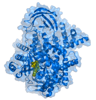
Phosphoinositide 3-kinases (PI3Ks), also called phosphatidylinositol 3-kinases, are a family of enzymes involved in cellular functions such as cell growth, proliferation, differentiation, motility, survival and intracellular trafficking, which in turn are involved in cancer.

<span class="mw-page-title-main">Phosphatidylinositol 4,5-bisphosphate</span> Chemical compound

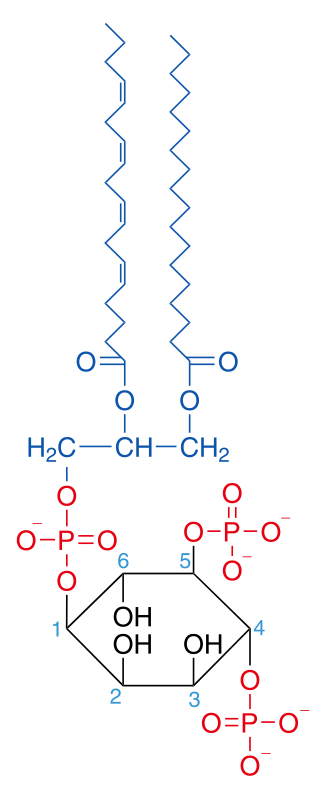
Phosphatidylinositol 4,5-bisphosphate or PtdIns(4,5)P2, also known simply as PIP2 or PI(4,5)P2, is a minor phospholipid component of cell membranes. PtdIns(4,5)P2 is enriched at the plasma membrane where it is a substrate for a number of important signaling proteins. PIP2 also forms lipid clusters that sort proteins.
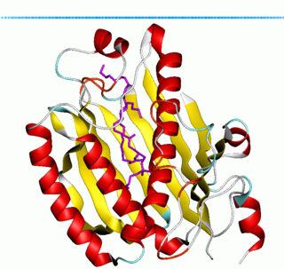
<span class="mw-page-title-main">Phosphatidylinositol 3-phosphate</span> Chemical compound

Phosphatidylinositol 3-phosphate (PtdIns3P) is a phospholipid found in cell membranes that helps to recruit a range of proteins, many of which are involved in protein trafficking, to the membranes. It is the product of both the class II and III phosphoinositide 3-kinases activity on phosphatidylinositol.

Phospholipase D (EC 3.1.4.4, lipophosphodiesterase II, lecithinase D, choline phosphatase, PLD; systematic name phosphatidylcholine phosphatidohydrolase) is an enzyme of the phospholipase superfamily that catalyses the following reaction

<span class="mw-page-title-main">Diacylglycerol kinase</span> Class of enzymes

Diacylglycerol kinase is a family of enzymes that catalyzes the conversion of diacylglycerol (DAG) to phosphatidic acid (PA), utilizing ATP as a source of the phosphate. In non-stimulated cells, DGK activity is low, allowing DAG to be used for glycerophospholipid biosynthesis, but on receptor activation of the phosphoinositide pathway, DGK activity increases, driving the conversion of DAG to PA. As both lipids are thought to function as bioactive lipid signaling molecules with distinct cellular targets, DGK therefore occupies an important position, effectively serving as a switch by terminating the signalling of one lipid while simultaneously activating signalling by another.

<span class="mw-page-title-main">FYVE domain</span>

In molecular biology the FYVE zinc finger domain is named after the four cysteine-rich proteins: Fab 1, YOTB, Vac 1, and EEA1, in which it has been found. FYVE domains bind phosphatidylinositol 3-phosphate, in a way dependent on its metal ion coordination and basic amino acids. The FYVE domain inserts into cell membranes in a pH-dependent manner. The FYVE domain has been connected to vacuolar protein sorting and endosome function.

<span class="mw-page-title-main">PX domain</span>

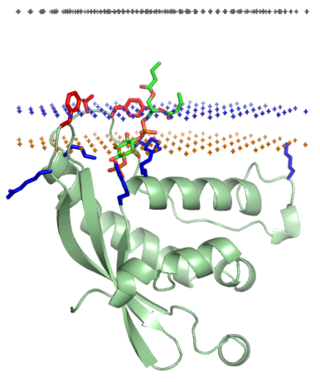
The PX domain is a phosphoinositide-binding structural domain involved in targeting of proteins to cell membranes.

<span class="mw-page-title-main">Phosphatidylinositol transfer protein</span>

Phosphatidylinositol transfer protein (PITP) or priming in exocytosis protein 3 (PEP3) is a ubiquitous cytosolic domain involved in transport of phospholipids from their site of synthesis in the endoplasmic reticulum and Golgi to other cell membranes.

<span class="mw-page-title-main">Phosphatidate phosphatase</span>

The enzyme phosphatidate phosphatase (PAP, EC 3.1.3.4) is a key regulatory enzyme in lipid metabolism, catalyzing the conversion of phosphatidate to diacylglycerol:

<span class="mw-page-title-main">Phospholipase C</span> Class of enzymes

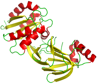
Phospholipase C (PLC) is a class of membrane-associated enzymes that cleave phospholipids just before the phosphate group (see figure). It is most commonly taken to be synonymous with the human forms of this enzyme, which play an important role in eukaryotic cell physiology, in particular signal transduction pathways. Phospholipase C's role in signal transduction is its cleavage of phosphatidylinositol 4,5-bisphosphate (PIP2) into diacyl glycerol (DAG) and inositol 1,4,5-trisphosphate (IP3), which serve as second messengers. Activators of each PLC vary, but typically include heterotrimeric G protein subunits, protein tyrosine kinases, small G proteins, Ca2+, and phospholipids.

<span class="mw-page-title-main">PI4KB</span> Protein-coding gene in the species Homo sapiens

Phosphatidylinositol 4-kinase beta is an enzyme that in humans is encoded by the PI4KB gene.

Membrane contact sites (MCS) are close appositions between two organelles. Ultrastructural studies typically reveal an intermembrane distance in the order of the size of a single protein, as small as 10 nm or wider, with no clear upper limit. These zones of apposition are highly conserved in evolution. These sites are thought to be important to facilitate signalling, and they promote the passage of small molecules, including ions, lipids and reactive oxygen species. MCS are important in the function of the endoplasmic reticulum (ER), since this is the major site of lipid synthesis within cells. The ER makes close contact with many organelles, including mitochondria, Golgi, endosomes, lysosomes, peroxisomes, chloroplasts and the plasma membrane. Both mitochondria and sorting endosomes undergo major rearrangements leading to fission where they contact the ER. Sites of close apposition can also form between most of these organelles most pairwise combinations. First mentions of these contact sites can be found in papers published in the late 1950s mainly visualized using electron microscopy (EM) techniques. Copeland and Dalton described them as “highly specialized tubular form of endoplasmic reticulum in association with the mitochondria and apparently in turn, with the vascular border of the cell”.

<span class="mw-page-title-main">Sec14</span>

Sec14 is a cytosolic protein found in yeast which plays a role in the regulation of several cellular functions, specifically those related to intracellular transport. Encoded by the Sec14 gene, Sec14p may transport phosphatidylinositol and phosphatidylcholine produced in the endoplasmic reticulum and the Golgi body to other cellular membranes. Additionally, Sec14p potentially plays a role in the localization of lipid raft proteins. Sec14p is an essential gene in yeast, and is homologous in function to phosphatidylinositol transfer protein in mammals. A conditional mutant with non-functional Sec14p presents with Berkeley bodies and deficiencies in protein secretion.

A FFAT motif is a protein sequence motif of six defined amino acids plus neighbouring residues that binds to proteins in the VAP protein family.

VAP proteins are conserved integral membrane proteins of the endoplasmic reticulum found in all eukaryotic cells. VAP stands for VAMP-associated protein, where VAMP stands for vesicle-associated membrane protein. Humans have two VAPs that consist of the essential Major Sperm Protein domain and linker plus transmembrane helix) to attach to the ER: VAPA and VAPB. A third VAP-like protein is Motile sperm domain containing 2 (MOSPD2), which has all the elements of VAP, and like them binds FFAT motifs, but has at its N-terminus a CRAL-TRIO domain that can bind and transfer lipids.

Membrane-mediated anesthesia or anaesthesia (UK) is a mechanism of action that involves an anesthetic exerting its effects through the lipid membrane. Established mechanism exists for both general and local anesthetics. The anesthetic binding site is within ordered lipids and binding disrupts the function of the ordered lipid. See Theories of general anaesthetic action for a broader discussion of purely theoretical mechanisms.

References

  1. "PITPNM3 Gene - PITPNM Family Member 3". GeneCards: The Human Gene Database. 4 October 2023. Retrieved 4 December 2023.
  2. 1 2 Cockcroft, Shamshad; Raghu, Padinjat (2016-11-25). "Topological organisation of the phosphatidylinositol 4,5-bisphosphate–phospholipase C resynthesis cycle: PITPs bridge the ER–PM gap". Biochemical Journal. 473 (23): 4289–4310. doi: 10.1042/bcj20160514c . ISSN   0264-6021. PMID   27888240.
  3. 1 2 3 Balla, Tamas (July 2013). "Phosphoinositides: Tiny Lipids With Giant Impact on Cell Regulation". Physiological Reviews. 93 (3): 1019–1137. doi:10.1152/physrev.00028.2012. ISSN   0031-9333. PMC   3962547 . PMID   23899561.
  4. 1 2 3 4 Quintanilla, Carlo Giovanni; Lee, Wan-Ru; Liou, Jen (2022-03-01). Olzmann, James (ed.). "Nir1 constitutively localizes at ER–PM junctions and promotes Nir2 recruitment for PIP 2 homeostasis". Molecular Biology of the Cell. 33 (3): br2. doi:10.1091/mbc.E21-07-0356. ISSN   1059-1524. PMC   9250379 . PMID   35020418. S2CID   245927652.
  5. 1 2 3 4 Chang, Chi-Lun; Liou, Jen (June 2015). "Phosphatidylinositol 4,5-Bisphosphate Homeostasis Regulated by Nir2 and Nir3 Proteins at Endoplasmic Reticulum-Plasma Membrane Junctions". Journal of Biological Chemistry. 290 (23): 14289–14301. doi: 10.1074/jbc.m114.621375 . ISSN   0021-9258. PMC   4505499 . PMID   25887399.
  6. Cockcroft, Shamshad; Lev, Sima (January 2020). "Mammalian PITPs at the Golgi and ER-Golgi Membrane Contact Sites". Contact. 3: 251525642096417. doi: 10.1177/2515256420964170 . ISSN   2515-2564. S2CID   226531182.
  7. Ile, Kristina E; Schaaf, Gabriel; Bankaitis, Vytas A (2006-10-18). "Phosphatidylinositol transfer proteins and cellular nanoreactors for lipid signaling". Nature Chemical Biology. 2 (11): 576–583. doi:10.1038/nchembio835. ISSN   1552-4450. PMID   17051233. S2CID   39526983.
  8. Matsumoto, Naoki; Nemoto-Sasaki, Yoko; Oka, Saori; Arai, Seisuke; Wada, Ikuo; Yamashita, Atsushi (July 2021). "Phosphorylation of human phospholipase A1 DDHD1 at newly identified phosphosites affects its subcellular localization". Journal of Biological Chemistry. 297 (1): 100851. doi: 10.1016/j.jbc.2021.100851 . ISSN   0021-9258. PMC   8234217 . PMID   34089703.
  9. Kim, SoHui; Kedan, Amir; Marom, Merav; Gavert, Nancy; Keinan, Omer; Selitrennik, Michael; Laufman, Orly; Lev, Sima (2013-07-30). "The phosphatidylinositol‐transfer protein Nir2 binds phosphatidic acid and positively regulates phosphoinositide signalling". EMBO Reports. 14 (10): 891–899. doi:10.1038/embor.2013.113. ISSN   1469-221X. PMC   3807235 . PMID   23897088.
  10. Varadi, Mihaly; Anyango, Stephen; Deshpande, Mandar; Nair, Sreenath; Natassia, Cindy; Yordanova, Galabina; Yuan, David; Stroe, Oana; Wood, Gemma; Laydon, Agata; Žídek, Augustin (2021-11-17). "AlphaFold Protein Structure Database: massively expanding the structural coverage of protein-sequence space with high-accuracy models". Nucleic Acids Research. 50 (D1): D439–D444. doi:10.1093/nar/gkab1061. ISSN   0305-1048. PMC   8728224 . PMID   34791371.
  11. Jumper, John; Evans, Richard; Pritzel, Alexander; Green, Tim; Figurnov, Michael; Ronneberger, Olaf; Tunyasuvunakool, Kathryn; Bates, Russ; Žídek, Augustin; Potapenko, Anna; Bridgland, Alex (2021-07-15). "Highly accurate protein structure prediction with AlphaFold". Nature. 596 (7873): 583–589. Bibcode:2021Natur.596..583J. doi:10.1038/s41586-021-03819-2. ISSN   0028-0836. PMC   8371605 . PMID   34265844.