Peter Nemes is a Hungarian-American chemist, who is active in the fields of bioanalytical chemistry, mass spectrometry, cell/developmental biology, neuroscience, and biochemistry.
Nemes has been an associate professor at the University of Maryland, College Park (UMD) since January 2018. Prior to his appointment there, he was an assistant professor at the Department of Chemistry at George Washington University (Washington, DC), where he taught bioanalytical chemistry.[1] Nemes graduated with summa cum laude with a Master's of Science (M.Sc.) from the Eötvös Loránd University in 2004. His original thesis research was conducted in the Department of Mass Spectrometry at the Hungarian Academy of Sciences, Budapest, Hungary. Under mentorship by Vekey Karoly, Nemes studied the formation of amino acid clusters in the gas phase upon electrospray ionization,1 such as the magic serine clusters that preferentially incorporate amino acids and sugars of certain chirality matching those enriched on Earth. During his PhD in Akos Vertes’ laboratory at the Department of Chemistry, The George Washington University (GWU; Washington, DC) between 2005 and 2009, he established the significance of spraying modes/regimes during electrospray ionization (ESI) mass spectrometry (MS) in efficient and soft ion generation2 and the confirmational state of proteins3. He also invented and patented laser ablation electrospray ionization (LAESI)4 mass spectrometry for in situ and in vivo analysis4 of tissues and single cells5 2- and 3-dimensional molecular imaging MS6 at ambient conditions for biological samples. He completed postdoctoral training in analytical neuroscience with Jonathan V. Sweedler at the University of Illinois Urbana–Champaign, IL. There, he developed capillary electrophoresis ESI MS instruments7 and built a unique matrix-assisted laser desorption ionization (MALDI–C60 secondary ionization mass spectrometry (SIMS) dual-ion source mass spectrometer8 to enable the analysis of small and large molecules in single cells.9
In 2011, Nemes became a Staff Fellow and then also Laboratory Leader at the US Food and Drug Administration (2011–2013). An independent investigator, Nemes developed a high-throughput approach10 based on Direct analysis in real time to enable the rapid differentiation of heparin from glycosaminoglycans, including authentic adulterated products confiscated by the FDA during the 2008 heparin crisis. Nemes also established the mass spectrometry facility at the White Oak Headquarters of the US FDA with several mass spectrometers to support regulatory science.
As a professor since 2013, Nemes has conducted cutting-edge research at the interface of bioanalytical instrumental chemistry and neurodevelopmental biology and taught courses in analytical chemistry and mass spectrometry. In Fall 2013, Nemes became an assistant professor at the Department of Chemistry at George Washington University (Washington, DC), where he taught analytical chemistry.[1] In January 2018, Nemes became an associate professor at the Department of Chemistry & Biochemistry, the University of Maryland, College Park (UMD), where he has been teaching instrumental analytical chemistry and biological mass spectrometry. Research in the Nemes Laboratory develops ultrasensitive and microanalytical platforms for high-resolution MS to study metabolic and proteomic processes with implications in cell and neurodevelopmental biology and health research. Using custom-built single-cell MS instruments, his research group has uncovered previously unknown metabolomic11 and proteomic12 differences between single embryonic cells that are fated to give rise to different types of tissues during vertebrate development. Their highly sensitive bottom-up proteomic approach enabled the detection intra-cell type cell heterogeneity in the embryo.13 Further, the group has also discovered molecules that are able to alter normal cell fate decisions in the embryo.11 The investigators next developed microprobe technologies that enabled the direct, in vivo analysis of these small14 and large15 molecules in cells in X. laevis embryos undergoing normal development. These results challenge basic understanding of molecular processes that are necessary for normal embryonic body and brain development and raise important implications to help understand, promote, and protect the health of humans and animals.16
Nemes has authored 46 peer-reviewed publications, 6 book chapters, and ~200 presentations. In 2015, Nemes was named a Beckman Young Investigator by the Arnold and Mabel Beckman Foundation and received the Arthur F. Findeis Award for Achievements by a Young Analytical Chemist by the Division of Analytical Chemistry of the American Chemical Society. In 2017, he received the DuPont Young Professor Award, the Robert J. Cotter New Investigator Award by the US Human Proteome Organization, and a Research Award from the American Society for Mass Spectrometry (ASMS). In 2018, Nemes was awarded the Georges Guiochon Faculty Fellowship from HPLC, Inct. Research in the Nemes Lab has been continuously funded by professional societies, companies, and federal funding agencies. Nemes holds a CAREER award from the Directorate of Biological Research of the National Science Foundation (NSF) and an Outstanding Research Award (R35) from the National Institute of General Medical Sciences (NIGMS).
1P. Nemes, G. Schlosser, and K. Vekey*, Amino acid cluster formation studied by electrospray ionization mass spectrometry, J. Mass Spectrom. 2005, 40, 43, https://doi.org/10.1002/jms.771
2P. Nemes, I. Marginean, and A. Vertes*, Spraying mode effect on droplet formation and ion chemistry in electrosprays, Anal. Chem. 2007, 79, 3105, https://doi.org/10.1021/ac062382i
3P. Nemes, S. Goyal, and A. Vertes*, Conformational and noncovalent complexation changes in proteins during electrospray ionization, Anal. Chem. 2008,80, 387–395, https://doi.org/10.1021/ac0714359
4P. Nemes and A. Vertes*, Laser ablation electrospray ionization for atmospheric pressure, in vivo, and imaging mass spectrometry, Anal. Chem. 2007, 79, 8098, https://doi.org/10.1021/ac071181r
5B. Shrestha, P. Nemes, and A. Vertes*, Ablation and analysis of small cell populations and single cells by consecutive laser pulses, Appl. Phys. A 2010, https://doi.org/10.1007/s00339-010-5781-2
6P. Nemes, A. A. Barton, and A. Vertes*, Three-dimensional imaging of metabolites in tissues under native conditions by laser ablation electrospray ionization mass spectrometry, Anal. Chem. 2009, 81, 6668, https://doi.org/10.1021/ac900745e
7P. Nemes, S. S. Rubakhin, J. Aerts, and J. V. Sweedler*, Qualitative and quantitative metabolomic investigation of single neurons by capillary electrophoresis electrospray ionization mass spectrometry, Nat. Protoc. 2013, 8, 783, https://doi.org/10.1038/nprot.2013.035
8E. J. Lanni, S. J. B. Dunham, P. Nemes, S. S. Rubakhin, J. V. Sweedler*, Biomolecular imaging with a C60-SIMS/MALDI dual ion source hybrid mass spectrometer: Instrumentation, matrix enhancement and single cell analysis, J. Am. Soc. Mass Spectrom. 2014, 11, 1897–1907, https://doi.org/10.1007/s13361-014-0978-9
9S. S. Rubakhin, E. V. Romanova, P. Nemes, and J. V. Sweedler, Profiling metabolites and peptides in single cells, Nat. Methods 2011, 8, S20–S29, https://doi.org/10.1038/nmeth.1549
10P. Nemes*, W. J. Hoover, and D. A. Keire, High-throughput differentiation of heparin from other glycosaminoglycans by pyrolysis mass spectrometry, Anal. Chem. 2013, 85, 7405–7412, https://doi.org/10.1021/ac401318q
12C. Lombard-Banek, S. A. Moody, and P. Nemes*, Single-cell mass spectrometry for discovery proteomics: quantifying translational cell heterogeneity in the 16-cell frog (Xenopus) embryo, Angew. Chem. Int. Ed. 2016, 55, 2454, https://doi.org/10.1002/anie.201510411
13C. Lombard-Banek, Sally A. Moody, and P. Nemes*, Label-free quantification of proteins in single embryonic cells with neural fate in the cleavage-stage frog (Xenopus laevis) embryo using capillary electrophoresis electrospray ionization high resolution mass spectrometry (CE-ESI-HRMS), Mol. Cell. Prot. 2016, 15, 2756, https://doi.org/10.1074/mcp.M115.057760
14R. M. Onjiko, E. P. Portero, S. A. Moody, and P. Nemes*,In situ microprobe single-cell capillary electrophoresis mass spectrometry: Metabolic reorganization in single differentiating cells in the live vertebrate (X. laevis) embryo, Anal. Chem. 2017, 89, 7069, https://doi.org/10.1021/acs.analchem.7b00880
15C. Lombard-Banek, S. A. Moody, M. Chiara Manzini, and P. Nemes*, Microsampling capillary electrophoresis mass spectrometry enables single-cell proteomics in complex tissues: developing cell clones in live Xenopus laevis and zebrafish embryos, Anal. Chem. 2019, 91, 4797, https://doi.org/10.1021/acs.analchem.9b00345
16C. Lombard-Banek, E. P. Portero, R. M. Onjiko, and P. Nemes*, New-generation mass spectrometry expands the toolbox of cell and developmental biology, genesis 2016, 55, e23012, https://doi.org/10.1002/dvg.23012
2008 International Research Fellowship Award, the Dimitris N. Chorafas Foundation (Luzern, Switzerland)[3]
2007 Young Investigator Travel Award, the Mass Spectrometry Discussion Group
Related Research Articles
An ion source is a device that creates atomic and molecular ions. Ion sources are used to form ions for mass spectrometers, optical emission spectrometers, particle accelerators, ion implanters and ion engines.
Electrospray ionization (ESI) is a technique used in mass spectrometry to produce ions using an electrospray in which a high voltage is applied to a liquid to create an aerosol. It is especially useful in producing ions from macromolecules because it overcomes the propensity of these molecules to fragment when ionized. ESI is different from other ionization processes since it may produce multiple-charged ions, effectively extending the mass range of the analyser to accommodate the kDa-MDa orders of magnitude observed in proteins and their associated polypeptide fragments.
Desorption electrospray ionization (DESI) is an ambient ionization technique that can be coupled to mass spectrometry (MS) for chemical analysis of samples at atmospheric conditions. Coupled ionization sources-MS systems are popular in chemical analysis because the individual capabilities of various sources combined with different MS systems allow for chemical determinations of samples. DESI employs a fast-moving charged solvent stream, at an angle relative to the sample surface, to extract analytes from the surfaces and propel the secondary ions toward the mass analyzer. This tandem technique can be used to analyze forensics analyses, pharmaceuticals, plant tissues, fruits, intact biological tissues, enzyme-substrate complexes, metabolites and polymers. Therefore, DESI-MS may be applied in a wide variety of sectors including food and drug administration, pharmaceuticals, environmental monitoring, and biotechnology.
Laser spray ionization refers to one of several methods for creating ions using a laser interacting with a spray of neutral particles or ablating material to create a plume of charged particles. The ions thus formed can be separated by m/z with mass spectrometry. Laser spray is one of several ion sources that can be coupled with liquid chromatography-mass spectrometry for the detection of larger molecules.
Matrix-assisted laser desorption electrospray ionization (MALDESI) was first introduced in 2006 as a novel ambient ionization technique which combines the benefits of electrospray ionization (ESI) and matrix-assisted laser desorption/ionization (MALDI). An infrared (IR) or ultraviolet (UV) laser can be utilized in MALDESI to resonantly excite an endogenous or exogenous matrix. The term 'matrix' refers to any molecule that is present in large excess and absorbs the energy of the laser, thus facilitating desorption of analyte molecules. The original MALDESI design was implemented using common organic matrices, similar to those used in MALDI, along with a UV laser. The current MALDESI source employs endogenous water or a thin layer of exogenously deposited ice as the energy-absorbing matrix where O-H symmetric and asymmetric stretching bonds are resonantly excited by a mid-IR laser.
Capillary electrophoresis–mass spectrometry (CE–MS) is an analytical chemistry technique formed by the combination of the liquid separation process of capillary electrophoresis with mass spectrometry. CE–MS combines advantages of both CE and MS to provide high separation efficiency and molecular mass information in a single analysis. It has high resolving power and sensitivity, requires minimal volume and can analyze at high speed. Ions are typically formed by electrospray ionization, but they can also be formed by matrix-assisted laser desorption/ionization or other ionization techniques. It has applications in basic research in proteomics and quantitative analysis of biomolecules as well as in clinical medicine. Since its introduction in 1987, new developments and applications have made CE-MS a powerful separation and identification technique. Use of CE–MS has increased for protein and peptides analysis and other biomolecules. However, the development of online CE–MS is not without challenges. Understanding of CE, the interface setup, ionization technique and mass detection system is important to tackle problems while coupling capillary electrophoresis to mass spectrometry.
Ambient ionization is a form of ionization in which ions are formed in an ion source outside the mass spectrometer without sample preparation or separation. Ions can be formed by extraction into charged electrospray droplets, thermally desorbed and ionized by chemical ionization, or laser desorbed or ablated and post-ionized before they enter the mass spectrometer.
Instrumental analysis is a field of analytical chemistry that investigates analytes using scientific instruments.
In mass spectrometry, liquid junction interface is an ion source or set-up that couples peripheric devices, such as capillary electrophoresis, to mass spectrometry.
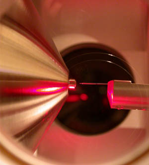
Laser ablation electrospray ionization (LAESI) is an ambient ionization method for mass spectrometry that combines laser ablation from a mid-infrared (mid-IR) laser with a secondary electrospray ionization (ESI) process. The mid-IR laser is used to generate gas phase particles which are then ionized through interactions with charged droplets from the ESI source. LAESI was developed in Professor Akos Vertes lab by Peter Nemes in 2007 and it was marketed commercially by Protea Biosciences, Inc until 2017. Fiber-LAESI for single-cell analysis approach was developed by Bindesh Shrestha in Professor Vertes lab in 2009. LAESI is a novel ionization source for mass spectrometry (MS) that has been used to perform MS imaging of plants, tissues, cell pellets, and even single cells. In addition, LAESI has been used to analyze historic documents and untreated biofluids such as urine and blood. The technique of LAESI is performed at atmospheric pressure and therefore overcomes many of the obstacles of traditional MS techniques, including extensive and invasive sample preparation steps and the use of high vacuum. Because molecules and aerosols are ionized by interacting with an electrospray plume, LAESI's ionization mechanism is similar to SESI and EESI techniques.
Renato Zenobi is a Swiss chemist. He is Professor of Chemistry at ETH Zurich. Throughout his career, Zenobi has contributed to the field of analytical chemistry.
Richard Dale Smith is a chemist and a Battelle Fellow and chief scientist within the biological sciences division, as well as the director of proteomics research at the Pacific Northwest National Laboratory (PNNL). Smith is also director of the NIH Proteomics Research Resource for Integrative Biology, an adjunct faculty member in the chemistry departments at Washington State University and the University of Utah, and an affiliate faculty member at the University of Idaho and the Department of Molecular Microbiology & Immunology, Oregon Health & Science University. He is the author or co-author of approximately 1100 peer-reviewed publications and has been awarded 70 US patents.
Extractive electrospray ionization (EESI) is a spray-type, ambient ionization source in mass spectrometry that uses two colliding aerosols, one of which is generated by electrospray. In standard EESI, syringe pumps provide the liquids for both an electrospray and a sample spray. In neutral desorption EESI (ND-EESI), the liquid for the sample aerosol is provided by a flow of nitrogen.
In cell biology, single-cell analysis and subcellular analysis refer to the study of genomics, transcriptomics, proteomics, metabolomics, and cell–cell interactions at the level of an individual cell, as opposed to more conventional methods which study bulk populations of many cells.
Electrostatic spray ionization (ESTASI) is an ambient ionization method for mass spectrometry (MS) analysis of samples located on a flat or porous surface, or inside a microchannel. It was developed in 2011 by Professor Hubert H. Girault’s group at the École Polytechnique Fédérale de Lausanne (EPFL) in Switzerland. In a typical ESTASI process, a droplet of a protic solvent containing analytes is deposited on a sample area of interest which itself is mounted to an insulating substrate. Under this substrate and right below the droplet, an electrode is placed and connected with a pulsed high voltage (HV) to electrostatically charge the droplet during pulsing. When the electrostatic pressure is larger than the surface tension, droplets and ions are sprayed. ESTASI is a contactless process based on capacitive coupling. One advantage of ESTASI is, that the electrode and sample droplet act contact-less avoiding thereby any oxidation or reduction of the sample compounds at the electrode surface, which often happens during standard electrospray ionization (ESI). ESTASI is a powerful new ambient ionization technique that has already found many applications in the detection of different analytes, such as organic molecules, peptides and proteins with molecule weight up to 70 kDa. Furthermore, it was used to couple MS with various separation techniques including capillary electrophoresis and gel isoelectric focusing, and it was successfully applied under atmospheric pressure to the direct analysis of samples with only few preparation steps.
Jonathan V Sweedler is an American chemist specializing in bioanalytical chemistry, neurochemistry and cell to cell biology and behavior. He is the James R. Eiszner Family Endowed Chair in Chemistry at the University of Illinois at Urbana-Champaign. Additionally, he holds a faculty appointment in the Beckman Institute. He is also an Elected Fellow to the American Chemical Society, for which he is also the society's Editor in Chief for the journal Analytical Chemistry.
John Michael Ramsey is an American analytical chemist at the University of North Carolina at Chapel Hill. He currently holds the position of Minnie N. Goldby Distinguished Professor of Chemistry. His current research with the university focuses on microscale and nanoscale devices such as microchip electrospray, microscale Ion trap mass spectrometers, and microfluidic point of care devices. He is ranked #2 in the "Giants of Nano" field on The Analytical Scientist Power List.
Gary Glish is an American analytical chemist at the University of North Carolina at Chapel Hill. He is a leading researcher in the fields of mass spectrometry, ion chemistry, and biomolecule analysis.
Probe electrospray ionization (PESI) is an electrospray-based ambient ionization technique which is coupled with mass spectrometry for sample analysis. Unlike traditional mass spectrometry ion sources which must be maintained in a vacuum, ambient ionization techniques permit sample ionization under ambient conditions, allowing for the high-throughput analysis of samples in their native state, often with minimal or no sample pre-treatment. The PESI ion source simply consists of a needle to which a high voltage is applied following sample pick-up, initiating electrospray directly from the solid needle.
Jonas Bergquist is a Swedish analytical chemist and clinical neuroscientist, professor and inspector equitandi at Uppsala University, distinguished professor in precision medicine at Binzhou Medical University and adjunct professor of pathology at University of Utah.
This article needs additional or more specific categories. Please help out by adding categories to it so that it can be listed with similar articles.(February 2021)
This page is based on this Wikipedia article Text is available under the CC BY-SA 4.0 license; additional terms may apply. Images, videos and audio are available under their respective licenses.