Plant-specific insert

Last updated
Solanum tuberosum Aspartic Protease 3 Plant-Specific Insert
PDB 3RFI.png
Structure of the saposin-like domain of a plant aspartic proteinase [1]
Identifiers
Organism Solanum tuberosum
SymbolStAP_PSI
PDB 3RFI
UniProt Q6B9W9

The plant-specific insert (PSI) or plant-specific sequence (PSS) [2] is an independent domain, exclusively found in plants, consisting of approximately 100 residues, found on the C-terminal lobe on some aspartic proteases (AP) called phytepsins. [3] The PSI, as an independent entity separate from its parent AP, is homologous to saposin and belongs to the saposin-like protein family (SAPLIP). [4]

Contents

Although the PSI is grouped along proteins in the SAPLIP family, the PSI does not contain a proper saposin-like domain. This is due to a circular permutation of the N- and C-termini of the PSI, in which the termini are "swapped". This has led to the PSI being termed a "swaposin" (a play-on-words of "swap" and "saposin") [5] although the tertiary structure still remains homologous to saposin and other members of the SAPLIP family. [4]

Structure

Suggested relationship between saposin and swaposin. They could have evolved from a similar gene. Both consist of four alpha helices with the order of helices being permuted relative to each other. Saposin Swaposin.svg
Suggested relationship between saposin and swaposin. They could have evolved from a similar gene. Both consist of four alpha helices with the order of helices being permuted relative to each other.

Among plants, APs between different species are generally homologous exhibiting high sequence identity whilst maintaining a similar tertiary structure to pepsin. [2] As such, plant APs preserve the zymogenic form of the mature AP common to other aspartic proteases in which the zymogen is kept inactive until removal of the prosegment from the active cleft.

Processing

The zymogenic form of plant APs contain the primary sequence of the PSI, although not all plant APs contain a PSI region. [4] The PSI itself is composed of approximately 100 residues and is found in the C-terminal primary structure of zymogenic plant APs, forming an independent domain from the characteristic bilobal tertiary structure of aspartic proteases. In the case of Procardosin A, the zymogenic form of Cardosin A (the major AP found in cardoon), the PSI is first removed before activation of the mature enzyme occurs in which the prosegment is cleaved during proteolytic processing. [2]

Tertiary structure

The crystal structure of prophytepsin (from barley) was the first known structure to elucidate the molecular topology of any PSI. [6] The crystal structure of prophytepsin derived PSI revealed that the overall length and position of the helices found in PSI is conserved with those of NK-lysin, another member of the SAPLIP group. [3] [6] [7] Moreover, sequence alignment between NK-lysin and PSI reveals that the relative positions of the disulfide bridges are also conserved, a common trait among SAPLIPS. [4]

Several crystal structures exist, containing the coordinates of the zymogenic parent AP and the SAPLIP domain of the PSI, for cardoon and barley. [3] [6] In these crystal structures, the PSI domain takes on a “closed” tertiary structure similar to that of NK-lysin and saposin. The X-ray crystallographic structure of potato PSI, recombinantly expressed separately from its parent AP, revealed a tertiary structure similar to the open structure of saposin C and forms a homodimer at pH 7.4. [1]

The first helix on the N-terminal end of the open structure of potato PSI also shows similarity in its tertiary structure to the hemagglutinin fusion peptide, exhibiting a similar boomerang motif. As well, this helix shares the overall helix-kink-helix shape with hemagglutinin induced by the presence of a tryptophan; this revelation is important as it suggests a reason for the N-terminal side helix interaction with phospholipid bilayers. That is, it is implied that this helix-kink-helix motif is critical for the fusogenic and membrane interactions of this helix. [3]

Function

The underlying function of proteins belonging to the SAPLIP group is to interact with membrane bilayers, either by perturbation (without permeabilization), permeabilization of the membrane or binding to the membrane. [3] [8] [9] Notable members of the SAPLIP family include granulysin (antimicrobial), [10] pulmonary surfactant-associated protein B (pulmonary surfactant regulation) [11] and the saposins (sphingolipid degradation) of which SAPLIPs are named after.

Specific to the PSI, it has been shown that PSI is involved in mediating interactions of the PSI, both alone and in combination with the PSI’s parent enzyme, with phospholipid membranes at acidic pH (~pH 4.5). Specifically, the PSI is involved in vacuolar targeting and membrane perturbation; this enables both the storage and movement of the AP into protein storage compartments within vacuoles contained in both leaves and roots of barley and cardoon. [2] [3] This is similar to the function of the SapB domain in human AOAH. [12]

Like other members of the SAPLIP family, the PSI confers antimicrobial activity. Potato PSI overexpressed separately in A. thalina increases resistance to the pathogen Botrytis cinerea , both by its own antifungal activity and by its ability to induce plant defenses. [13] Potato PSI also has selective cytotoxic activity against pathogens and cancer cells (but not human T cells, RBC, or plant cells). It retains its antimicrobial activity when attached back into the parent AP. [14]

Atomic force microscopy experimentation on potato PSI expressed separately from its parent AP has revealed that anionic phospholipid membranes are rearranged by PSI in a similar fashion to that observed with saposin C. [1] As well, PSI from potato has also been shown to exhibit Michaelis-Menten-like kinetics, elucidated from large unilamellar vesicle (LUV) disruption assays in a dose-dependent manner, a feature unique among SAPLIPs. [1] The Michaelis-Menten-like kinetics coupled with the PSIs independent function from its parent AP has thus led to the revelation that the PSI is the first known example of an “enzyme within an enzyme”. [1]

See also

Related Research Articles

In biology and biochemistry, protease inhibitors, or antiproteases, are molecules that inhibit the function of proteases. Many naturally occurring protease inhibitors are proteins.

Plasmepsin

Plasmepsins are a class of at least 10 enzymes produced by the Plasmodium falciparum parasite. There are ten different isoforms of these proteins and ten genes coding them respectively in Plasmodium. It has been suggested that the plasmepsin family is smaller in other human Plasmodium species. Expression of Plm I, II, IV, V, IX, X and HAP occurs in the erythrocytic cycle, and expression of Plm VI, VII, VIII, occurs in the exoerythrocytic cycle. Through their haemoglobin-degrading activity, they are an important cause of symptoms in malaria sufferers. Consequently, this family of enzymes is a potential target for antimalarial drugs.

Glucocerebrosidase

β-Glucocerebrosidase is an enzyme with glucosylceramidase activity that is needed to cleave, by hydrolysis, the beta-glucosidic linkage of the chemical glucocerebroside, an intermediate in glycolipid metabolism that is abundant in cell membranes. It is localized in the lysosome, where it remains associated with the lysosomal membrane. β-Glucocerebrosidase is 497 amino acids in length and has a molecular weight of 59,700 Daltons.

Aspergillopepsin I is an enzyme. This enzyme catalyses the following chemical reaction

Cystatin group of endogenous cysteine proteinase inhibitors

The cystatins are a family of cysteine protease inhibitors which share a sequence homology and a common tertiary structure of an alpha helix lying on top of an anti-parallel beta sheet. The family is subdivided as described below.

Aspartic protease class of enzymes

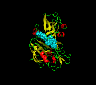
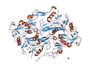
Aspartic proteases are a catalytic type of protease enzymes that use an activated water molecule bound to one or more aspartate residues for catalysis of their peptide substrates. In general, they have two highly conserved aspartates in the active site and are optimally active at acidic pH. Nearly all known aspartyl proteases are inhibited by pepstatin.

Thermolysin class of enzymes

Thermolysin is a thermostable neutral metalloproteinase enzyme produced by the Gram-positive bacteria Bacillus thermoproteolyticus. It requires one zinc ion for enzyme activity and four calcium ions for structural stability. Thermolysin specifically catalyzes the hydrolysis of peptide bonds containing hydrophobic amino acids. However thermolysin is also widely used for peptide bond formation through the reverse reaction of hydrolysis. Thermolysin is the most stable member of a family of metalloproteinases produced by various Bacillus species. These enzymes are also termed 'neutral' proteinases or thermolysin -like proteinases (TLPs).

Prosaposin protein-coding gene in the species Homo sapiens

Prosaposin, also known as PSAP, is a protein which in humans is encoded by the PSAP gene.

Proteinase K

In molecular biology Proteinase K is a broad-spectrum serine protease. The enzyme was discovered in 1974 in extracts of the fungus Engyodontium album. Proteinase K is able to digest hair (keratin), hence, the name "Proteinase K". The predominant site of cleavage is the peptide bond adjacent to the carboxyl group of aliphatic and aromatic amino acids with blocked alpha amino groups. It is commonly used for its broad specificity. This enzyme belongs to Peptidase family S8 (subtilisin). The molecular weight of Proteinase K is 28,900 daltons.

Saposin protein domain type of protein domain

The saposin domains refers to two evolutionally-conserved protein domains found in saposin and related proteins (SAPLIP). Saposins are small lysosomal proteins that serve as activators of various lysosomal lipid-degrading enzymes. They probably act by isolating the lipid substrate from the membrane surroundings, thus making it more accessible to the soluble degradative enzymes. All mammalian saposins are synthesized as a single precursor molecule (prosaposin) which contains four Saposin-B domains, yielding the active saposins after proteolytic cleavage, and two Saposin-A domains that are removed in the activation reaction.

Cathepsin E protein-coding gene in the species Homo sapiens

Cathepsin E is an enzyme that in humans is encoded by the CTSE gene. The enzyme is also known as slow-moving proteinase, erythrocyte membrane aspartic proteinase, SMP, EMAP, non-pepsin proteinase, cathepsin D-like acid proteinase, cathepsin E-like acid proteinase, cathepsin D-type proteinase) is an enzyme.

Nepenthesin is an aspartic protease of plant origin that has so far been identified in the pitcher secretions of Nepenthes and in the leaves of Drosera peltata. It is similar to pepsin, but differs in that it also cleaves on either side of Asp residues and at Lys┼Arg. While more pH and temperature stable than porcine pepsin A, it is considerably less stable in urea or guanidine hydrochloride. It is the only known protein with such a stability profile.

Kazal domain InterPro Domain

The Kazal domain is an evolutionary conserved protein domain usually indicative of serine protease inhibitors. However, kazal-like domains are also seen in the extracellular part of agrins, which are not known to be protease inhibitors.

CDC48 N-terminal domain

In molecular biology, the CDC48 N-terminal domain is a protein domain found in AAA ATPases including cell division protein 48 (CDC48), VCP-like ATPase and N-ethylmaleimide sensitive fusion protein. It is a substrate recognition domain which binds polypeptides, prevents protein aggregation, and catalyses refolding of permissive substrates. It is composed of two equally sized subdomains. The amino-terminal subdomain (CDC48_N) forms a double-psi beta-barrel whose pseudo-twofold symmetry is mirrored by an internal sequence repeat of 42 residues. The carboxy-terminal subdomain (CDC48_2) forms a novel six-stranded beta-clam fold. Together these subdomains form a kidney-shaped structure, in close agreement with results from electron microscopy. CDC48_N is related to numerous proteins including prokaryotic transcription factors, metabolic enzymes, the protease cofactors UFD1 and PrlF, and aspartic proteinases.

Streptogrisin B is an enzyme. This enzyme catalyses the following chemical reaction

Aspergillopepsin II

Aspergilloglutamic peptidase, also called aspergillopepsin II is a proteolytic enzyme. The enzyme was previously thought be an aspartic protease, but it was later shown to be a glutamic protease with a catalytic Glu residue at the active site, and was therefore renamed aspergilloglutamic peptidase.

Rhizopuspepsin is an enzyme. This enzyme catalyses the following chemical reaction

Mucorpepsin is an enzyme. This enzyme catalyses the following chemical reaction

Rhodotorulapepsin is an enzyme. This enzyme catalyses the following chemical reaction

Scytalidopepsin B

Scytalidocarboxyl peptidase B, also known as Scytalidoglutamic peptidase and Scytalidopepsin B is a proteolytic enzyme. It was previously thought to be an aspartic protease, but determination of its molecular structure showed it to belong a novel group of proteases, glutamic protease.

References

  1. 1 2 3 4 5 Bryksa BC, Bhaumik P, Magracheva E, De Moura DC, Kurylowicz M, Zdanov A, Dutcher JR, Wlodawer A, Yada RY (2011). "Structure and mechanism of the saposin-like domain of a plant aspartic proteinase". J Biol Chem. 286 (32): 28265–75. doi:10.1074/jbc.M111.252619. PMC   3151071 . PMID   21676875.
  2. 1 2 3 4 Asuman Mutlu & Susannah Gal (1999). "Plant aspartic proteinases: enzymes on the way to a function". Physiologia Plantarum. 105 (3): 569–576. doi:10.1034/j.1399-3054.1999.105324.x.
  3. 1 2 3 4 5 6 Egas C, Lavoura N, Resende R, Brito RM, Pires E, Maria de Lima CP, Faro C (2000). "The Saposin-like Domain of the Plant Aspartic Proteinase PrecursorIs a Potent Inducer of Vesicle Leakage". J Biol Chem. 275 (49): 38190–38196. doi: 10.1074/jbc.M006093200 . PMID   10982803.
  4. 1 2 3 4 Heike BRUHN (2005). "A short guided tour through functional and structural features of saposin-like proteins". Biochem J. 389 (15): 249–257. doi:10.1042/BJ20050051. PMC   1175101 . PMID   15992358.
  5. 1 2 Ponting CP, Russell RB (May 1995). "Swaposins: circular permutations within genes encoding saposin homologues". Trends in Biochemical Sciences. 20 (5): 179–80. doi:10.1016/S0968-0004(00)89003-9. PMID   7610480.
  6. 1 2 3 Kervinen J, Tobin GJ, Costa J, Waugh DS, Wlodawer A, Zdanov A (1999). "Crystal structure of plant aspartic proteinase prophytepsin: inactivation and vacuolar targeting". EMBO J. 18 (14): 3947–3955. doi:10.1093/emboj/18.14.3947. PMC   1171470 . PMID   10406799.
  7. Isaura Simões & Carlos Faro (2004). "Structure and function of plant aspartic proteinases". Eur J Biochem. 271 (11): 2067–2075. doi:10.1111/j.1432-1033.2004.04136.x. hdl: 10316/8129 . PMID   15153096.
  8. Bruhn H (2005). "A short guided tour through functional and structural features of saposin-like proteins". Biochem J. 389 (15): 249–257. doi:10.1042/BJ20050051. PMC   1175101 . PMID   15992358.
  9. Kolter T, Sandhoff K (2005). "Principles of lysosomal membrane digestion: stimulation of sphingolipid degradation by sphingolipid activator proteins and anionic lysosomal lipids". Annu Rev Cell Dev Biol. 21: 81–103. doi:10.1146/annurev.cellbio.21.122303.120013. PMID   16212488.
  10. Anderson DH, Sawaya MR, Cascio D, Ernst W, Modlin R, Krensky A, Eisenberg D (2003). "Granulysin crystal structure and a structure-derived lytic mechanism". J. Mol. Biol. 325 (2): 355–365. CiteSeerX   10.1.1.327.5540 . doi:10.1016/S0022-2836(02)01234-2. PMID   12488100.
  11. Gordon LM, Lee KY, Lipp MM, Zasadzinski JA, Walther FJ, Sherman MA, Waring AJ (2000). "Conformational mapping of the N-terminal segment of surfactant protein B in lipid using 13C-enhanced Fourier transform infrared spectroscopy". J Pept Res. 55 (4): 330–347. doi:10.1034/j.1399-3011.2000.00693.x. PMID   10798379.
  12. Staab JF, Ginkel DL, Rosenberg GB, Munford RS (1994). "A saposin-like domain influences the intracellular localization, stability, and catalytic activity of human acyloxyacyl hydrolase". J. Biol. Chem. 269 (38): 23736–42. PMID   8089145.
  13. Frey, María Eugenia; D'Ippolito, Sebastián; Pepe, Alfonso; Daleo, Gustavo Raúl; Guevara, María Gabriela (May 2018). "Transgenic expression of plant-specific insert of potato aspartic proteases (StAP-PSI) confers enhanced resistance to Botrytis cinerea in Arabidopsis thaliana". Phytochemistry. 149: 1–11. doi:10.1016/j.phytochem.2018.02.004. PMID   29428248.
  14. Muñoz, Fernando; Caracciolo, Pablo C.; Daleo, Gustavo; Abraham, Gustavo A.; Guevara, M. Gabriela (September 2014). "Evaluation of in vitro cytotoxic activity of mono-PEGylated StAP3 (Solanum tuberosum aspartic protease 3) forms". Biotechnology Reports. 3: 1–7. doi:10.1016/j.btre.2014.05.007. PMC   5466107 . PMID   28626641.