Prosaposin

Last updated
PSAP
Protein PSAP PDB 1m12.png
Available structures
PDB Ortholog search: PDBe RCSB
Identifiers
Aliases PSAP , GLBA, SAP1, prosaposin, SAP2, PSAPD, PARK24
External IDs OMIM: 176801 MGI: 97783 HomoloGene: 37680 GeneCards: PSAP
Orthologs
SpeciesHumanMouse
Entrez
Ensembl
UniProt
RefSeq (mRNA)

NM_002778
NM_001042465
NM_001042466

RefSeq (protein)

NP_001035930
NP_001035931
NP_002769
NP_002769.1

Location (UCSC) Chr 10: 71.82 – 71.85 Mb Chr 10: 60.11 – 60.14 Mb
PubMed search [3] [4]
Wikidata
View/Edit Human View/Edit Mouse

Prosaposin, also known as PSAP, is a protein which in humans is encoded by the PSAP gene. [5]

This highly conserved glycoprotein is a precursor for 4 cleavage products: saposins A, B, C, and D. Saposin is an acronym for Sphingolipid Activator PrO[S]teINs. [6] Each domain of the precursor protein is approximately 80 amino acid residues long with nearly identical placement of cysteine residues and glycosylation sites. Saposins A-D localize primarily to the lysosomal compartment where they facilitate the catabolism of glycosphingolipids with short oligosaccharide groups. The precursor protein exists both as a secretory protein and as an integral membrane protein and has neurotrophic activities. [5]

Saposins A–D are required for the hydrolysis of certain sphingolipids by specific lysosomal hydrolases. [7]

Family members

Structure

Every saposin contains about 80 amino acid residues and has six equally placed cysteines, two prolines, and a glycosylation site (two in saposin A, one each in saposins B, C, and D). [7] Since saposins characteristics of extreme heat-stability, abundance of disulfide linkages, and resistance to most proteases, they are assumed to be extremely compact and rigidly disulfide-linked molecules. Each saposin has an α-helical structure that is seen as being important for stimulation because this structure is maximal at a pH of 4.5; which is optimal for many lysosomal hydrolases. [7] This helical structure is seen in all (especially with the first region), but saposin has been predicted to have β-sheet configuration due to it first 24 amino acids of the N-end. [9]

Function

They probably act by isolating the lipid substrate from the membrane surroundings, thus making it more accessible to the soluble degradative enzymes. which contains four Saposin-B domains, yielding the active saposins after proteolytic cleavage, and two Saposin-A domains that are removed in the activation reaction. The Saposin-B domains also occur in other proteins, many of them active in the lysis of membranes. [14] [15]

Clinical significance

Mutations in this gene have been associated with Gaucher disease, Tay–Sachs disease, and metachromatic leukodystrophy. [6]

See also

Related Research Articles

Metachromatic leukodystrophy (MLD) is a lysosomal storage disease which is commonly listed in the family of leukodystrophies as well as among the sphingolipidoses as it affects the metabolism of sphingolipids. Leukodystrophies affect the growth and/or development of myelin, the fatty covering which acts as an insulator around nerve fibers throughout the central and peripheral nervous systems. MLD involves cerebroside sulfate accumulation. Metachromatic leukodystrophy, like most enzyme deficiencies, has an autosomal recessive inheritance pattern.

<span class="mw-page-title-main">Angiogenin</span> Protein-coding gene in the species Homo sapiens

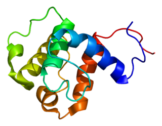
Angiogenin (ANG) also known as ribonuclease 5 is a small 123 amino acid protein that in humans is encoded by the ANG gene. Angiogenin is a potent stimulator of new blood vessels through the process of angiogenesis. Ang hydrolyzes cellular RNA, resulting in modulated levels of protein synthesis and interacts with DNA causing a promoter-like increase in the expression of rRNA. Ang is associated with cancer and neurological disease through angiogenesis and through activating gene expression that suppresses apoptosis.

<span class="mw-page-title-main">Arylsulfatase A</span>

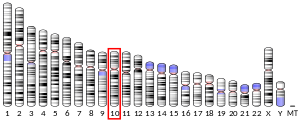
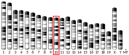
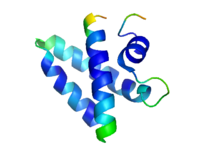
Arylsulfatase A is an enzyme that breaks down sulfatides, namely cerebroside 3-sulfate into cerebroside and sulfate. In humans, arylsulfatase A is encoded by the ARSA gene.

<span class="mw-page-title-main">Heat shock factor</span> Transcription factor

In molecular biology, heat shock factors (HSF), are the transcription factors that regulate the expression of the heat shock proteins. A typical example is the heat shock factor of Drosophila melanogaster.

<span class="mw-page-title-main">ARF1</span> Protein-coding gene in the species Homo sapiens

ADP-ribosylation factor 1 is a protein that in humans is encoded by the ARF1 gene.

<span class="mw-page-title-main">GPR135</span> Protein-coding gene in the species Homo sapiens

Probable G-protein coupled receptor 135 is a protein that in humans is encoded by the GPR135 gene.

<span class="mw-page-title-main">Saposin protein domain</span>

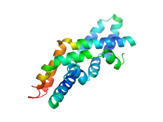
The saposin domains refers to two evolutionally-conserved protein domains found in saposin and related proteins (SAPLIP). Saposins are small lysosomal proteins that serve as activators of various lysosomal lipid-degrading enzymes. They probably act by isolating the lipid substrate from the membrane surroundings, thus making it more accessible to the soluble degradative enzymes. All mammalian saposins are synthesized as a single precursor molecule (prosaposin) which contains four Saposin-B domains, yielding the active saposins after proteolytic cleavage, and two Saposin-A domains that are removed in the activation reaction.

<span class="mw-page-title-main">PRKAA2</span> Protein-coding gene in the species Homo sapiens

5'-AMP-activated protein kinase catalytic subunit alpha-2 is an enzyme that in humans is encoded by the PRKAA2 gene.

<span class="mw-page-title-main">CUL2</span> Protein-coding gene in the species Homo sapiens

Cullin-2 is a protein that in humans is encoded by the CUL2 gene.

<span class="mw-page-title-main">RGS19</span> Protein-coding gene in the species Homo sapiens

Regulator of G-protein signaling 19 is a protein that in humans is encoded by the RGS19 gene.

<span class="mw-page-title-main">PASK</span> Protein-coding gene in the species Homo sapiens

PAS domain-containing serine/threonine-protein kinase is an enzyme that in humans is encoded by the PASK gene.

<span class="mw-page-title-main">UBR1</span> Mammalian protein found in Homo sapiens

The human gene UBR1 encodes the enzyme ubiquitin-protein ligase E3 component n-recognin 1.

<span class="mw-page-title-main">AP3M1</span> Protein-coding gene in the species Homo sapiens

AP-3 complex subunit mu-1 is a protein that in humans is encoded by the AP3M1 gene.

<span class="mw-page-title-main">ELK3</span> Protein-coding gene in humans

ETS domain-containing protein Elk-3 is a protein that in humans is encoded by the ELK3 gene.

<span class="mw-page-title-main">CLN8</span> Protein-coding gene in the species Homo sapiens

Protein CLN8 is a protein that in humans is encoded by the CLN8 gene.

<span class="mw-page-title-main">MAP2K1IP1</span> Protein-coding gene in the species Homo sapiens

Mitogen-activated protein kinase scaffold protein 1 is a scaffold protein that in humans is encoded by the MAPKSP1 gene.

<span class="mw-page-title-main">GNG5</span> Protein-coding gene in the species Homo sapiens

Guanine nucleotide-binding protein G(I)/G(S)/G(O) subunit gamma-5 is a protein that in humans is encoded by the GNG5 gene.

<span class="mw-page-title-main">PDE8A</span> Protein-coding gene in the species Homo sapiens

High affinity cAMP-specific and IBMX-insensitive 3',5'-cyclic phosphodiesterase 8A is an enzyme that in humans is encoded by the PDE8A gene. Work by Sebastiaan Bol et al. showed that 5 different transcript variants and their corresponding isoforms are expressed in human macrophages, and suggests that this protein may be required by HIV-1 for its replication.

Richard I. Morimoto is a Japanese American molecular biologist. He is the Bill and Gayle Cook Professor of Biology and Director of the Rice Institute for Biomedical Research at Northwestern University.

<span class="mw-page-title-main">Plant-specific insert</span>

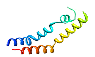
The plant-specific insert (PSI) or plant-specific sequence (PSS) is an independent domain, exclusively found in plants, consisting of approximately 100 residues, found on the C-terminal lobe on some aspartic proteases (AP) called phytepsins. The PSI, as an independent entity separate from its parent AP, is homologous to saposin and belongs to the saposin-like protein family (SAPLIP).

References

  1. 1 2 3 GRCh38: Ensembl release 89: ENSG00000197746 - Ensembl, May 2017
  2. 1 2 3 GRCm38: Ensembl release 89: ENSMUSG00000004207 - Ensembl, May 2017
  3. "Human PubMed Reference:". National Center for Biotechnology Information, U.S. National Library of Medicine.
  4. "Mouse PubMed Reference:". National Center for Biotechnology Information, U.S. National Library of Medicine.
  5. 1 2 "Entrez Gene: PSAP prosaposin (variant Gaucher disease and variant metachromatic leukodystrophy)".
  6. 1 2 Morimoto S, Yamamoto Y, O'Brien JS, Kishimoto Y (May 1990). "Distribution of saposin proteins (sphingolipid activator proteins) in lysosomal storage and other diseases". Proc. Natl. Acad. Sci. U.S.A. 87 (9): 3493–7. Bibcode:1990PNAS...87.3493M. doi: 10.1073/pnas.87.9.3493 . PMC   53927 . PMID   2110365.
  7. 1 2 3 4 Kishimoto Y, Hiraiwa M, O'Brien JS (September 1992). "Saposins: structure, function, distribution, and molecular genetics". J. Lipid Res. 33 (9): 1255–67. doi: 10.1016/S0022-2275(20)40540-1 . PMID   1402395.
  8. Morimoto S, Martin BM, Yamamoto Y, Kretz KA, O'Brien JS, Kishimoto Y (May 1989). "Saposin A: second cerebrosidase activator protein". Proc. Natl. Acad. Sci. U.S.A. 86 (9): 3389–93. Bibcode:1989PNAS...86.3389M. doi: 10.1073/pnas.86.9.3389 . PMC   287138 . PMID   2717620.
  9. 1 2 O'Brien JS, Kishimoto Y (March 1991). "Saposin proteins: structure, function, and role in human lysosomal storage disorders". FASEB J. 5 (3): 301–8. doi:10.1096/fasebj.5.3.2001789. PMID   2001789. S2CID   40251569.
  10. HUGO Gene Nomenclature Committee, "GM2A", HGNC database, retrieved 2016-03-13.
  11. Ahn VE, Leyko P, Alattia JR, Chen L, Privé GG (August 2006). "Crystal structures of saposins A and C". Protein Sci. 15 (8): 1849–57. doi:10.1110/ps.062256606. PMC   2242594 . PMID   16823039.
  12. Ahn VE, Faull KF, Whitelegge JP, Fluharty AL, Privé GG (January 2003). "Crystal structure of saposin B reveals a dimeric shell for lipid binding". Proc. Natl. Acad. Sci. U.S.A. 100 (1): 38–43. Bibcode:2003PNAS..100...38A. doi: 10.1073/pnas.0136947100 . PMC   140876 . PMID   12518053.
  13. 1 2 Rossmann M, Schultz-Heienbrok R, Behlke J, Remmel N, Alings C, Sandhoff K, Saenger W, Maier T (May 2008). "Crystal structures of human saposins C and D: implications for lipid recognition and membrane interactions". Structure. 16 (5): 809–17. doi: 10.1016/j.str.2008.02.016 . PMID   18462685.
  14. Ponting CP (1994). "Acid sphingomyelinase possesses a domain homologous to its activator proteins: saposins B and D". Protein Sci. 3 (2): 359–361. doi:10.1002/pro.5560030219. PMC   2142785 . PMID   8003971.
  15. Hofmann K, Tschopp J (1996). "Cytotoxic T cells: more weapons for new targets?". Trends Microbiol. 4 (3): 91–94. doi:10.1016/0966-842X(96)81522-8. PMID   8868085.

Further reading