Prefoldin

Last updated
Prefoldin
Prefoldin 3D Structure.jpg
This is the crystal structure of an archaeal prefoldin taken from the archaeon Methanobacterium thermoautotrophicum. The image was produced by Siegerts, R., Scheufler, C., Moarefi, I. using X-Ray diffraction. The heterohexameric protein complex contains two alpha subunits, and four beta subunits. ( PDB: 1FXK )
Identifiers
SymbolPrefoldin
InterPro IPR009053
CATH 1fxk
SCOP2 1fxk / SCOPe / SUPFAM

Prefoldin (GimC) is a superfamily of proteins used in protein folding complexes. It is classified as a heterohexameric molecular chaperone in both archaea and eukarya, including humans. A prefoldin molecule works as a transfer protein in conjunction with a molecule of chaperonin to form a chaperone complex and correctly fold other nascent proteins. One of prefoldin's main uses in eukarya is the formation of molecules of actin for use in the eukaryotic cytoskeleton.

Contents

Purpose and uses

Prefoldin is one family of chaperone proteins found in the domains of eukarya and archaea. Prefoldin acts in combination with other molecules to promote protein folding in cells where there are many other competing pathways for folding. [1] Chaperone proteins perform non-covalent assembly of other polypeptide-containing structures in vivo. They are implicated in the folding of most other proteins.

In archaea, prefoldins are believed to function in combination with group II chaperonins [2] in de novo protein folding. In eukarya however, prefoldins have acquired a more specific function: they are used to establish correct tubular assembly for many tubular proteins, such as actin. [3] Actin accounts for 5-10% of all protein found in eukaryotic cells, which therefore means that prefoldin is quite prevalent in the cells. Actin is made of two strings of beads wound round each other and is one of the three main parts of the cytoskeleton of eukaryotic cells. [4] Prefoldin bonds specifically to cytosolic chaperonin protein. This complex of prefoldin and chaperonin then forms molecules of actin in the cytosol. The prefoldin acts as a transporter molecule that transports bound, unfolded target proteins to the chaperonin (C-CPN) molecule. [3]

For example, the prefoldin that is used in the formation of actin also transfers α or β tubulin to a cytosolic chaperonin. The prefoldin, however, does not form a ternary complex with tubulin and chaperonin. Once the tubulins are in contact with the chaperonin, the prefoldin automatically lets go and leaves the active site, due to its high affinity for the chaperonin molecule. Once the prefoldin is in contact with the chaperonin protein, it loses its affinity for the unfolded target protein.

Prefoldin is triggered only to bind to nonnative target proteins in the cytosol so that it will only bind to unfolded proteins. Unlike many other molecular chaperones, prefoldin does not use chemical energy, in the form of adenosine triphosphate (ATP), to promote protein folding. [5]

Discovery

Prefoldin was found by the laboratory of Nicholas J. Cowan from the Department of Biochemistry at the New York University Medical Center. It was discovered using chromatography. Unfolded labeled β-actin from bovine testes was put into solution. This solution contained an excess of cytosolic chaperonin (C-CPN), a eukaryotic chaperone protein necessary for actin folding. After gel filtration of the actin, the actin complex, consisting of actin and its bonded proteins, began to form and the molecular weight of the complex was observed. Gel electrophoresis was used to analyze the protein complex, the complex formed a single band that was excised and ran on an SDS gel. It resolved into five bands, therefore proving that a heterooligomeric protein is used to bind to unfolded actin. [3]

An archaeal homolog of prefoldin that also functions as a molecular chaperone has been identified. [6] Eukaryotic prefoldin likely evolved from archaea, as it is not present (or has been lost) from bacteria.

Structure

Prefoldin is a hetero hexameric protein consisting of two α subunits and four β subunits. [7] [2] The beta subunits contain 120 amino acid residues each, while the α subunits contain 140 amino acid residues each. [2] Each subunit was found to have a width of 8.4 nm in the archaea Methanococcus thermoautrophicum. [2] The height was calculated at 1.8-2.6 nm. [2] The subunits are arranged by hydrophobic interactions with two β barrels at the center and coiled-coil α helices protruding down from them as if it were a jellyfish.

The lower "tentacles" of the jellyfish shape is the interface between prefoldin and chaperonin. [8]

Related Research Articles

<span class="mw-page-title-main">Microtubule</span> Polymer of tubulin that forms part of the cytoskeleton

Microtubules are polymers of tubulin that form part of the cytoskeleton and provide structure and shape to eukaryotic cells. Microtubules can be as long as 50 micrometres, as wide as 23 to 27 nm and have an inner diameter between 11 and 15 nm. They are formed by the polymerization of a dimer of two globular proteins, alpha and beta tubulin into protofilaments that can then associate laterally to form a hollow tube, the microtubule. The most common form of a microtubule consists of 13 protofilaments in the tubular arrangement.

<span class="mw-page-title-main">Actin</span> Family of proteins

Actin is a family of globular multi-functional proteins that form microfilaments in the cytoskeleton, and the thin filaments in muscle fibrils. It is found in essentially all eukaryotic cells, where it may be present at a concentration of over 100 μM; its mass is roughly 42 kDa, with a diameter of 4 to 7 nm.

<span class="mw-page-title-main">Tubulin</span> Superfamily of proteins that make up microtubules

Tubulin in molecular biology can refer either to the tubulin protein superfamily of globular proteins, or one of the member proteins of that superfamily. α- and β-tubulins polymerize into microtubules, a major component of the eukaryotic cytoskeleton. Microtubules function in many essential cellular processes, including mitosis. Tubulin-binding drugs kill cancerous cells by inhibiting microtubule dynamics, which are required for DNA segregation and therefore cell division.

<span class="mw-page-title-main">Chaperonin</span> InterPro Family

HSP60, also known as chaperonins (Cpn), is a family of heat shock proteins originally sorted by their 60kDa molecular mass. They prevent misfolding of proteins during stressful situations such as high heat, by assisting protein folding. HSP60 belong to a large class of molecules that assist protein folding, called molecular chaperones.

<span class="mw-page-title-main">DNA clamp</span>

A DNA clamp, also known as a sliding clamp, is a protein complex that serves as a processivity-promoting factor in DNA replication. As a critical component of the DNA polymerase III holoenzyme, the clamp protein binds DNA polymerase and prevents this enzyme from dissociating from the template DNA strand. The clamp-polymerase protein–protein interactions are stronger and more specific than the direct interactions between the polymerase and the template DNA strand; because one of the rate-limiting steps in the DNA synthesis reaction is the association of the polymerase with the DNA template, the presence of the sliding clamp dramatically increases the number of nucleotides that the polymerase can add to the growing strand per association event. The presence of the DNA clamp can increase the rate of DNA synthesis up to 1,000-fold compared with a nonprocessive polymerase.

<span class="mw-page-title-main">T-complex 1</span> Protein-coding gene in the species Homo sapiens

In humans, the gene T-complex 1, a.k.a. TCP1, encodes the protein TCP-1, a.k.a. T-complex protein 1 subunit alpha.

<span class="mw-page-title-main">CCT5 (gene)</span> Protein-coding gene in the species Homo sapiens

T-complex protein 1 subunit epsilon is a protein that in humans is encoded by the CCT5 gene.

<span class="mw-page-title-main">PFDN1</span> Protein-coding gene in the species Homo sapiens

Prefoldin subunit 1 is a protein that in humans is encoded by the PFDN1 gene.

<span class="mw-page-title-main">Prefoldin subunit 6</span> Protein-coding gene in the species Homo sapiens

Prefoldin subunit 6 is a protein that in humans is encoded by the PFDN6 gene.

<span class="mw-page-title-main">CCT7</span> Protein-coding gene in the species Homo sapiens

T-complex protein 1 subunit eta is a protein that in humans is encoded by the CCT7 gene.

<span class="mw-page-title-main">CCT6A</span> Protein-coding gene in the species Homo sapiens

T-complex protein 1 subunit zeta is a protein that in humans is encoded by the CCT6A gene.

<span class="mw-page-title-main">CCT2 (gene)</span> Protein-coding gene in humans

T-complex protein 1 subunit beta is a protein that in humans is encoded by the CCT2 gene.

<span class="mw-page-title-main">PFDN5</span> Protein-coding gene in the species Homo sapiens

Prefoldin subunit 5 is a protein that in humans is encoded by the PFDN5 gene.

<span class="mw-page-title-main">CCT3</span> Protein-coding gene in the species Homo sapiens

T-complex protein 1 subunit gamma is a protein that in humans is encoded by the CCT3 gene.

<span class="mw-page-title-main">PFDN2</span> Protein-coding gene in the species Homo sapiens

Prefoldin subunit 2 is a protein that in humans is encoded by the PFDN2 gene.

<span class="mw-page-title-main">PFDN4</span> Protein-coding gene in the species Homo sapiens

Prefoldin subunit 4 is a protein that in humans is encoded by the PFDN4 gene.

<span class="mw-page-title-main">CCT6B</span> Protein-coding gene in the species Homo sapiens

T-complex protein 1 subunit zeta-2 is a protein that in humans is encoded by the CCT6B gene.

Proteostasis is the dynamic regulation of a balanced, functional proteome. The proteostasis network includes competing and integrated biological pathways within cells that control the biogenesis, folding, trafficking, and degradation of proteins present within and outside the cell. Loss of proteostasis is central to understanding the cause of diseases associated with excessive protein misfolding and degradation leading to loss-of-function phenotypes, as well as aggregation-associated degenerative disorders. Therapeutic restoration of proteostasis may treat or resolve these pathologies.

Chaperones, also called molecular chaperones, are proteins that assist other proteins in assuming their three-dimensional fold, which is necessary for protein function. However, the fold of a protein is sensitive to environmental conditions, such as temperature and pH, and thus chaperones are needed to keep proteins in their functional fold across various environmental conditions. Chaperones are an integral part of a cell's protein quality control network by assisting in protein folding and are ubiquitous across diverse biological taxa. Since protein folding, and therefore protein function, is susceptible to environmental conditions, chaperones could represent an important cellular aspect of biodiversity and environmental tolerance by organisms living in hazardous conditions. Chaperones also affect the evolution of proteins in general, as many proteins fundamentally require chaperones to fold or are naturally prone to misfolding, and therefore mitigates protein aggregation.

<span class="mw-page-title-main">TRiC (complex)</span> Multiprotein complex used in cellular proteostasis

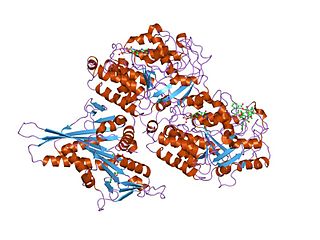
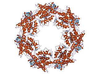
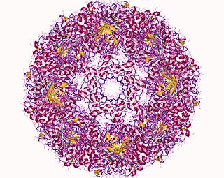
T-complex protein Ring Complex (TRiC), otherwise known as Chaperonin Containing TCP-1 (CCT), is a multiprotein complex and the chaperonin of eukaryotic cells. Like the bacterial GroEL, the TRiC complex aids in the folding of ~10% of the proteome, and actin and tubulin are some of its best known substrates. TRiC is an example of a biological machine that folds substrates within the central cavity of its barrel-like assembly using the energy from ATP hydrolysis.

References

  1. Oxford Dictionary of Biochemistry and Molecular Biology. Oxford: Oxford University Press. 1997.
  2. 1 2 3 4 5 Whitehead TA, Boonyaratanakornkit BB, Höllrigl V, Clark DS (April 2007). "A filamentous molecular chaperone of the prefoldin family from the deep-sea hyperthermophile Methanocaldococcus jannaschii". Protein Science. 16 (4): 626–34. doi:10.1110/ps.062599907. PMC   2203346 . PMID   17384227.
  3. 1 2 3 Vainberg IE, Lewis SA, Rommelaere H, Ampe C, Vandekerckhove J, Klein HL, Cowan NJ (May 1998). "Prefoldin, a chaperone that delivers unfolded proteins to cytosolic chaperonin". Cell. 93 (5): 863–73. doi: 10.1016/s0092-8674(00)81446-4 . PMID   9630229.
  4. Fruton JS (1999). Protein, Enzymes, Genes; The interplay of chemistry and Biology. New Haven: Yale University Press.
  5. Pockley A (2005). Molecular Chaperones and Cell Signaling. Cambridge: Cambridge University Press.
  6. Leroux MR, Fändrich M, Klunker D, Siegers K, Lupas AN, Brown JR, Schiebel E, Dobson CM, Hartl FU (December 1999). "MtGimC, a novel archaeal chaperone related to the eukaryotic chaperonin cofactor GimC/prefoldin". The EMBO Journal. 18 (23): 6730–43. doi:10.1093/emboj/18.23.6730. PMC   1171735 . PMID   10581246.
  7. Siegert R, Leroux MR, Scheufler C, Hartl FU, Moarefi I (November 2000). "Structure of the molecular chaperone prefoldin: unique interaction of multiple coiled coil tentacles with unfolded proteins". Cell. 103 (4): 621–32. doi: 10.1016/s0092-8674(00)00165-3 . PMID   11106732.
  8. Sahlan M, Zako T, Yohda M (April 2018). "Prefoldin, a jellyfish-like molecular chaperone: functional cooperation with a group II chaperonin and beyond". Biophysical Reviews. 10 (2): 339–345. doi:10.1007/s12551-018-0400-0. PMC   6420498 . PMID   29427249.