Protein A

Last updated
Protein A, Ig-binding domain
Protein A 1DEE 1L6X.png
Structure of a domain of protein A as a three-helix bundle binding to the heavy variable chain of a VH3 human Fab (left). [1] Minimized protein A bound to Fc fragment of Rituximab (right). [2] Uniprot P02976 .
Identifiers
SymbolSpA
Pfam PF02216
InterPro IPR003132
SCOP2 1DEE / SCOPe / SUPFAM
Available protein structures:
Pfam   structures / ECOD  
PDB RCSB PDB; PDBe; PDBj
PDBsum structure summary
PDB 1dee , 1l6x

Protein A is a 42 kDa surface protein originally found in the cell wall of the bacteria Staphylococcus aureus . It is encoded by the spa gene and its regulation is controlled by DNA topology, cellular osmolarity, and a two-component system called ArlS-ArlR. It has found use in biochemical research because of its ability to bind immunoglobulins. It is composed of five homologous Ig-binding domains that fold into a three-helix bundle. Each domain is able to bind proteins from many mammalian species, most notably IgGs. It binds the heavy chain within the Fc region of most immunoglobulins and also within the Fab region in the case of the human VH3 family. Through these interactions in serum, where IgG molecules are bound in the wrong orientation (in relation to normal antibody function), the bacteria disrupts opsonization and phagocytosis. [3]

Contents

History

As a by-product of his work on type-specific staphylococcus antigens, Verwey reported in 1940 that a protein fraction prepared from extracts of these bacteria non-specifically precipitated rabbit antisera raised against different staphylococcus types. [4] In 1958, Jensen confirmed Verwey's finding and showed that rabbit pre-immunization sera as well as normal human sera bound to the active component in the staphylococcus extract; he designated this component Antigen A (because it was found in fraction A of the extract) but thought it was a polysaccharide. [5] The misclassification of the protein was the result of faulty tests, [6] but it was not long thereafter (1962) that Löfkvist and Sjöquist corrected the error and confirmed that Antigen A was in fact a surface protein on the bacterial wall of certain strains of S. aureus. [7] The Bergen group from Norway named the protein "Protein A" after the antigen fraction isolated by Jensen. [8]

Protein A antibody binding

It has been shown via crystallographic refinement that the primary binding site for protein A is on the Fc region, between the CH2 and CH3 domains. [9] In addition, protein A has been shown to bind human IgG molecules containing IgG F(ab')2 fragments from the human VH3 gene family. [10]

Protein A can bind with strong affinity to the Fc portion of immunoglobulin of certain species as shown in the below table. [11]

SpeciesSubclassBinding
HumanIgAvariable
IgDweak or none
IgEweak or none
IgG1strong
IgG2strong
IgG3weak or none
IgG4strong
IgMvariable
Avian egg yolkIgYweak or none
Bovinemedium
Caninemedium
Goatweak or none
Guinea pigIgG1strong
Hamsterweak
Horsemedium
Koalaweak or none
Llamaweak or none
Monkey (rhesus)strong
MurineIgG1weak
IgG2astrong
IgG2medium to strong
IgG3medium
IgMvariable
Pigmedium to strong
Rabbitstrong
RatIgG1weak or none
IgG2aweak or none
IgG2bweak or none
IgG3weak
Sheepweak or none

Other antibody binding proteins

In addition to protein A, other immunoglobulin-binding bacterial proteins such as protein G, protein A/G and protein L are all commonly used to purify, immobilize or detect immunoglobulins.

Role in pathogenesis

As a pathogen, Staphylococcus aureus utilizes protein A, along with a host of other proteins and surface factors, to aid its survival and virulence. To this end, protein A plays a multifaceted role:

  1. By binding the Fc portion of antibodies, protein A renders them inaccessible to the opsonins, thus impairing phagocytosis of the bacteria via immune cell attack.
  2. Protein A facilitates the adherence of S. aureus to human von Willebrand factor (vWF)-coated surfaces, thus increasing the bacteria's infectiousness at the site of skin penetration.
  3. Protein A can inflame lung tissue by binding to tumor necrosis factor 1 (TNFR-1) receptors. This interaction has been shown to play a key role in the pathogenesis of staphylococcal pneumonia.
  4. Protein A has been shown to cripple humoral (antibody-mediated) immunity which in turn means that individuals can be repeatedly infected with S. aureus since they cannot mount a strong antibody response.
  5. Protein A has been shown to promote the formation of biofilms both when the protein is covalently linked to the bacterial cell wall as well as in solution. [12]

Protein A helps inhibit phagocytic engulfment and acts as an immunological disguise. Higher levels of protein A in different strains of S. aureus have been associated with nasal carriage of this bacteria. [13]

Mutants of S. aureus lacking protein A are more efficiently phagocytosed in vitro, and mutants in infection models have diminished virulence. [14]

Production

Protein A is produced and purified in industrial fermentation for use in immunology, biological research and industrial applications (see below). Natural (or native) protein A can be cultured in Staphylococcus aureus and contains the five homologous antibody binding regions described above and a C-terminal region for cell wall attachment. Today, protein A is more commonly produced recombinantly in Escherichia coli. ( Brevibacillus has also been shown to be an effective host. [15] ) Recombinant versions of protein A also contain the five homologous antibody binding domains but may vary in other parts of the structure in order to facilitate coupling to porous substrates. [16] Engineered versions of the protein are also available, the first of which was rProtein A, B4, C-CYS. [17] Engineered versions are multimers (typically tetramers, pentamers or hexamers) of a single domain which has been modified to improve usability in industrial applications.

Research

Protein A is often coupled to other molecules such as a fluorescent dye, enzymes, biotin, colloidal gold or radioactive iodine without affecting the antibody binding site. Examples including protein A–gold (PAG) stain is used in immunogold labelling, fluorophore coupled protein A for immunofluorescence, and DNA docking strand coupled protein A for DNA-PAINT imaging. [18] It is also widely utilized coupled to magnetic, latex and agarose beads.

Protein A is often immobilized onto a solid support and used as reliable method for purifying total IgG from crude protein mixtures such as serum or ascites fluid, or coupled with one of the above markers to detect the presence of antibodies. The first example of protein A being coupled to a porous bead for purification of IgG was published in 1972. [19] Immunoprecipitation studies with protein A conjugated to beads are also commonly used to purify proteins or protein complexes indirectly through antibodies against the protein or protein complex of interest.

Role in industrial purification of antibodies

This process flow diagram shows how monoclonal antibodies are typically purified at industrial scale. Platform Purification of Antibodies using Protein A.jpg
This process flow diagram shows how monoclonal antibodies are typically purified at industrial scale.

The first reference in the literature to a commercially available protein A chromatography resin appeared in 1976. [20] Today, chromatographic separation using protein A immobilized on porous substrates is the most widely established method for purifying monoclonal antibodies (mAbs) from harvest cell culture supernatant. [21] The choice of protein A as the preferred method is due to the high purity and yield which are easily and reliably achieved. This forms the basis for a general antibody purification "platform" which simplifies manufacturing operations and reduces the time and effort required to develop purification processes. [22] A typical mAb purification process is shown at right. Albeit the long history of protein A chromatography for the production of antibodies, the process is still being improved today. Continuous chromatography, more precisely periodic counter-current chromatography, enormously increases the productivity of the purification step.

Related Research Articles

<span class="mw-page-title-main">Antibody</span> Protein(s) forming a major part of an organisms immune system

An antibody (Ab) is the secreted form of a B cell receptor; the term immunoglobulin can refer to either the membrane-bound form or the secreted form of the B cell receptor, but they are, broadly speaking, the same protein, and so the terms are often treated as synonymous. Antibodies are large, Y-shaped proteins belonging to the immunoglobulin superfamily which are used by the immune system to identify and neutralize foreign objects such as bacteria and viruses, including those that cause disease. Antibodies can recognize virtually any size antigen with diverse chemical compositions from molecules. Each antibody recognizes one or more specific antigens. This term literally means "antibody generator", as it is the presence of an antigen that drives the formation of an antigen-specific antibody. Each tip of the "Y" of an antibody contains a paratope that specifically binds to one particular epitope on an antigen, allowing the two molecules to bind together with precision. Using this mechanism, antibodies can effectively "tag" a microbe or an infected cell for attack by other parts of the immune system, or can neutralize it directly.

<span class="mw-page-title-main">CD32</span> Surface receptor glycoprotein

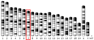
CD32, also known as FcγRII or FCGR2, is a surface receptor glycoprotein belonging to the Ig gene superfamily. CD32 can be found on the surface of a variety of immune cells. CD32 has a low-affinity for the Fc region of IgG antibodies in monomeric form, but high affinity for IgG immune complexes. CD32 has two major functions: cellular response regulation, and the uptake of immune complexes. Cellular responses regulated by CD32 include phagocytosis, cytokine stimulation, and endocytic transport. Dysregulated CD32 is associated with different forms of autoimmunity, including systemic lupus erythematosus. In humans, there are three major CD32 subtypes: CD32A, CD32B, and CD32C. While CD32A and CD32C are involved in activating cellular responses, CD32B is inhibitory.

<span class="mw-page-title-main">Monoclonal antibody</span> Antibodies from clones of the same blood cell

A monoclonal antibody is an antibody produced from a cell lineage made by cloning a unique white blood cell. All subsequent antibodies derived this way trace back to a unique parent cell.

<span class="mw-page-title-main">Superantigen</span> Antigen which strongly activates the immune system

Superantigens (SAgs) are a class of antigens that result in excessive activation of the immune system. Specifically they cause non-specific activation of T-cells resulting in polyclonal T cell activation and massive cytokine release. Superantigens act by binding to the MHC proteins on antigen-presenting cells (APCs) and to the TCRs on their adjacent helper T-cells, bringing the signaling molecules together, and thus leading to the activation of the T-cells, regardless of the peptide displayed on the MHC molecule. SAgs are produced by some pathogenic viruses and bacteria most likely as a defense mechanism against the immune system. Compared to a normal antigen-induced T-cell response where 0.0001-0.001% of the body's T-cells are activated, these SAgs are capable of activating up to 20% of the body's T-cells. Furthermore, Anti-CD3 and Anti-CD28 antibodies (CD28-SuperMAB) have also shown to be highly potent superantigens.

<span class="mw-page-title-main">Classical complement pathway</span> Aspect of the immune system

The classical complement pathway is one of three pathways which activate the complement system, which is part of the immune system. The classical complement pathway is initiated by antigen-antibody complexes with the antibody isotypes IgG and IgM.

Affinity chromatography is a method of separating a biomolecule from a mixture, based on a highly specific macromolecular binding interaction between the biomolecule and another substance. The specific type of binding interaction depends on the biomolecule of interest; antigen and antibody, enzyme and substrate, receptor and ligand, or protein and nucleic acid binding interactions are frequently exploited for isolation of various biomolecules. Affinity chromatography is useful for its high selectivity and resolution of separation, compared to other chromatographic methods.

<span class="mw-page-title-main">Single-chain variable fragment</span> Fragment

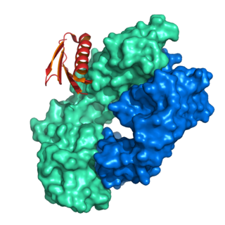
A single-chain variable fragment (scFv) is not actually a fragment of an antibody, but instead is a fusion protein of the variable regions of the heavy (VH) and light chains (VL) of immunoglobulins, connected with a short linker peptide of ten to about 25 amino acids. The linker is usually rich in glycine for flexibility, as well as serine or threonine for solubility, and can either connect the N-terminus of the VH with the C-terminus of the VL, or vice versa. This protein retains the specificity of the original immunoglobulin, despite removal of the constant regions and the introduction of the linker. The image to the right shows how this modification usually leaves the specificity unaltered.

<span class="mw-page-title-main">Antibody-dependent cellular cytotoxicity</span> Cell-mediated killing of other cells mediated by antibodies

Antibody-dependent cellular cytotoxicity (ADCC), also referred to as antibody-dependent cell-mediated cytotoxicity, is a mechanism of cell-mediated immune defense whereby an effector cell of the immune system kills a target cell, whose membrane-surface antigens have been bound by specific antibodies. It is one of the mechanisms through which antibodies, as part of the humoral immune response, can act to limit and contain infection.

<span class="mw-page-title-main">Fragment crystallizable region</span> Tail region of an antibody

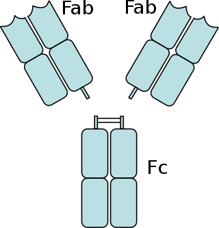
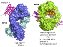
The fragment crystallizable region is the tail region of an antibody that interacts with cell surface receptors called Fc receptors and some proteins of the complement system. This region allows antibodies to activate the immune system, for example, through binding to Fc receptors. In IgG, IgA and IgD antibody isotypes, the Fc region is composed of two identical protein fragments, derived from the second and third constant domains of the antibody's two heavy chains; IgM and IgE Fc regions contain three heavy chain constant domains in each polypeptide chain. The Fc regions of IgGs bear a highly conserved N-glycosylation site. Glycosylation of the Fc fragment is essential for Fc receptor-mediated activity. The N-glycans attached to this site are predominantly core-fucosylated diantennary structures of the complex type. In addition, small amounts of these N-glycans also bear bisecting GlcNAc and α-2,6 linked sialic acid residues.

<span class="mw-page-title-main">CD22</span> Lectin molecule

CD22, or cluster of differentiation-22, is a molecule belonging to the SIGLEC family of lectins. It is found on the surface of mature B cells and to a lesser extent on some immature B cells. Generally speaking, CD22 is a regulatory molecule that prevents the overactivation of the immune system and the development of autoimmune diseases.

MSCRAMM adhesin proteins mediate the initial attachment of bacteria to host tissue, providing a critical step to establish infection.

<span class="mw-page-title-main">Hemolysin</span> Molecule destroying the membrane of red blood cells

Hemolysins or haemolysins are lipids and proteins that cause lysis of red blood cells by disrupting the cell membrane. Although the lytic activity of some microbe-derived hemolysins on red blood cells may be of great importance for nutrient acquisition, many hemolysins produced by pathogens do not cause significant destruction of red blood cells during infection. However, hemolysins are often capable of lysing red blood cells in vitro.

<span class="mw-page-title-main">Isotype (immunology)</span>

In immunology, antibodies are classified into several types called isotypes or classes. The variable (V) regions near the tip of the antibody can differ from molecule to molecule in countless ways, allowing it to specifically target an antigen . In contrast, the constant (C) regions only occur in a few variants, which define the antibody's class. Antibodies of different classes activate distinct effector mechanisms in response to an antigen . They appear at different stages of an immune response, differ in structural features, and in their location around the body.

<span class="mw-page-title-main">Protein L</span>

Protein L was first isolated from the surface of bacterial species Peptostreptococcus magnus and was found to bind immunoglobulins through L chain interaction, from which the name was suggested. It consists of 719 amino acid residues. The molecular weight of protein L purified from the cell walls of Peptostreptoccus magnus was first estimated as 95kD by SDS-PAGE in the presence of reducing agent 2-mercaptoethanol, while the molecular weight was determined to 76kD by gel chromatography in the presence of 6 M guanidine HCl. Protein L does not contain any interchain disulfide loops, nor does it consist of disulfide-linked subunits. It is an acidic molecule with a pI of 4.0. Unlike protein A and protein G, which bind to the Fc region of immunoglobulins (antibodies), protein L binds antibodies through light chain interactions. Since no part of the heavy chain is involved in the binding interaction, Protein L binds a wider range of antibody classes than protein A or G. Protein L binds to representatives of all antibody classes, including IgG, IgM, IgA, IgE and IgD. Single chain variable fragments (scFv) and Fab fragments also bind to protein L.

Protein A/G is a recombinant fusion protein that combines IgG binding domains of both protein A and protein G. Protein A/G contains four Fc binding domains from protein A and two from protein G, yielding a final mass of 50,460 daltons. The binding of protein A/G is less pH-dependent than protein A, but otherwise has the additive properties of protein A and G.

Small modular immunopharmaceuticals, or SMIPs for short, are artificial proteins that are intended for use as pharmaceutical drugs. They are largely built from parts of antibodies (immunoglobulins), and like them have a binding site for antigens that could be used for monoclonal antibody therapy. SMIPs have similar biological half-life and, being smaller than antibodies, are reasoned to have better tissue penetration properties. They were invented by Trubion and are now being developed by Emergent BioSolutions, which acquired Trubion in 2010.

A bispecific monoclonal antibody is an artificial protein that can simultaneously bind to two different types of antigen or two different epitopes on the same antigen. Naturally occurring antibodies typically only target one antigen. BsAbs can be manufactured in several structural formats. BsAbs can be designed to recruit and activate immune cells, to interfere with receptor signaling and inactivate signaling ligands, and to force association of protein complexes. BsAbs have been explored for cancer immunotherapy, drug delivery, and Alzeimer's disease.

<span class="mw-page-title-main">Affitin</span>

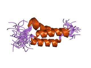
Affitins are artificial proteins with the ability to selectively bind antigens. They are structurally derived from the DNA binding protein Sac7d, found in Sulfolobus acidocaldarius, a microorganism belonging to the archaeal domain. By randomizing the amino acids on the binding surface of Sac7d and subjecting the resulting protein library to rounds of ribosome display, the affinity can be directed towards various targets, such as peptides, proteins, viruses, and bacteria.

<span class="mw-page-title-main">SpAB protein domain</span>

In molecular biology, the domain B, refers to the immunoglobulin-binding domain found in the Staphylococcus aureus virulence factor protein A (SpA). Hence, it is abbreviated to SpAB.

Recombinant antibodies are antibody fragments produced by using recombinant antibody coding genes. They mostly consist of a heavy and light chain of the variable region of immunoglobulin. Recombinant antibodies have many advantages in both medical and research applications, which make them a popular subject of exploration and new production against specific targets. The most commonly used form is the single chain variable fragment (scFv), which has shown the most promising traits exploitable in human medicine and research. In contrast to monoclonal antibodies produced by hybridoma technology, which may lose the capacity to produce the desired antibody over time or the antibody may undergo unwanted changes, which affect its functionality, recombinant antibodies produced in phage display maintain high standard of specificity and low immunogenicity.

References

  1. Graille M, Stura EA, Corper AL, Sutton BJ, Taussig MJ, Charbonnier JB, Silverman GJ (May 2000). "Crystal structure of a Staphylococcus aureus protein A domain complexed with the Fab fragment of a human IgM antibody: structural basis for recognition of B-cell receptors and superantigen activity". Proceedings of the National Academy of Sciences of the United States of America. 97 (10): 5399–404. Bibcode:2000PNAS...97.5399G. doi: 10.1073/pnas.97.10.5399 . PMC   25840 . PMID   10805799.
  2. Idusogie EE, Presta LG, Gazzano-Santoro H, Totpal K, Wong PY, Ultsch M, et al. (April 2000). "Mapping of the C1q binding site on rituxan, a chimeric antibody with a human IgG1 Fc". Journal of Immunology. 164 (8): 4178–84. doi: 10.4049/jimmunol.164.8.4178 . PMID   10754313.
  3. Keener AB, Thurlow LT, Kang S, Spidale NA, Clarke SH, Cunnion KM, et al. (February 2017). "Staphylococcus aureus Protein A Disrupts Immunity Mediated by Long-Lived Plasma Cells". Journal of Immunology. 198 (3): 1263–1273. doi:10.4049/jimmunol.1600093. PMC   5266639 . PMID   28031339.
  4. Verwey WF (April 1940). "A type-specific antigenic protein derived from the staphylococcus". The Journal of Experimental Medicine. 71 (5): 635–44. doi:10.1084/jem.71.5.635. PMC   2135093 . PMID   19870987.
  5. Jensen, K (1958). "A normally occurring staphylococcus antibody in human serum". Acta Pathol. Microbiol. Scand. 44 (4): 421–428. doi:10.1111/j.1699-0463.1958.tb01093.x. PMID   17504410.
  6. Dixon, Frank J. (Aug 11, 1982). ADVANCES IN IMMUNOLOGY . Academic Press. p.  158. ISBN   9780120224333.
  7. Löfkvist T, Sjöquist J (November 1962). "Chemical and serological analysis of antigen preparations from staphylococcus aureus". Acta Pathologica et Microbiologica Scandinavica. 56 (3): 295–304. doi:10.1111/j.1699-0463.1962.tb04908.x.
  8. Grov A, Myklestad B, Oeding P (1964). "Immunochemical studies on antigen preparations from staphylococcus aureus. 1. Isolation and chemical characterization of Antigen A". Acta Pathologica et Microbiologica Scandinavica. 61 (4): 588–96. doi:10.1111/apm.1964.61.4.588. PMID   14185494.
  9. Deisenhofer J (April 1981). "Crystallographic refinement and atomic models of a human Fc fragment and its complex with fragment B of protein A from Staphylococcus aureus at 2.9- and 2.8-A resolution". Biochemistry. 20 (9): 2361–70. doi:10.1021/bi00512a001. PMID   7236608.
  10. Sasso EH, Silverman GJ, Mannik M (September 1991). "Human IgA and IgG F(ab')2 that bind to staphylococcal protein A belong to the VHIII subgroup". Journal of Immunology. 147 (6): 1877–83. doi: 10.4049/jimmunol.147.6.1877 . PMID   1909733. S2CID   35812099.
  11. Affinity Chromatography (PDF). Vol. 1: Antibodies (AF ed.). GE Healthcare. 2016. p. 48.
  12. "A Pathogen's Swiss Army Knife". Small Things Considered. Retrieved 2016-08-25.
  13. Muthukrishnan G, Quinn GA, Lamers RP, Diaz C, Cole AL, Chen S, Cole AM (April 2011). "Exoproteome of Staphylococcus aureus reveals putative determinants of nasal carriage". Journal of Proteome Research. 10 (4): 2064–78. doi:10.1021/pr200029r. PMC   3070068 . PMID   21338050.
  14. Goodyear CS, Silverman GJ (May 2003). "Death by a B cell superantigen: In vivo VH-targeted apoptotic supraclonal B cell deletion by a Staphylococcal Toxin". The Journal of Experimental Medicine. 197 (9): 1125–39. doi:10.1084/jem.20020552. PMC   2193973 . PMID   12719481.
  15. Kosugi A, JP, Yajima K, JP (November 18, 2014), United States Patent: 8889389 - Process for producing protein A-like protein with use of Brevibacillus genus bacterium , retrieved 2016-08-26
  16. "usp31nf26s1_c130". www.uspbpep.com. General Chapters: PROTEIN A QUALITY ATTRIBUTES. Retrieved 2016-08-26.
  17. Hober S (June 12, 2012), United States Patent: 8198404 - Mutated immunoglobulin-binding protein , retrieved 2016-08-26
  18. Schlichthaerle T, Ganji M, Auer A, Kimbu Wade O, Jungmann R (April 2019). "Bacterially Derived Antibody Binders as Small Adapters for DNA-PAINT Microscopy". ChemBioChem. 20 (8): 1032–1038. doi:10.1002/cbic.201800743. hdl: 21.11116/0000-0003-E68E-A . PMID   30589198. S2CID   58547594.
  19. Hjelm H, Hjelm K, Sjöquist J (November 1972). "Protein A from Staphylococcus aureus. Its isolation by affinity chromatography and its use as an immunosorbent for isolation of immunoglobulins". FEBS Letters. 28 (1): 73–6. doi: 10.1016/0014-5793(72)80680-X . PMID   4630462. S2CID   8733702.
  20. Skvaril, F. (1976-10-01). "The question of specificity in binding human IgG subclasses to protein A-sepharose". Immunochemistry. 13 (10): 871–872. doi:10.1016/0019-2791(76)90188-9. PMID   12109.
  21. Shukla AA, Hubbard B, Tressel T, Guhan S, Low D (March 2007). "Downstream processing of monoclonal antibodies--application of platform approaches". Journal of Chromatography. B, Analytical Technologies in the Biomedical and Life Sciences. Polyclonal and Monoclonal Antibody Production, Purification, Process and Product Analytics. 848 (1): 28–39. doi:10.1016/j.jchromb.2006.09.026. PMID   17046339.
  22. Liu HF, Ma J, Winter C, Bayer R (2010-09-01). "Recovery and purification process development for monoclonal antibody production". mAbs. 2 (5): 480–99. doi:10.4161/mabs.2.5.12645. PMC   2958570 . PMID   20647768.