Related Research Articles

Aortic stenosis is the narrowing of the exit of the left ventricle of the heart, such that problems result. It may occur at the aortic valve as well as above and below this level. It typically gets worse over time. Symptoms often come on gradually with a decreased ability to exercise often occurring first. If heart failure, loss of consciousness, or heart related chest pain occur due to AS the outcomes are worse. Loss of consciousness typically occurs with standing or exercising. Signs of heart failure include shortness of breath especially when lying down, at night, or with exercise, and swelling of the legs. Thickening of the valve without causing obstruction is known as aortic sclerosis.

A heart valve is a biological one-way valve that allows blood to flow in one direction through the chambers of the heart. Four valves are usually present in a mammalian heart and together they determine the pathway of blood flow through the heart. A heart valve opens or closes according to differential blood pressure on each side.

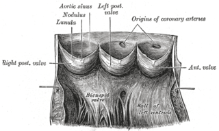
The aortic valve is a valve in the heart of humans and most other animals, located between the left ventricle and the aorta. It is one of the four valves of the heart and one of the two semilunar valves, the other being the pulmonary valve. The aortic valve normally has three cusps or leaflets, although in 1–2% of the population it is found to congenitally have two leaflets. The aortic valve is the last structure in the heart the blood travels through before stopping the flow through the systemic circulation.
The mitral valve, also known as the bicuspid valve or left atrioventricular valve, is one of the four heart valves. It has two cusps or flaps and lies between the left atrium and the left ventricle of the heart. The heart valves are all one-way valves allowing blood flow in just one direction. The mitral valve and the tricuspid valve are known as the atrioventricular valves because they lie between the atria and the ventricles.

Echocardiography, also known as cardiac ultrasound, is the use of ultrasound to examine the heart. It is a type of medical imaging, using standard ultrasound or Doppler ultrasound. The visual image formed using this technique is called an echocardiogram, a cardiac echo, or simply an echo.

Aortic regurgitation (AR), also known as aortic insufficiency (AI), is the leaking of the aortic valve of the heart that causes blood to flow in the reverse direction during ventricular diastole, from the aorta into the left ventricle. As a consequence, the cardiac muscle is forced to work harder than normal.

Interventional cardiology is a branch of cardiology that deals specifically with the catheter based treatment of structural heart diseases. Andreas Gruentzig is considered the father of interventional cardiology after the development of angioplasty by interventional radiologist Charles Dotter.

Bicuspid aortic valve (BAV) is a form of heart disease in which two of the leaflets of the aortic valve fuse during development in the womb resulting in a two-leaflet (bicuspid) valve instead of the normal three-leaflet (tricuspid) valve. BAV is the most common cause of heart disease present at birth and affects approximately 1.3% of adults. Normally, the mitral valve is the only bicuspid valve and this is situated between the heart's left atrium and left ventricle. Heart valves play a crucial role in ensuring the unidirectional flow of blood from the atrium to the ventricles, or from the ventricle to the aorta or pulmonary trunk. BAV is normally inherited.

A transesophageal echocardiogram, or TEE, is an alternative way to perform an echocardiogram. A specialized probe containing an ultrasound transducer at its tip is passed into the patient's esophagus. This allows image and Doppler evaluation which can be recorded. It is commonly used during cardiac surgery and is an excellent modality for assessing the aorta, although there are some limitations.
Aneurysm of the aortic sinus, also known as the sinus of Valsalva, is a rare abnormality of the aorta, the largest artery in the body. The aorta normally has three small pouches that sit directly above the aortic valve, and an aneurysm of one of these sinuses is a thin-walled swelling. Aneurysms may affect the right (65–85%), non-coronary (10–30%), or rarely the left coronary sinus. These aneurysms may not cause any symptoms but if large can cause shortness of breath, palpitations or blackouts. Aortic sinus aneurysms can burst or rupture into adjacent cardiac chambers, which can lead to heart failure if untreated.

Mitral regurgitation (MR), also known as mitral insufficiency or mitral incompetence, is a form of valvular heart disease in which the mitral valve is insufficient and does not close properly when the heart pumps out blood. It is the abnormal leaking of blood backwards – regurgitation from the left ventricle, through the mitral valve, into the left atrium, when the left ventricle contracts. Mitral regurgitation is the most common form of valvular heart disease.
Aortic valve replacement is a procedure whereby the failing aortic valve of a patient's heart is replaced with an artificial heart valve. The aortic valve may need to be replaced because:
A transthoracic echocardiogram (TTE) is the most common type of echocardiogram, which is a still or moving image of the internal parts of the heart using ultrasound. In this case, the probe is placed on the chest or abdomen of the subject to get various views of the heart. It is used as a non-invasive assessment of the overall health of the heart, including a patient's heart valves and degree of heart muscle contraction. The images are displayed on a monitor for real-time viewing and then recorded.

Valvular heart disease is any cardiovascular disease process involving one or more of the four valves of the heart. These conditions occur largely as a consequence of aging, but may also be the result of congenital (inborn) abnormalities or specific disease or physiologic processes including rheumatic heart disease and pregnancy.
Aortic valve repair or aortic valve reconstruction is the reconstruction of both form and function of a dysfunctional aortic valve. Most frequently it is used for the treatment of aortic regurgitation. It can also become necessary for the treatment of aortic aneurysm, less frequently for congenital aortic stenosis.
The following outline is provided as an overview of and topical guide to cardiology, the branch of medicine dealing with disorders of the human heart. The field includes medical diagnosis and treatment of congenital heart defects, coronary artery disease, heart failure, valvular heart disease and electrophysiology. Physicians who specialize in cardiology are called cardiologists.
Cardiothoracic anesthesiology is a subspeciality of the medical practice of anesthesiology, devoted to the preoperative, intraoperative, and postoperative care of adult and pediatric patients undergoing cardiothoracic surgery and related invasive procedures.
Cardiac imaging refers to minimally invasive imaging of the heart using ultrasound, magnetic resonance imaging (MRI), computed tomography (CT), or nuclear medicine (NM) imaging with PET or SPECT. These cardiac techniques are otherwise referred to as echocardiography, Cardiac MRI, Cardiac CT, Cardiac PET and Cardiac SPECT including myocardial perfusion imaging.
Transcatheter pulmonary valve replacement (TPVR), also known as percutaneous pulmonary valve implantation (PPVI), is the replacement of the pulmonary valve via catheterization through a vein. It is a significantly less invasive procedure in comparison to open heart surgery and is commonly used to treat conditions such as pulmonary atresia.
Mitral annular calcification (MAC) is a multifactorial chronic degenerative process in which calcium with lipid is deposited (calcified) in the annular fibrosa ring of the heart's mitral valve. MAC was first discovered and described in 1908 by M. Bonninger in the journal Deutsche Medizinische Wochenschrift. In the majority of cases, affected patients are asymptomatic and the condition is only noted incidentally on echocardiography or computed tomography (CT) scans. However, mitral annular calcification remains clinically significant because while in many cases the calcification is limited to the annulus and proximal leaflet bases, it may also extend further into the valve structure. This may potentially cause mitral regurgitation (MR) or more rarely mitral stenosis (MS), which may produce the classic symptoms of these conditions over time. In addition, calcification of the annulus can inhibit electrical conduction of the AV node, consequently causing various degrees of heart block. While MAC does not usually necessitate treatment independently, the degree of calcification present in the annulus is an important factor in choosing the most appropriate treatment modality for several conditions that do require intervention, particularly those that cause symptomatic obstruction of left ventricular outflow (LVOT).
References
- 1 2 3 4 Hurwitz, L. E., & Roberts, W. C. (1973). Quadricuspid semilunar valve. The American Journal of Cardiology, 31(5), 623–626.
- 1 2 Tutarel, O. (2004). The quadricuspid aortic valve: a comprehensive review. The Journal of Heart Valve Disease, 13(4), 534-537.
- 1 2 3 Zhu, J., Zhang, J., Wu, S., Zhang, Y., Ding, F., & Mei, J. (2013). Congenital quadricuspid aortic valve associated with aortic insufficiency and mitral regurgitation. Journal of Cardiothoracic Surgery, 8(1), 87.
- 1 2 3 4 5 Jagannath, A. D., Johri, A. M., Liberthson, R., Larobina, M., Passeri, J., Tighe, D., & Agnihotri, A. K. (2011). Quadricuspid aortic valve: a report of 12 cases and a review of the literature. Echocardiography, 28(9), 1035-1040.
- 1 2 3 Godefroid, O., Colles, P., Vercauteren, S., Louagie, Y., & Marchandise, B. (2006). Quadricuspid aortic valve: a rare etiology of aortic regurgitation. European Journal of Echocardiography, 7(2), 168-170.
- 1 2 Douglas, H., Moore, M., & Purvis, J. (2012). Comprehensive assessment of a quadricuspid aortic valve and coronary arteries by multidetector cardiac CT. Heart, 98(24), 1838-1838.
- ↑ Janosi, R. A., Lind, T., Buck, T., & Erbel, R. (2013). Quadricuspid Aortic Valve Revealed by Real-Time, 3-Dimensional Transesophageal Echocardiography. Texas Heart Institute Journal, 40(2), 207.
- ↑ Song, M. G., Yang, H. S., Lee, D. H., Shin, J. K., Chee, H. K., & Kim, J. S. (2014). Mid-term results in patients having tricuspidization of the quadricuspid aortic valve. Journal of cardiothoracic surgery, 9(1), 29.