Related Research Articles
In physiology, nociception, also nocioception; from Latin nocere 'to harm/hurt') is the sensory nervous system's process of encoding noxious stimuli. It deals with a series of events and processes required for an organism to receive a painful stimulus, convert it to a molecular signal, and recognize and characterize the signal to trigger an appropriate defensive response.

The brainstem is the posterior stalk-like part of the brain that connects the cerebrum with the spinal cord. In the human brain the brainstem is composed of the midbrain, the pons, and the medulla oblongata. The midbrain is continuous with the thalamus of the diencephalon through the tentorial notch, and sometimes the diencephalon is included in the brainstem.

The grey columns are three regions of the somewhat ridge-shaped mass of grey matter in the spinal cord. These regions present as three columns: the anterior grey column, the posterior grey column, and the lateral grey column, all of which are visible in cross-section of the spinal cord.

A nociceptor is a sensory neuron that responds to damaging or potentially damaging stimuli by sending "possible threat" signals to the spinal cord and the brain. The brain creates the sensation of pain to direct attention to the body part, so the threat can be mitigated; this process is called nociception.

The spinothalamic tract is a nerve tract in the anterolateral system in the spinal cord. This tract is an ascending sensory pathway to the thalamus. From the ventral posterolateral nucleus in the thalamus, sensory information is relayed upward to the somatosensory cortex of the postcentral gyrus.

The periaqueductal gray (PAG), also known as the central gray, is a brain region that plays a critical role in autonomic function, motivated behavior and behavioural responses to threatening stimuli. PAG is also the primary control center for descending pain modulation. It has enkephalin-producing cells that suppress pain.

The reticular formation is a set of interconnected nuclei in the brainstem that spans from the lower end of the medulla oblongata to the upper end of the midbrain. The neurons of the reticular formation make up a complex set of neural networks in the core of the brainstem. It is not anatomically well defined, because it includes neurons located in different parts of the brain.
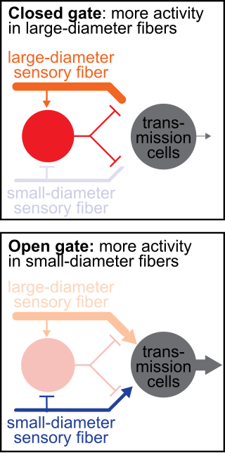
The gate control theory of pain asserts that non-painful input closes the nerve "gates" to painful input, which prevents pain sensation from traveling to the central nervous system.

The vestibulospinal tract is a neural tract in the central nervous system. Specifically, it is a component of the extrapyramidal system and is classified as a component of the medial pathway. Like other descending motor pathways, the vestibulospinal fibers of the tract relay information from nuclei to motor neurons. The vestibular nuclei receive information through the vestibulocochlear nerve about changes in the orientation of the head. The nuclei relay motor commands through the vestibulospinal tract. The function of these motor commands is to alter muscle tone, extend, and change the position of the limbs and head with the goal of supporting posture and maintaining balance of the body and head.

The nucleus raphe magnus (NRM) is one of the seven raphe nuclei. It is situated in the pons in the brainstem, just rostral to the nucleus raphe obscurus.

The apex of the posterior grey column, one of the three grey columns of the spinal cord, is capped by a V-shaped or crescentic mass of translucent, gelatinous neuroglia, termed the substantia gelatinosa of Rolando, which contains both neuroglia cells, and small neurons. The gelatinous appearance is due to an abundance of neuropil with a very low concentration of myelinated fibers. It extends the entire length of the spinal cord and into the medulla oblongata where it becomes the spinal trigeminal nucleus.

The posterior thoracic nucleus, is a group of interneurons found in the medial part of lamina VII, also known as the intermediate zone, of the spinal cord. It is located from the cervical segment C8 to lumbar segment L3 of the spinal cord and is an important structure for proprioception of the lower limb.
The Rexed laminae comprise a system of ten layers of grey matter (I–X), identified in the early 1950s by Bror Rexed to label portions of the grey columns of the spinal cord.

Alpha (α) motor neurons (also called alpha motoneurons), are large, multipolar lower motor neurons of the brainstem and spinal cord. They innervate extrafusal muscle fibers of skeletal muscle and are directly responsible for initiating their contraction. Alpha motor neurons are distinct from gamma motor neurons, which innervate intrafusal muscle fibers of muscle spindles.
The gigantocellular reticular nucleus is the (efferent/motor) medial zone of the reticular formation of the caudal pons and rostral medulla oblongata. It consists of a substantial number of giant neurons, but also contains small and medium sized neurons.
The spinoreticular tract is a partially decussating (crossed-over) four-neuron sensory pathway of the central nervous system. The tract transmits slow nociceptive/pain information from the spinal cord to reticular formation which in turn relays the information to the thalamus via reticulothalamic fibers as well as to other parts of the brain. Most (85%) second-order axons arising from sensory C first-order fibers ascend in the spinoreticular tract - it is consequently responsible for transmiting "slow", dull, poorly-localised pain. By projecting to the reticular activating system (RAS), the tract also mediates arousal/alertness in response to noxious (harmful) stimuli. The tract is phylogenetically older than the spinothalamic ("neospinothalamic") tract.
The hypothalamospinal tract is an unmyelinated non-decussated descending nerve tract that arises in the hypothalamus and projects to the brainstem and spinal cord to synapse with pre-ganglionic autonomic neurons.
The spinohypothalamic tract or spinohypothalamic fibers is a sensory fiber tract projecting from the spinal cord to the hypothalamus directly to mediate reflex autonomic and endocrine responses to painful stimuli (the hypothalamus receives additional indirect nociceptive projections from the reticular formation, and periaqueductal gray. The fibers of this tract synapse with hypothalamic neurons which in turn give rise to the hypothalamospinal tract that mediates the response of the autonomic nervous system to pain.
The spinomesencephalic pathway, spinomesencephalic tract or spino-quadrigeminal system of Mott, includes a number of ascending tracts in the spinal cord, including the spinotectal tract. The spinomesencephalic tract is one of the ascending tracts in the anterolateral system of the spinal cord that projects to various parts of the midbrain. It is involved in the processing of pain and visceral sensations.
References
- 1 2 3 4 Tan, Sheryl; Faull, Richard L. M.; Curtis, Maurice A. (April 2023). "The tracts, cytoarchitecture, and neurochemistry of the spinal cord". The Anatomical Record. 306 (4): 777–819. doi:10.1002/ar.25079.
- 1 2 3 4 5 Kiernan, John A.; Rajakumar, Nagalingam (2013). Barr's The Human Nervous System: An Anatomical Viewpoint (10th ed.). Philadelphia: Wolters Kluwer Lippincott Williams & Wilkins. pp. 154, 291–293. ISBN 978-1-4511-7327-7.
- ↑ Donkelaar, Hans J. ten; Kachlík, David; Tubbs, R. Shane. An Illustrated Terminologia Neuroanatomica: A Concise Encyclopedia of Human Neuroanatomy. Springer. p. 418. ISBN 978-3-319-64789-0.
- ↑ "lateral raphespinal tract".
- 1 2 3 4 Patestas, Maria A.; Gartner, Leslie P. (2016). A Textbook of Neuroanatomy (2nd ed.). Hoboken, New Jersey: Wiley-Blackwell. pp. 224–225, 310–311. ISBN 978-1-118-67746-9.