SOBER1

Last updated
AtSOBER1.png
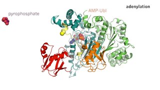
Crystal structure of Arabidopsis thaliana SOBER1, PDB code 6avv. Alpha helices are in red, beta strands in gold, catalytic site residues in black.
Identifiers
SymbolSOBER1
InterPro IPR029058
CATH 6avv
SCOP 6avv
SUPERFAMILY 6avv

SOBER1 is an enzyme that catalyzes the biochemical reaction of deacetylation. [1] The SOBER (Suppressor of AvrBsT-elicited resistance) 1 protein is conserved in plants and it suppresses the plant's ability to carry out the hypersensitive response against infection by certain pathogenic effector proteins from the YopJ family. [2] SOBER1 belongs to the protein superfamily of α/β hydrolases and possesses a canonical serine/histidine/aspartate catalytic triad to carry out the deacetylation reaction. There have been contradicting reports about SOBER1's potential phospholipase activity, with one study claiming phospholipase A2 activity of the protein [3] and another study being unable to reproduce this result. [1]

Enzyme biological molecule

Enzymes are macromolecular biological catalysts. Enzymes accelerate chemical reactions. The molecules upon which enzymes may act are called substrates and the enzyme converts the substrates into different molecules known as products. Almost all metabolic processes in the cell need enzyme catalysis in order to occur at rates fast enough to sustain life. Metabolic pathways depend upon enzymes to catalyze individual steps. The study of enzymes is called enzymology and a new field of pseudoenzyme analysis has recently grown up, recognising that during evolution, some enzymes have lost the ability to carry out biological catalysis, which is often reflected in their amino acid sequences and unusual 'pseudocatalytic' properties.

Enzyme catalysis catalysis of chemical reactions by specialized proteins known as enzymes

Enzyme catalysis is the increase in the rate of a chemical reaction by the active site of a protein. The protein catalyst (enzyme) may be part of a multi-subunit complex, and/or may transiently or permanently associate with a Cofactor. Catalysis of biochemical reactions in the cell is vital due to the very low reaction rates of the uncatalysed reactions at room temperature and pressure. A key driver of protein evolution is the optimization of such catalytic activities via protein dynamics.

Protein biological molecule consisting of chains of amino acid residues

Proteins are large biomolecules, or macromolecules, consisting of one or more long chains of amino acid residues. Proteins perform a vast array of functions within organisms, including catalysing metabolic reactions, DNA replication, responding to stimuli, providing structure to cells and organisms, and transporting molecules from one location to another. Proteins differ from one another primarily in their sequence of amino acids, which is dictated by the nucleotide sequence of their genes, and which usually results in protein folding into a specific three-dimensional structure that determines its activity.

Contents

Relationship to acyl-protein thioesterases

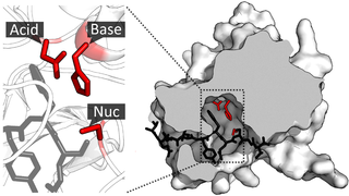
Members of the SOBER1 family are considered closely related to acyl-protein thioesterases, judged by their protein structure. [2] However, a change in their amino acid sequence renders SOBER1's biochemical properties into a deacetylase; in particular the hydrophobic tunnel, which is found in acyl-protein thioesterases, is impaired by additional amino acids in the lid structure of SOBER1, creating a new surface for binding of the acetyl group. [1]

Acyl-protein thioesterase

Acyl-protein thioesterases are enzymes that cleave off lipid modifications on proteins, located on the sulfur atom of cysteine residues linked via a thioester bond. Acyl-protein thioesterases are part of the α/β hydrolase superfamily of proteins and have a conserved catalytic triad. For that reason, acyl-protein thioesterases are also able to hydrolyze oxygen-linked ester bonds.

Protein structure three-dimensional arrangement of atoms in an amino acid-chain molecule

Protein structure is the three-dimensional arrangement of atoms in an amino acid-chain molecule. Proteins are polymers – specifically polypeptides – formed from sequences of amino acids, the monomers of the polymer. A single amino acid monomer may also be called a residue indicating a repeating unit of a polymer. Proteins form by amino acids undergoing condensation reactions, in which the amino acids lose one water molecule per reaction in order to attach to one another with a peptide bond. By convention, a chain under 30 amino acids is often identified as a peptide, rather than a protein. To be able to perform their biological function, proteins fold into one or more specific spatial conformations driven by a number of non-covalent interactions such as hydrogen bonding, ionic interactions, Van der Waals forces, and hydrophobic packing. To understand the functions of proteins at a molecular level, it is often necessary to determine their three-dimensional structure. This is the topic of the scientific field of structural biology, which employs techniques such as X-ray crystallography, NMR spectroscopy, and dual polarisation interferometry to determine the structure of proteins.

Protein primary structure linear sequence of amino acids in a peptide or protein

Protein primary structure is the linear sequence of amino acids in a peptide or protein. By convention, the primary structure of a protein is reported starting from the amino-terminal (N) end to the carboxyl-terminal (C) end. Protein biosynthesis is most commonly performed by ribosomes in cells. Peptides can also be synthesized in the laboratory. Protein primary structures can be directly sequenced, or inferred from DNA sequences.

Mechanism of SOBER1 deacetylase function, highlighting the additional amino acids in the protein lid. Nature Communications8(1):2201, Creative Commons Attribution 4.0 International License, https://creativecommons.org/licenses/by/4.0/ SOBER1 mechanism.png
Mechanism of SOBER1 deacetylase function, highlighting the additional amino acids in the protein lid. Nature Communications8(1):2201, Creative Commons Attribution 4.0 International License, https://creativecommons.org/licenses/by/4.0/

Targets

So far, the following proteins have been identified as SOBER1 targets: AvrBsT; ACIP1.

A biological target is anything within a living organism to which some other entity is directed and/or binds, resulting in a change in its behavior or function. Examples of common classes of biological targets are proteins and nucleic acids. The definition is context-dependent, and can refer to the biological target of a pharmacologically active drug compound, the receptor target of a hormone, or some other target of an external stimulus. Biological targets are most commonly proteins such as enzymes, ion channels, and receptors.

See also

In chemistry, acylation is the process of adding an acyl group to a compound. The compound providing the acyl group is called the acylating agent.

Catalytic triad

A catalytic triad is a set of three coordinated amino acids that can be found in the active site of some enzymes. Catalytic triads are most commonly found in hydrolase and transferase enzymes. An Acid-Base-Nucleophile triad is a common motif for generating a nucleophilic residue for covalent catalysis. The residues form a charge-relay network to polarise and activate the nucleophile, which attacks the substrate, forming a covalent intermediate which is then hydrolysed to release the product and regenerate free enzyme. The nucleophile is most commonly a serine or cysteine amino acid, but occasionally threonine or even selenocysteine. The 3D structure of the enzyme brings together the triad residues in a precise orientation, even though they may be far apart in the sequence.

The compendium of protein lysine acetylation (CPLA) database contains the sites of experimentally identified lysine acetylation sites.

Related Research Articles

Protease enzyme, type of hydrolase

A protease is an enzyme that helps proteolysis: protein catabolism by hydrolysis of peptide bonds. Proteases have evolved multiple times, and different classes of protease can perform the same reaction by completely different catalytic mechanisms. Proteases can be found in all forms of life and viruses.
Serine protease enzyme, type of peptidase

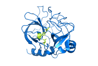
Serine proteases are enzymes that cleave peptide bonds in proteins, in which serine serves as the nucleophilic amino acid at the (enzyme's) active site. They are found ubiquitously in both eukaryotes and prokaryotes. Serine proteases fall into two broad categories based on their structure: chymotrypsin-like (trypsin-like) or subtilisin-like. In humans, they are responsible for coordinating various physiological functions, including digestion, immune response, blood coagulation and reproduction.

Acetylation describes a reaction that introduces an acetyl functional group into a chemical compound. Deacetylation is the removal of an acetyl group.

A metalloproteinase, or metalloprotease, is any protease enzyme whose catalytic mechanism involves a metal. An example of this would be meltrin which plays a significant role in the fusion of muscle cells during embryo development, in a process known as myogenesis.

Phospholipase D (PLD) is an enzyme of the phospholipase superfamily. Phospholipases occur widely, and can be found in a wide range of organisms, including bacteria, yeast, plants, animals, and viruses. Phospholipase D’s principal substrate is phosphatidylcholine, which it hydrolyzes to produce the signal molecule phosphatidic acid (PA), and soluble choline. Plants contain numerous genes that encode various PLD isoenzymes, with molecular weights ranging from 90-125 kDa. Mammalian cells encode two isoforms of phospholipase D: PLD1 and PLD2. Phospholipase D is an important player in many physiological processes, including membrane trafficking, cytoskeletal reorganization, receptor-mediated endocytosis, exocytosis, and cell migration. Through these processes, it has been further implicated in the pathophysiology of multiple diseases: in particular the progression of Parkinson’s and Alzheimer’s, as well as various cancers.

Serine hydrolases are one of the largest known enzyme classes comprising approximately ~200 enzymes or 1% of the genes in the human proteome. A defining characteristic of these enzymes is the presence of a nucleophilic serine in their active site, which is used for the hydrolysis of substrates. Catalysis proceeds by the formation of an acyl-enzyme intermediate through this serine, followed by water/hydroxide-induced saponification of the intermediate and regeneration of the enzyme. Unlike other non-catalytic serines, the nucleophilic serine of these hydrolases is typically activated by a proton relay involving a catalytic triad consisting of the serine, an acidic residue and a basic residue, although variations on this mechanism exist.

ACOT2 protein-coding gene in the species Homo sapiens

Acyl-CoA thioesterase 2, also known as ACOT2, is an enzyme which in humans is encoded by the ACOT2 gene.

ACOT4 protein-coding gene in the species Homo sapiens

Acyl-coenzyme A thioesterase 4 is an enzyme that in humans is encoded by the ACOT4 gene.

ACOT11 protein-coding gene in the species Homo sapiens

Acyl-coenzyme A thioesterase 11 also known as StAR-related lipid transfer protein 14 (STARD14) is an enzyme that in humans is encoded by the ACOT11 gene. This gene encodes a protein with acyl-CoA thioesterase activity towards medium (C12) and long-chain (C18) fatty acyl-CoA substrates which relies on its StAR-related lipid transfer domain. Expression of a similar murine protein in brown adipose tissue is induced by cold exposure and repressed by warmth. Expression of the mouse protein has been associated with obesity, with higher expression found in obesity-resistant mice compared with obesity-prone mice. Alternative splicing results in two transcript variants encoding different isoforms.

N-Acylphosphatidylethanolamines (NAPEs) are hormones released by the small intestine into the bloodstream when it processes fat. NAPEs travel to the hypothalamus in the brain and suppress appetite. This mechanism could be relevant for treating obesity.

N-acyl phosphatidylethanolamine phospholipase D (NAPE-PLD) is an enzyme that catalyzes the release of N-acylethanolamine (NAE) from N-acyl-phosphatidylethanolamine (NAPE). This is a major part of the process that converts ordinary lipids into chemical signals like anandamide and oleoylethanolamine. In humans, the NAPE-PLD protein is encoded by the NAPEPLD gene.

Threonine protease

Threonine proteases are a family of proteolytic enzymes harbouring a threonine (Thr) residue within the active site. The prototype members of this class of enzymes are the catalytic subunits of the proteasome, however the acyltransferases convergently evolved the same active site geometry and mechanism.

ACOT6 protein-coding gene in the species Homo sapiens

Acyl-CoA thioesterase 6 is a protein that in humans is encoded by the ACOT6 gene. The protein, also known as C14orf42, is an enzyme with thioesterase activity.

LYPLAL1 protein-coding gene in the species Homo sapiens

Lysophospholipase-like 1 is a protein in humans that is encoded by the LYPLAL1 gene. The protein is a α/β-hydrolase of uncharacterized metabolic function. Genome-wide association studies in humans have linked the gene to fat distribution and waist-to-hip ratio. The protein's enzymatic function is unclear. LYPLAL1 was reported to act as a triglyceride lipase in adipose tissue and another study suggested that the protein may play a role in the depalmitoylation of calcium-activated potassium channels. However, LYPLAL1 does not depalmitoylate the oncogene Ras and a structural and enzymatic study concluded that LYPLAL1 is generally unable to act as a lipase and is instead an esterase that prefers short-chain substrates, such as acetyl groups. Structural comparisons have suggested that LYPLAL1 might be a protein deacetylase, but this has not been experimentally tested.

Acyl-CoA thioesterase 9 protein-coding gene in the species Homo sapiens

Acyl-CoA thioesterase 9 is a protein that is encoded by the human ACOT9 gene. It is a member of the acyl-CoA thioesterase superfamily, which is a group of enzymes that hydrolyze Coenzyme A esters. There is no known function, however it has been shown to act as a long-chain thioesterase at low concentrations, and a short-chain thioesterase at high concentrations.

Ketoacyl synthase

Ketoacyl synthases (KSs) catalyze the condensation reaction of acyl-CoA or acyl-acyl ACP with malonyl-CoA to form 3-ketoacyl-CoA or with malonyl-ACP to form 3-ketoacyl-ACP. This reaction is a key step in the fatty acid synthesis cycle, as the resulting acyl chain is two carbon atoms longer than before. KSs exist as individual enzymes, as they do in type II fatty acid synthesis and type II polyketide synthesis, or as domains in large multidomain enzymes, such as type I fatty acid synthases (FASs) and polyketide synthases (PKSs). KSs are divided into five families: KS1, KS2, KS3, KS4, and KS5.

ACOT13 protein-coding gene in the species Homo sapiens

Acyl-CoA thioesterase 13 is a protein that in humans is encoded by the ACOT13 gene. This gene encodes a member of the thioesterase superfamily. In humans, the protein co-localizes with microtubules and is essential for sustained cell proliferation.

ACOT1 protein-coding gene in the species Homo sapiens

Acyl-CoA thioesterase 1 is a protein that in humans is encoded by the ACOT1 gene.

References

  1. 1 2 3 Bürger M, Willige BC, Chory J (December 2017). "A hydrophobic anchor mechanism defines a deacetylase family that suppresses host response against YopJ effectors". Nature Communications. 8 (1): 2201. doi:10.1038/s41467-017-02347-w. PMC   5736716 . PMID   29259199.
  2. 1 2 Cunnac S, Wilson A, Nuwer J, Kirik A, Baranage G, Mudgett MB (February 2007). "A conserved carboxylesterase is a SUPPRESSOR OF AVRBST-ELICITED RESISTANCE in Arabidopsis". The Plant Cell. 19 (2): 688–705. doi:10.1105/tpc.106.048710. PMC   1867326 . PMID   17293566.
  3. Kirik A, Mudgett MB (December 2009). "SOBER1 phospholipase activity suppresses phosphatidic acid accumulation and plant immunity in response to bacterial effector AvrBsT". Proceedings of the National Academy of Sciences of the United States of America. 106 (48): 20532–7. doi:10.1073/pnas.0903859106. PMC   2787154 . PMID   19918071.