SUCLG1

Last updated
SUCLG1
Protein SUCLG1 PDB 1euc.png
Identifiers
Aliases SUCLG1 , GALPHA, MTDPS9, SUCLA1, succinate-CoA ligase alpha subunit, succinate-CoA ligase GDP/ADP-forming subunit alpha
External IDs OMIM: 611224 MGI: 1927234 HomoloGene: 55785 GeneCards: SUCLG1
Orthologs
SpeciesHumanMouse
Entrez
Ensembl
UniProt
RefSeq (mRNA)

NM_003849

NM_019879

RefSeq (protein)

NP_003840

NP_063932

Location (UCSC) Chr 2: 84.42 – 84.46 Mb Chr 6: 73.23 – 73.25 Mb
PubMed search [3] [4]
Wikidata
View/Edit Human View/Edit Mouse

Succinyl-CoA ligase [GDP-forming] subunit alpha, mitochondrial is an enzyme that in humans is encoded by the SUCLG1 gene. [5] [6]

Contents

Structure

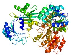
The enzyme encoded by SUCLG1 can exist in either a phosphorylated form or a dephosphorylated form. In the dephosphorylated structure, a phosphate ion works in coordination with a histidine residue in the active site and the two alpha helices, one contributed by each subunit of the alphabeta-dimer to stabilize the structure. One of the alpha helices contains amino acids, the modification of which result in conformational changes that accommodate either the bound phosphoryl group or the free phosphate ion. [7]

Function

This gene encodes the alpha subunit of the heterodimeric enzyme succinate coenzyme A ligase. This enzyme is targeted to the mitochondria and catalyzes the conversion of succinyl CoA and ADP or GDP to succinate and ATP or GTP. Mutations in this gene are the cause of the metabolic disorder fatal infantile lactic acidosis and mitochondrial DNA depletion. [8] [9]

Clinical significance

Succinate-CoA ligase deficiency is responsible for encephalomyopathy with mitochondrial DNA depletion and mild methylmalonic aciduria. Mutations in SUCLG1 lead to complete absence of SUCLG1 protein and are responsible for a very severe disorder with antenatal manifestations. Furthermore, it is shown that in the absence of SUCLG1 protein, no SUCLA2 protein is found in fibroblasts by western blot analysis. This result is consistent with a degradation of SUCLA2 when its heterodimer partner, SUCLG1, is absent. [10] As mitochondrial DNA depletion in muscle is not a constant finding in SUCLG1 patients, diagnosis should not be based on it; additionally, it may be that alternative physiopathological mechanisms may be considered to explain the combined respiratory chain deficiency observed in these patients. [9]

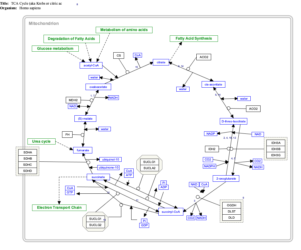
Interactive pathway map

Click on genes, proteins and metabolites below to link to respective articles. [§ 1]

[[File:
TCACycle WP78.png Go to articleGo to articleGo to articleGo to articleGo to articleGo to articleGo to articleGo to articleGo to articleGo to articleGo to articleGo to articleGo to articleGo to articleGo to articleGo to articleGo to articleGo to articleGo to articleGo to articleGo to articleGo to articleGo to articleGo to articleGo to articleGo to articleGo to articleGo to articleGo to articleGo to articleGo to articleGo to articleGo to articleGo to articleGo to articleGo to articleGo to articleGo to articleGo to articleGo to articleGo to articleGo to HMDBGo to articleGo to articleGo to articleGo to articleGo to articleGo to articleGo to articleGo to articleGo to articleGo to article
[[
]]
[[
]]
[[
]]
[[
]]
[[
]]
[[
]]
[[
]]
[[
]]
[[
]]
[[
]]
[[
]]
[[
]]
[[
]]
[[
]]
[[
]]
[[
]]
[[
]]
[[
]]
[[
]]
[[
]]
[[
]]
[[
]]
[[
]]
[[
]]
[[
]]
[[
]]
[[
]]
[[
]]
[[
]]
[[
]]
[[
]]
[[
]]
[[
]]
[[
]]
[[
]]
[[
]]
[[
]]
[[
]]
[[
]]
[[
]]
[[
]]
[[
]]
[[
]]
[[
]]
[[
]]
[[
]]
[[
]]
[[
]]
[[
]]
[[
]]
[[
]]
[[
]]
[[
]]
[[
]]
[[
]]
[[
]]
[[
]]
[[
]]
[[
]]
[[
]]
[[
]]
[[
]]
[[
]]
[[
]]
TCACycle WP78.png Go to articleGo to articleGo to articleGo to articleGo to articleGo to articleGo to articleGo to articleGo to articleGo to articleGo to articleGo to articleGo to articleGo to articleGo to articleGo to articleGo to articleGo to articleGo to articleGo to articleGo to articleGo to articleGo to articleGo to articleGo to articleGo to articleGo to articleGo to articleGo to articleGo to articleGo to articleGo to articleGo to articleGo to articleGo to articleGo to articleGo to articleGo to articleGo to articleGo to articleGo to articleGo to HMDBGo to articleGo to articleGo to articleGo to articleGo to articleGo to articleGo to articleGo to articleGo to articleGo to article
|alt=TCACycle_WP78 edit]]
TCACycle_WP78 edit
  1. The interactive pathway map can be edited at WikiPathways: "TCACycle_WP78".

Related Research Articles

<span class="mw-page-title-main">Citric acid cycle</span> Metabolic pathway

The citric acid cycle (CAC)—also known as the Krebs cycle or the TCA cycle (tricarboxylic acid cycle)—is a series of chemical reactions to release stored energy through the oxidation of acetyl-CoA derived from carbohydrates, fats, and proteins. The Krebs cycle is used by organisms that respire (as opposed to organisms that ferment) to generate energy, either by anaerobic respiration or aerobic respiration. In addition, the cycle provides precursors of certain amino acids, as well as the reducing agent NADH, that are used in numerous other reactions. Its central importance to many biochemical pathways suggests that it was one of the earliest components of metabolism and may have originated abiogenically. Even though it is branded as a 'cycle', it is not necessary for metabolites to follow only one specific route; at least three alternative segments of the citric acid cycle have been recognized.

A nucleoside triphosphate is a nucleoside containing a nitrogenous base bound to a 5-carbon sugar, with three phosphate groups bound to the sugar. They are the molecular precursors of both DNA and RNA, which are chains of nucleotides made through the processes of DNA replication and transcription. Nucleoside triphosphates also serve as a source of energy for cellular reactions and are involved in signalling pathways.

<span class="mw-page-title-main">Mitochondrial matrix</span> Space within the inner membrane of the mitochondrion

In the mitochondrion, the matrix is the space within the inner membrane. The word "matrix" stems from the fact that this space is viscous, compared to the relatively aqueous cytoplasm. The mitochondrial matrix contains the mitochondrial DNA, ribosomes, soluble enzymes, small organic molecules, nucleotide cofactors, and inorganic ions.[1] The enzymes in the matrix facilitate reactions responsible for the production of ATP, such as the citric acid cycle, oxidative phosphorylation, oxidation of pyruvate, and the beta oxidation of fatty acids.

<span class="mw-page-title-main">SDHD</span> Protein-coding gene in the species Homo sapiens

Succinate dehydrogenase [ubiquinone] cytochrome b small subunit, mitochondrial (CybS), also known as succinate dehydrogenase complex subunit D (SDHD), is a protein that in humans is encoded by the SDHD gene. Names previously used for SDHD were PGL and PGL1. Succinate dehydrogenase is an important enzyme in both the citric acid cycle and the electron transport chain.

Substrate-level phosphorylation is a metabolism reaction that results in the production of ATP or GTP by the transfer of a phosphate group from a substrate directly to ADP or GDP. Transferring from a higher energy (whether phosphate group attached or not) into a lower energy product. This process uses some of the released chemical energy, the Gibbs free energy, to transfer a phosphoryl (PO3) group to ADP or GDP from another phosphorylated compound. Occurs in glycolysis and in the citric acid cycle.

<span class="mw-page-title-main">Succinyl coenzyme A synthetase</span>

Succinyl coenzyme A synthetase is an enzyme that catalyzes the reversible reaction of succinyl-CoA to succinate. The enzyme facilitates the coupling of this reaction to the formation of a nucleoside triphosphate molecule from an inorganic phosphate molecule and a nucleoside diphosphate molecule. It plays a key role as one of the catalysts involved in the citric acid cycle, a central pathway in cellular metabolism, and it is located within the mitochondrial matrix of a cell.

<span class="mw-page-title-main">SDHB</span> Protein-coding gene in the species Homo sapiens

Succinate dehydrogenase [ubiquinone] iron-sulfur subunit, mitochondrial (SDHB) also known as iron-sulfur subunit of complex II (Ip) is a protein that in humans is encoded by the SDHB gene.

<span class="mw-page-title-main">HADHA</span> Protein-coding gene in the species Homo sapiens

Trifunctional enzyme subunit alpha, mitochondrial also known as hydroxyacyl-CoA dehydrogenase/3-ketoacyl-CoA thiolase/enoyl-CoA hydratase, alpha subunit is a protein that in humans is encoded by the HADHA gene. Mutations in HADHA have been associated with trifunctional protein deficiency or long-chain 3-hydroxyacyl-coenzyme A dehydrogenase deficiency.

<span class="mw-page-title-main">HADHB</span> Protein-coding gene in the species Homo sapiens

Trifunctional enzyme subunit beta, mitochondrial (TP-beta) also known as 3-ketoacyl-CoA thiolase, acetyl-CoA acyltransferase, or beta-ketothiolase is an enzyme that in humans is encoded by the HADHB gene.

<span class="mw-page-title-main">Succinate—CoA ligase (ADP-forming)</span>

In enzymology, a succinate-CoA ligase (ADP-forming) is an enzyme that catalyzes the chemical reaction

<span class="mw-page-title-main">Succinate—CoA ligase (GDP-forming)</span>

In enzymology, a succinate—CoA ligase (GDP-forming) is an enzyme that catalyzes the chemical reaction

<span class="mw-page-title-main">Cytochrome c oxidase subunit 2</span> Enzyme of the respiratory chain encoded by the mitochondrial genome

Cytochrome c oxidase subunit 2, also known as cytochrome c oxidase polypeptide II, is a protein that in humans is encoded by the MT-CO2 gene. Cytochrome c oxidase subunit II, abbreviated COXII, COX2, COII, or MT-CO2, is the second subunit of cytochrome c oxidase. It is also one of the three mitochondrial DNA (mtDNA) encoded subunits of respiratory complex IV.

<span class="mw-page-title-main">IDH3G</span> Protein-coding gene in the species Homo sapiens

Isocitrate dehydrogenase [NAD] subunit gamma, mitochondrial is an enzyme that in humans is encoded by the IDH3G gene.

<span class="mw-page-title-main">SUCLA2</span> Protein-coding gene in the species Homo sapiens

Succinyl-CoA ligase [ADP-forming] subunit beta, mitochondrial (SUCLA2), also known as ADP-forming succinyl-CoA synthetase (SCS-A), is an enzyme that in humans is encoded by the SUCLA2 gene on chromosome 13.

<span class="mw-page-title-main">PMPCA</span> Protein-coding gene in the species Homo sapiens

Mitochondrial-processing peptidase subunit alpha is an enzyme that in humans is encoded by the PMPCA gene. This gene PMPCA encoded a protein that is a member of the peptidase M16 family. This protein is located in the mitochondrial matrix and catalyzes the cleavage of the leader peptides of precursor proteins newly imported into the mitochondria, though it only functions as part of a heterodimeric complex.

<span class="mw-page-title-main">NDUFA12</span> Protein-coding gene in the species Homo sapiens

NADH dehydrogenase [ubiquinone] 1 alpha subcomplex subunit 12 is an enzyme that in humans is encoded by the NDUFA12 gene. The NDUFA12 protein is a subunit of NADH dehydrogenase (ubiquinone), which is located in the mitochondrial inner membrane and is the largest of the five complexes of the electron transport chain. Mutations in subunits of NADH dehydrogenase (ubiquinone), also known as Complex I, frequently lead to complex neurodegenerative diseases such as Leigh's syndrome that result from mitochondrial complex I deficiency.

<span class="mw-page-title-main">OGDH</span> Enzyme involved in Krebs cycle

Alpha-ketoglutarate dehydrogenase also known as 2-oxoglutarate dehydrogenase E1 component, mitochondrial is an enzyme that in humans is encoded by the OGDH gene.

<span class="mw-page-title-main">LIG3</span> Protein-coding gene in the species Homo sapiens

DNA ligase 3 is an enzyme that, in humans, is encoded by the LIG3 gene. The human LIG3 gene encodes ATP-dependent DNA ligases that seal interruptions in the phosphodiester backbone of duplex DNA.

<span class="mw-page-title-main">Mitochondrial DNA depletion syndrome</span> Medical condition

Mitochondrial DNA depletion syndrome, or Alper's disease, is any of a group of autosomal recessive disorders that cause a significant drop in mitochondrial DNA in affected tissues. Symptoms can be any combination of myopathic, hepatopathic, or encephalomyopathic. These syndromes affect tissue in the muscle, liver, or both the muscle and brain, respectively. The condition is typically fatal in infancy and early childhood, though some have survived to their teenage years with the myopathic variant and some have survived into adulthood with the SUCLA2 encephalomyopathic variant. There is currently no curative treatment for any form of MDDS, though some preliminary treatments have shown a reduction in symptoms.

<span class="mw-page-title-main">SUCLG2</span> Protein-coding gene in the species Homo sapiens

Succinyl-CoA ligase [GDP-forming] subunit beta, mitochondrial is an enzyme that in humans is encoded by the SUCLG2 gene on chromosome 3.

References

  1. 1 2 3 GRCh38: Ensembl release 89: ENSG00000163541 - Ensembl, May 2017
  2. 1 2 3 GRCm38: Ensembl release 89: ENSMUSG00000052738 - Ensembl, May 2017
  3. "Human PubMed Reference:". National Center for Biotechnology Information, U.S. National Library of Medicine.
  4. "Mouse PubMed Reference:". National Center for Biotechnology Information, U.S. National Library of Medicine.
  5. James M, Man NT, Edwards YH, Morris GE (Apr 1997). "The molecular basis for cross-reaction of an anti-dystrophin antibody with alpha-actinin". Biochimica et Biophysica Acta (BBA) - Molecular Basis of Disease. 1360 (2): 169–76. doi: 10.1016/s0925-4439(96)00076-2 . PMID   9128182.
  6. "Entrez Gene: SUCLG1 succinate-CoA ligase, GDP-forming, alpha subunit".
  7. Fraser ME, James MN, Bridger WA, Wolodko WT (Jun 2000). "Phosphorylated and dephosphorylated structures of pig heart, GTP-specific succinyl-CoA synthetase". Journal of Molecular Biology. 299 (5): 1325–39. doi:10.1006/jmbi.2000.3807. PMID   10873456.
  8. Ostergaard E (Apr 2008). "Disorders caused by deficiency of succinate-CoA ligase". Journal of Inherited Metabolic Disease. 31 (2): 226–9. doi:10.1007/s10545-008-0828-7. PMID   18392745. S2CID   12722653.
  9. 1 2 Valayannopoulos V, Haudry C, Serre V, Barth M, Boddaert N, Arnoux JB, Cormier-Daire V, Rio M, Rabier D, Vassault A, Munnich A, Bonnefont JP, de Lonlay P, Rötig A, Lebre AS (Jun 2010). "New SUCLG1 patients expanding the phenotypic spectrum of this rare cause of mild methylmalonic aciduria". Mitochondrion. 10 (4): 335–41. doi:10.1016/j.mito.2010.02.006. PMID   20197121.
  10. Rouzier C, Le Guédard-Méreuze S, Fragaki K, Serre V, Miro J, Tuffery-Giraud S, Chaussenot A, Bannwarth S, Caruba C, Ostergaard E, Pellissier JF, Richelme C, Espil C, Chabrol B, Paquis-Flucklinger V (Oct 2010). "The severity of phenotype linked to SUCLG1 mutations could be correlated with residual amount of SUCLG1 protein" (PDF). Journal of Medical Genetics. 47 (10): 670–6. doi:10.1136/jmg.2009.073445. PMID   20693550. S2CID   35860287.

Further reading