Septum transversum

Last updated
Septum transversum
Diaphragma-embryo.png
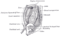
Diaphragm of embryo.
Gray1088.png
Liver with the septum transversum. Human embryo 3 mm. long.
Details
Carnegie stage 10
Precursor mesenchyme
Gives rise to diaphragm / Ventral mesentery
Identifiers
TE E5.2.0.4.0.0.2
Anatomical terminology

The septum transversum is a thick mass of cranial mesenchyme, formed in the embryo, that gives rise to parts of the thoracic diaphragm and the ventral mesentery of the foregut in the developed human being and other mammals.

Contents

Origins

The septum transversum originally arises as the most cranial part of the mesenchyme on day 22. [1] During craniocaudal folding, it assumes a position cranial to the developing heart at the level of the cervical vertebrae. [1] During subsequent weeks the dorsal end of the embryo grows much faster than its ventral counterpart resulting in an apparent descent of the ventrally located septum transversum. At week 8, it can be found at the level of the thoracic vertebrae. [1] [2]

Nerve supply

After successful craniocaudal folding the septum transversum picks up innervation from the adjacent ventral rami of spinal nerves C3, C4 and C5, thus forming the precursor of the phrenic nerve. During the descent of the septum, the phrenic nerve is carried along and assumes its descending pathway.

During embryonic development of the thoracic diaphragm, myoblast cells from the septum invade the other components of the diaphragm. They thus give rise to the motor and sensory innervation of the muscular diaphragm by the phrenic nerve.

Derivatives

The cranial part of the septum transversum gives rise to the central tendon of the diaphragm, [1] and is the origin of the myoblasts that invade the pleuroperitoneal folds resulting in the formation of the muscular diaphragm. [3]

The caudal part of the septum transversum is invaded by the hepatic diverticulum which divides within it to form the liver and thus gives rise to the ventral mesentery of the foregut, which in turn is the precursor of the lesser omentum, the visceral peritoneum of the liver and the falciform ligament.

Though not derived from the septum transversum, development of the liver is highly dependent upon signals originating here. Bone morphogenetic protein 2 (BMP-2), BMP-4 and BMP-7 produced from the septum transversum join fibroblast growth factor (FGF) signals from the cardiac mesoderm induce part of the foregut to differentiate towards a hepatic fate. [4]

Additional images

Related Research Articles

Mesoderm Middle germ layer that forms muscle, bone, blood vessels and more

In all bilaterian animals, the mesoderm is one of the three primary germ layers in the very early embryo. The other two layers are the ectoderm and endoderm, with the mesoderm as the middle layer between them.

Rib

In vertebrate anatomy, ribs are the long curved bones which form the rib cage, part of the axial skeleton. In most tetrapods, ribs surround the chest, enabling the lungs to expand and thus facilitate breathing by expanding the chest cavity. They serve to protect the lungs, heart, and other internal organs of the thorax. In some animals, especially snakes, ribs may provide support and protection for the entire body.

Body cavity

A body cavity is any space or compartment, or potential space in the animal body. Cavities accommodate organs and other structures; cavities as potential spaces contain fluid.

Articles related to anatomy include:

Phrenic nerve

The phrenic nerve is a mixed motor/sensory nerve which originates from the C3-C5 spinal nerves in the neck. The nerve is important for breathing because it provides exclusive motor control of the diaphragm, the primary muscle of respiration. In humans, the right and left phrenic nerves are primarily supplied by the C4 spinal nerve, but there is also contribution from the C3 and C5 spinal nerves. From its origin in the neck, the nerve travels downward into the chest to pass between the heart and lungs towards the diaphragm.

Thoracic diaphragm Sheet of internal skeletal muscle

The thoracic diaphragm, or simply the diaphragm, is a sheet of internal skeletal muscle in humans and other mammals that extends across the bottom of the thoracic cavity. The diaphragm separates the thoracic cavity, containing the heart and lungs, from the abdominal cavity and performs an important function in respiration: as the diaphragm contracts, the volume of the thoracic cavity increases, creating a negative pressure there, which draws air into the lungs.

Mesentery Contiguous fold of tissues that supports the intestines

The mesentery is an organ that attaches the intestines to the posterior abdominal wall in humans and is formed by the double fold of peritoneum. It helps in storing fat and allowing blood vessels, lymphatics, and nerves to supply the intestines, among other functions.

Paramesonephric duct

Paramesonephric ducts are paired ducts of the embryo that run down the lateral sides of the urogenital ridge and terminate at the sinus tubercle in the primitive urogenital sinus. In the female, they will develop to form the fallopian tubes, uterus, cervix, and the upper one-third of the vagina.

Celiac artery

The coeliacartery, also known as the coeliac trunk, or truncus coeliacus, is the first major branch of the abdominal aorta. It is about 1.25 cm in length. Branching from the aorta at thoracic vertebra 12 (T12) in humans, it is one of three anterior/ midline branches of the abdominal aorta.

Pharyngeal arch Embryonic precursor structures in vertebrates

The pharyngeal arches, also known as visceral arches, are structures seen in the embryonic development of vertebrates that are recognisable precursors for many structures. In fish, the arches are known as the branchial arches, or gill arches.

Falciform ligament

The falciform ligament is a ligament that attaches the liver to the front body wall, and separates the liver into the left medial lobe and right lateral lobe. The falciform ligament, from Latin 'sickle-shaped', is a broad and thin fold of peritoneum, its base being directed downward and backward and its apex upward and forward. The falciform ligament droops down from the hilum of the liver.

Foregut Anterior part of the gastrointestinal tract

The foregut is the anterior part of the alimentary canal, from the mouth to the duodenum at the entrance of the bile duct. Beyond the stomach, the foregut is attached to the abdominal walls by mesentery. The foregut arises from the endoderm, developing from the folding primitive gut, and is developmentally distinct from the midgut and hindgut. Although the term “foregut” is typically used in reference to the anterior section of the primitive gut, components of the adult gut can also be described with this designation. Pain in the epigastric region, just below the intersection of the ribs, typically refers to structures in the adult foregut.

Vitelline veins

The vitelline veins are veins that drain blood from the yolk sac and the gut tube during gestation.

Lateral plate mesoderm

Lateral plate mesoderm is a type of mesoderm that is found at the periphery of the embryo.

Subclavian nerve

The subclavian nerve or nerve to the subclavius is small branch of the upper trunk of the brachial plexus where C5 and C6 join. It contains axons derived from the ventral rami of the fifth (C5) and sixth (C6) cervical nerves. The subclavian nerve provides motor innervation to the subclavius muscle.

Human embryonic development

Human embryonic development, or human embryogenesis, refers to the development and formation of the human embryo. It is characterised by the processes of cell division and cellular differentiation of the embryo that occurs during the early stages of development. In biological terms, the development of the human body entails growth from a one-celled zygote to an adult human being. Fertilisation occurs when the sperm cell successfully enters and fuses with an egg cell (ovum). The genetic material of the sperm and egg then combine to form a single cell called a zygote and the germinal stage of development commences. Embryonic development in the human, covers the first eight weeks of development; at the beginning of the ninth week the embryo is termed a fetus. Human embryology is the study of this development during the first eight weeks after fertilisation. The normal period of gestation (pregnancy) is about nine months or 40 weeks.

Hepatic diverticulum

The hepatic diverticulum is a primordial cellular extension of the embryonic foregut endoderm that gives rise to the parenchyma of the liver and the [[bile duct]. It typically differentiates from the endoderm in the third or fourth week of gestation and is reabsorbed in tubular structures of the septum transversum by the eighth week.

The face and neck development of the human embryo refers to the development of the structures from the third to eighth week that give rise to the future head and neck. They consist of three layers, the ectoderm, mesoderm and endoderm, which form the mesenchyme, neural crest and neural placodes. The paraxial mesoderm forms structures named somites and somitomeres that contribute to the development of the floor of the brain and voluntary muscles of the craniofacial region. The lateral plate mesoderm consists of the laryngeal cartilages. The three tissue layers give rise to the pharyngeal apparatus, formed by six pairs of pharyngeal arches, a set of pharyngeal pouches and pharyngeal grooves, which are the most typical feature in development of the head and neck. The formation of each region of the face and neck is due to the migration of the neural crest cells which come from the ectoderm. These cells determine the future structure to develop in each pharyngeal arch. Eventually, they also form the neurectoderm, which forms the forebrain, midbrain and hindbrain, cartilage, bone, dentin, tendon, dermis, pia mater and arachnoid mater, sensory neurons, and glandular stroma.

The development of the digestive system concerns the epithelium of the digestive system and the parenchyma of its derivatives, which originate from the endoderm. Connective tissue, muscular components, and peritoneal components originate in the mesoderm. Different regions of the gut tube such as the esophagus, stomach, duodenum, etc. are specified by a retinoic acid gradient that causes transcription factors unique to each region to be expressed. Differentiation of the gut and its derivatives depends upon reciprocal interactions between the gut endoderm and its surrounding mesoderm. Hox genes in the mesoderm are induced by a Hedgehog signaling pathway secreted by gut endoderm and regulate the craniocaudal organization of the gut and its derivatives. The gut system extends from the oropharyngeal membrane to the cloacal membrane and is divided into the foregut, midgut, and hindgut.

Pulmonary pleurae Serous membrane that lines the wall of thoracic cavity and the surface of lung

The pulmonary pleurae are the two layers of the invaginated sac surrounding each lung and attaching to the thoracic cavity. The visceral pleura is the delicate membrane that covers the surface of each lung, and dips into the fissures between the lobes of the lung. The parietal pleura is the outer membrane which is attached to the inner surface of the thoracic cavity. It also separates the pleural cavity from the mediastinum. The parietal pleura is innervated by the intercostal nerves and the phrenic nerve.

References

  1. 1 2 3 4 Mitchell, Barry; Sharma, Ram (2009-01-01), Mitchell, Barry; Sharma, Ram (eds.), "Chapter 3 - The body cavities and the diaphragm", Embryology (Second Edition), Churchill Livingstone, pp. 15–18, doi:10.1016/b978-0-7020-3225-7.50006-3, ISBN   978-0-7020-3225-7 , retrieved 2020-12-05
  2. Moore, Keith L. (2003). The developing human : clinically oriented embryology. Persaud, T. V. N. (7th [ed.] ed.). Philadelphia, Pa.: Saunders. ISBN   978-0-8089-2265-0. OCLC   49526919.
  3. Moore, N. Anthony. (2007). Gross and developmental anatomy. Roy, William A. (2nd ed ed.). St. Louis: Mosby. ISBN   0-323-04551-0. OCLC   70220003.CS1 maint: extra text (link)
  4. Carlson, Bruce M. (2004). Human embryology and developmental biology. Carlson, Bruce M. (3rd ed ed.). [St. Louis, Mo.]: Mosby. ISBN   0-323-01487-9. OCLC   60346934.CS1 maint: extra text (link)