Development of the digestive system

Last updated

The development of the digestive system in the human embryo concerns the epithelium of the digestive system and the parenchyma of its derivatives, which originate from the endoderm. Connective tissue, muscular components, and peritoneal components originate in the mesoderm. Different regions of the gut tube such as the esophagus, stomach, duodenum, etc. are specified by a retinoic acid gradient that causes transcription factors unique to each region to be expressed. Differentiation of the gut and its derivatives depends upon reciprocal interactions between the gut endoderm and its surrounding mesoderm. Hox genes in the mesoderm are induced by a Hedgehog signaling pathway secreted by gut endoderm and regulate the craniocaudal organization of the gut and its derivatives. The gut system extends from the oropharyngeal membrane to the cloacal membrane and is divided into the foregut, midgut, and hindgut. [1]

Contents

Body cavities

At the end of the third week, the neural tube, which is a fold of one of the layers of the trilaminar germ disc, called the ectoderm, appears. This layer elevates and closes dorsally, while the gut tube rolls up and closes ventrally to create a "tube on top of a tube." The mesoderm, which is another layer of the trilaminar germ disc, holds the tubes together and the lateral plate mesoderm, the middle layer of the germ disc, splits to form a visceral layer associated with the gut and a parietal layer, which along with the overlying ectoderm, forms the lateral body wall. The space between the visceral and parietal layers of lateral plate mesoderm is the primitive body cavity. When the lateral body wall folds, it moves ventrally and fuses at the midline. The body cavity closes, except in the region of the connecting stalk. Here, the gut tube maintains an attachment to the yolk sac. The yolk sac is a membranous sac attached to the embryo, which provides nutrients and functions as the circulatory system of the very early embryo. [1]

The lateral body wall folds, pulling the amnion in with it so that the amnion surrounds the embryo and extends over the connecting stalk, which becomes the umbilical cord, which connects the fetus with the placenta. If the ventral body wall fails to close, ventral body wall defects can result, such as ectopia cordis, a congenital malformation in which the heart is abnormally located outside the thorax. Another defect is gastroschisis, a congenital defect in the anterior abdominal wall through which the abdominal contents freely protrude. Another possibility is bladder exstrophy, in which part of the urinary bladder is present outside the body. In normal circumstances, the parietal mesoderm will form the parietal layer of serous membranes lining the outside (walls) of the peritoneal, pleural, and pericardial cavities. The visceral layer will form the visceral layer of the serous membranes covering the lungs, heart, and abdominal organs. These layers are continuous at the root of each organ as the organs lie in their respective cavities. The peritoneum, a serum membrane that forms the lining of the abdominal cavity, forms in the gut layers and in places mesenteries extend from the gut as double layers of peritoneum. Mesenteries provide a pathway for vessels, nerves, and lymphatics to the organs. Initially, the gut tube from the caudal end of the foregut to the end of the hindgut is suspended from the dorsal body wall by dorsal mesentery. Ventral mesentery, derived from the septum transversum, exists only in the region of the terminal part of the esophagus, the stomach, and the upper portion of the duodenum. [2]

Rotation and herniation

Diagram showing the process by which the intestine rotates and herniates during normal development. From panel A to B (left-sided views), the midgut loop rotates 90deg in a counterclockwise direction (caudal-to-rostral view), so that its position changes from midsagittal (A) to transverse (B1). The small intestine forms loops (B2) and slides back into the abdomen (B3) during resolution of the hernia. Meanwhile, the cecum moves from the left to the right side, which represents the additional 180deg counterclockwise rotation of the intestine (C, central view). Intestinal rotation and herniation.png
Diagram showing the process by which the intestine rotates and herniates during normal development. From panel A to B (left-sided views), the midgut loop rotates 90° in a counterclockwise direction (caudal-to-rostral view), so that its position changes from midsagittal (A) to transverse (B1). The small intestine forms loops (B2) and slides back into the abdomen (B3) during resolution of the hernia. Meanwhile, the cecum moves from the left to the right side, which represents the additional 180° counterclockwise rotation of the intestine (C, central view).

In the process of lengthening growth, the intestinal duct herniates and rotates. Herniation (Latin, meaning 'rupture') takes place at around 7½ weeks in the human embryo and refers to the retraction of the intestine from the extraembryonal navel coelom into the abdomen (panel B3).

The intestinal duct rotates by 90° (counterclockwise when viewing from tail-to-head) around the boy axis (see panel B1) in the same direction as the heart. According to the Axial Twist theory, the rotation is part of a complex twist that involves the entire body in all vertebrate animals. During this twist, the anterior head region rotates 90° counterclockwise, the body 90° clockwise, but sparing the heart and bowels. Accordingly, the embryo turns on its side. The forebrain becomes turned around with respect to the while the heart and bowels (which do not take part in the twist) become turned counterclockwise with respect to the body. [4] [5]

Folding occurs in a typical manner as shown in the diagram (panel B2-3, C). The folding is a result of the elongation of the duct.

Diaphragm and thoracic cavity

The diaphragm divides the body cavity into the thoracic cavity and the abdominal cavity. It develops from four components: the septum transversum (central tendon), the pleuroperitoneal membranes, the dorsal mesentery of the esophagus, and muscular components from somites at cervical levels three to five (C3–5) of the body wall. Since the septum transversum is located initially opposite cervical segments of three to five, and since muscle cells for the diaphragm originate from somites at these segments, the phrenic nerve, which innervates the diaphragm, also arises from these segments of the spinal cord (C3, 4, and 5). The thoracic cavity is divided into the pericardial cavity and two pleural cavities for the lungs by the pleuropericardial membranes. [6]

Divisions of the gut tube

As a result of the cephalocaudal and lateral folding of the embryo, a portion of the endoderm-lined yolk sac cavity is incorporated into the embryo to form the primitive gut. In the cephalic and caudal parts of the embryo, the primitive gut forms a tube, the foregut and hindgut, respectively. The middle part, the midgut, remains temporally connected to the yolk sac by means of the vitelline duct. [6]

Foregut

The foregut gives rise to the esophagus, the trachea, lung buds, the stomach, and the duodenum proximal to the entrance of the bile duct. In addition, the liver, pancreas, and biliary apparatus develop as outgrowths of the endodermal epithelium of the upper part of the duodenum. Since the upper part of the foregut is divided by the tracheoesophageal septum into the esophagus posteriorly and the trachea and lung buds anteriorly, deviation of the septum may result in abnormal openings between the trachea and esophagus. The epithelial liver cords and biliary system growing out into the septum transversum differentiate into parenchyma. Hematopoietic cells (present in the liver in greater numbers before birth than afterward), Kupffer cells, and connective tissue cells originate in the mesoderm. The pancreas develops from a ventral bud and a dorsal bud that later fuse to form the definitive pancreas. Sometimes, the two parts surround the duodenum (annular pancreas), causing constriction of the gut. [7]

Midgut

The midgut forms the primary intestinal loop, from which originates the distal duodenum to the entrance of the bile duct. The loop continues to the junction of the proximal two-thirds of the transverse colon with the distal third. At its apex, the primary loop remains temporarily in open connection with the yolk sac through the vitelline duct. During the sixth week, the loop grows so rapidly that it protrudes into the umbilical cord (physiological herniation). In the 10th week, it returns into the abdominal cavity. While these processes are occurring, the midgut loop rotates 270° counterclockwise. Common abnormalities at this stage of development include remnants of the vitelline duct, failure of the midgut to return to the abdominal cavity, malrotation, stenosis, and duplication of parts. [6]

Hindgut

The hindgut gives rise to the region from the distal third of the transverse colon to the upper part of the anal canal. The distal part of the anal canal originates from the ectoderm. The hindgut enters the posterior region of the cloaca (future anorectal canal), and the allantois enters the anterior region (future urogenital sinus). The urorectal septum divides the two regions and breakdown of the cloacal membrane covering this area provides communication to the exterior for the anus and urogenital sinus. The upper part of the anal canal is derived from endoderm of the hindgut. The lower part (one-third) is derived from ectoderm around the proctodeum. Ectoderm, in the region of the proctodeum on the surface of part of the cloaca, proliferates and invaginates to create the anal pit. Subsequently, degeneration of the cloacal membrane establishes continuity between the upper and lower parts of the anal canal. Abnormalities in the size of the posterior region of the cloaca shift the entrance of the anus anteriorly, causing rectovaginal and rectourethral fistulas and atresias. [8]

Molecular regulation

Regional specification of the gut tube into different components occurs during the time that the lateral body folds are bringing the two sides of the tube together. Different regions of the gut tube are initiated by retinoic acid (RA) from the pharynx to the colon. This RA causes transcription factors to be expressed in different regions of the gut tube. Thus, SOX2 specifies the esophagus and stomach; PDX1 specifies the duodenum; CDXC specifies the small intestine; CDXA specifies the large intestine and rectum. [9]

The differentiation of the gut and its derivatives depends upon reciprocal interactions between the gut endoderm (epithelium) and its surrounding mesoderm (an epithelial-mesenchymal interaction). Hox genes in the mesoderm are induced by SHH secreted by gut endoderm and regulate the craniocaudal organization of the gut and its derivatives. Once the mesoderm is specified by this code, it instructs the endoderm to form components of the mid- and hindgut regions, such as the small intestine, caecum, colon, and cloaca. [1]

Mesentery

Portions of the gut tube and its derivatives are suspended from the dorsal and ventral body wall by mesenteries, double layers of peritoneum that enclose an organ and connect it to the body wall. Such organs are called intraperitoneal, whereas organs that lie against the posterior body wall and are covered by peritoneum on their anterior surface only are considered retroperitoneal. So, mesenteries are double layers of peritoneum that pass from one organ to another or from an organ to the body wall as a peritoneal ligament. Mesenteries provide pathways for vessels, nerves, and lymphatic structures to and from abdominal viscera. [6]

Initially the foregut, midgut, and hindgut are in extensive contact with the mesenchyme of the posterior abdominal wall. By the fifth week, the connecting tissue bridge has narrowed, and the caudal part of the foregut, the midgut, and a major part of the hindgut are suspended from the abdominal wall by the dorsal mesentery, which extends from the lower end of the esophagus to the cloacal region of the hindgut. In the region of the stomach, it forms the dorsal mesogastrium or greater omentum. In the region of the duodenum, it forms the dorsal mesoduodenum; and in the region of the colon, it forms the dorsal mesocolon. Dorsal mesentery, of the jejunal and ileal loops, forms the mesentery proper. [6]

The ventral mesentery, located in the region of the terminal part of the esophagus, the stomach and the upper part of the duodenum, is derived from the septum transversum. Growth of the liver into the mesenchyme of the septum transversum divides the ventral mesentery into the lesser omentum, extending from the lower portion of the esophagus, the stomach, and the upper portion of the duodenum to the liver and the falciform ligament, extending from the liver to the ventral body wall. [6]

See also

Related Research Articles

<span class="mw-page-title-main">Mesoderm</span> Middle germ layer of embryonic development

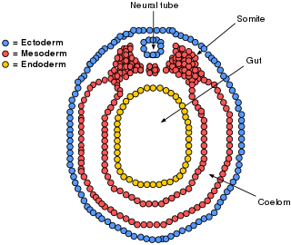
The mesoderm is the middle layer of the three germ layers that develops during gastrulation in the very early development of the embryo of most animals. The outer layer is the ectoderm, and the inner layer is the endoderm.

<span class="mw-page-title-main">Peritoneum</span> Serous membrane that forms lining of abdominal cavity or coelom

The peritoneum is the serous membrane forming the lining of the abdominal cavity or coelom in amniotes and some invertebrates, such as annelids. It covers most of the intra-abdominal organs, and is composed of a layer of mesothelium supported by a thin layer of connective tissue. This peritoneal lining of the cavity supports many of the abdominal organs and serves as a conduit for their blood vessels, lymphatic vessels, and nerves.

<span class="mw-page-title-main">Body cavity</span> Internal space within a multicellular organism

A body cavity is any space or compartment, or potential space, in an animal body. Cavities accommodate organs and other structures; cavities as potential spaces contain fluid.

<span class="mw-page-title-main">Coelom</span> The main body cavity in many animals

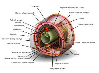
The coelom is the main body cavity in many animals and is positioned inside the body to surround and contain the digestive tract and other organs. In some animals, it is lined with mesothelium. In other animals, such as molluscs, it remains undifferentiated. In the past, and for practical purposes, coelom characteristics have been used to classify bilaterian animal phyla into informal groups.

<span class="mw-page-title-main">Mesentery</span> Contiguous fold of tissues that supports the intestines

The mesentery is an organ that attaches the intestines to the posterior abdominal wall and is formed by the double fold of peritoneum. It helps in storing fat and allowing blood vessels, lymphatics, and nerves to supply the intestines, among other functions.

<span class="mw-page-title-main">Omphalocele</span> Rare abdominal wall defect in which internal organs remain outside of the abdomen in a sac

Omphalocele or omphalocoele also called exomphalos, is a rare abdominal wall defect. Beginning at the 6th week of development, rapid elongation of the gut and increased liver size reduces intra abdominal space, which pushes intestinal loops out of the abdominal cavity. Around 10th week, the intestine returns to the abdominal cavity and the process is completed by the 12th week. Persistence of intestine or the presence of other abdominal viscera in the umbilical cord results in an omphalocele.

A germ layer is a primary layer of cells that forms during embryonic development. The three germ layers in vertebrates are particularly pronounced; however, all eumetazoans produce two or three primary germ layers. Some animals, like cnidarians, produce two germ layers making them diploblastic. Other animals such as bilaterians produce a third layer between these two layers, making them triploblastic. Germ layers eventually give rise to all of an animal's tissues and organs through the process of organogenesis.

<span class="mw-page-title-main">Yolk sac</span> Membranous sac attached to an embryo

The yolk sac is a membranous sac attached to an embryo, formed by cells of the hypoblast layer of the bilaminar embryonic disc. This is alternatively called the umbilical vesicle by the Terminologia Embryologica (TE), though yolk sac is far more widely used. In humans, the yolk sac is important in early embryonic blood supply, and much of it is incorporated into the primordial gut during the fourth week of embryonic development.

<span class="mw-page-title-main">Serous membrane</span> Smooth coating lining contents & inner walls of body cavities

The serous membrane is a smooth tissue membrane of mesothelium lining the contents and inner walls of body cavities, which secrete serous fluid to allow lubricated sliding movements between opposing surfaces. The serous membrane that covers internal organs is called a visceral membrane; while the one that covers the cavity wall is called the parietal membrane. Between the two opposing serosal surfaces is often a potential space, mostly empty except for the small amount of serous fluid.

The primitive node is the organizer for gastrulation in most amniote embryos. In birds it is known as Hensen's node, and in amphibians it is known as the Spemann-Mangold organizer. It is induced by the Nieuwkoop center in amphibians, or by the posterior marginal zone in amniotes including birds.

<span class="mw-page-title-main">Septum transversum</span>

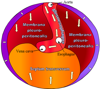
The septum transversum is a thick mass of cranial mesenchyme, formed in the embryo, that gives rise to parts of the thoracic diaphragm and the ventral mesentery of the foregut in the developed human being and other mammals.

<span class="mw-page-title-main">Foregut</span> Anterior part of the gastrointestinal tract

The foregut in humans is the anterior part of the alimentary canal, from the distal esophagus to the first half of the duodenum, at the entrance of the bile duct. Beyond the stomach, the foregut is attached to the abdominal walls by mesentery. The foregut arises from the endoderm, developing from the folding primitive gut, and is developmentally distinct from the midgut and hindgut. Although the term “foregut” is typically used in reference to the anterior section of the primitive gut, components of the adult gut can also be described with this designation. Pain in the epigastric region, just below the intersection of the ribs, typically refers to structures in the adult foregut.

<span class="mw-page-title-main">Midgut</span> Embryonic structure from which most of the human intestines develop

The midgut is the portion of the human embryo from which most of the intestines develop. After it bends around the superior mesenteric artery, it is called the "midgut loop". It comprises the portion of the alimentary canal from the end of the foregut at the opening of the bile duct to the hindgut, about two-thirds of the way through the transverse colon.

<span class="mw-page-title-main">Lateral plate mesoderm</span>

The lateral plate mesoderm is the mesoderm that is found at the periphery of the embryo. It is to the side of the paraxial mesoderm, and further to the axial mesoderm. The lateral plate mesoderm is separated from the paraxial mesoderm by a narrow region of intermediate mesoderm. The mesoderm is the middle layer of the three germ layers, between the outer ectoderm and inner endoderm.

<span class="mw-page-title-main">Bilaminar embryonic disc</span>

The bilaminar embryonic disc, bilaminar blastoderm or embryonic disc is the distinct two-layered structure of cells formed in an embryo. In the development of the human embryo this takes place by day eight. It is formed when the inner cell mass, also known as the embryoblast, forms a bilaminar disc of two layers, an upper layer called the epiblast and a lower layer called the hypoblast, which will eventually form into fetus. These two layers of cells are stretched between two fluid-filled cavities at either end: the primitive yolk sac and the amniotic sac.

<span class="mw-page-title-main">Cloaca (embryology)</span> Structure in the embryo

The cloaca is a structure in the development of the urinary and reproductive organs.

<span class="mw-page-title-main">Human embryonic development</span> Development and formation of the human embryo

Human embryonic development, or human embryogenesis, is the development and formation of the human embryo. It is characterised by the processes of cell division and cellular differentiation of the embryo that occurs during the early stages of development. In biological terms, the development of the human body entails growth from a one-celled zygote to an adult human being. Fertilization occurs when the sperm cell successfully enters and fuses with an egg cell (ovum). The genetic material of the sperm and egg then combine to form the single cell zygote and the germinal stage of development commences. Embryonic development in the human, covers the first eight weeks of development; at the beginning of the ninth week the embryo is termed a fetus. The eight weeks has 23 stages.

The development of the reproductive system is the part of embryonic growth that results in the sex organs and contributes to sexual differentiation. Due to its large overlap with development of the urinary system, the two systems are typically described together as the urogenital or genitourinary system.

<span class="mw-page-title-main">Mesentery (zoology)</span> Membrane inside the body cavity of an animal

In zoology, a mesentery is a membrane inside the body cavity of an animal. The term identifies different structures in different phyla: in vertebrates it is a double fold of the peritoneum enclosing the intestines; in other organisms it forms complete or incomplete partitions of the body cavity, whether that is the coelom or, as in the Anthozoa, the gastrovascular cavity.

References

  1. 1 2 3 Sadler TW, Sadler-Redmond SL (2012). LANGMAN Embriología médica. Vol. I (12 ed.). Philadelphia, PA: The Point.
  2. Tortora G, Derrickson B (2008). Principios de anatomía y fisiología. Vol. I (11 ed.). Buenos Aires: Panamericana.
  3. Soffers JH, Hikspoors JP, Mekonen HK, Koehler SE, Lamers WH (August 2015). "The growth pattern of the human intestine and its mesentery". BMC Developmental Biology. 15 (1): 31. doi: 10.1186/s12861-015-0081-x . PMC   4546136 . PMID   26297675.
  4. de Lussanet, Marc H.E.; Osse, Jan W.M. (2012). "An ancestral axial twist explains the contralateral forebrain and the optic chiasm in vertebrates". Animal Biology. 62 (2): 193–216. arXiv: 1003.1872 . doi:10.1163/157075611X617102. S2CID   7399128.
  5. de Lussanet, M.H.E. (2019). "Opposite asymmetries of face and trunk and of kissing and hugging, as predicted by the axial twist hypothesis". PeerJ. 7: e7096. doi: 10.7717/peerj.7096 . PMC   6557252 . PMID   31211022.
  6. 1 2 3 4 5 6 Moore K, Agur L (2009). Embriología Clínica Moore. Vol. XI (5th ed.). Madrid: Elsevier Health Sciences.
  7. Guyton A, Hall J (2011). Guyton y Hall Fisiología Médica. Vol. II (11a ed.). Madrid: Elsevier Health Sciences.
  8. Boron W, Boulpaep E (2012). Medical Physiology Boron. Vol. II (2nd ed.). Philadelphia: Elsevier Health Sciences.
  9. Barrett K, Barman S, Boitano S (2010). Ganong Fisiología médica. Vol. X (23rd ed.). New York: Mc Graw Hill.