
Psoriasis is a long-lasting, noncontagious autoimmune disease characterized by raised areas of abnormal skin. These areas are typically red, or purple on some people with darker skin, dry, itchy, and scaly. Psoriasis varies in severity from small, localized patches to complete body coverage. Injury to the skin can trigger psoriatic skin changes at that spot, which is known as the Koebner phenomenon.

Pityriasis lichenoides et varioliformis acuta (PLEVA) is a disease of the immune system. It is the more severe version of pityriasis lichenoides chronica. The disease is characterized by rashes and small lesions on the skin. The disease is more common in males and usually occurs in young adulthood, although it has been seen in every age group and every race. It is possible for the disease to go into remission for short periods of time or forever.
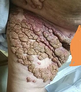
A skin condition, also known as cutaneous condition, is any medical condition that affects the integumentary system—the organ system that encloses the body and includes skin, hair, nails, and related muscle and glands. The major function of this system is as a barrier against the external environment.

Lichen planus (LP) is a chronic inflammatory and immune-mediated disease that affects the skin, nails, hair, and mucous membranes. It is not an actual lichen, and is only named that because it looks like one. It is characterized by polygonal, flat-topped, violaceous papules and plaques with overlying, reticulated, fine white scale, commonly affecting dorsal hands, flexural wrists and forearms, trunk, anterior lower legs and oral mucosa. Although there is a broad clinical range of LP manifestations, the skin and oral cavity remain as the major sites of involvement. The cause is unknown, but it is thought to be the result of an autoimmune process with an unknown initial trigger. There is no cure, but many different medications and procedures have been used in efforts to control the symptoms.

Keratoderma blennorrhagicum etymologically meaning keratinized (kerato-) skin (derma-) mucousy (blenno-) discharge (-rrhagia) are skin lesions commonly found on the palms and soles but which may spread to the scrotum, scalp and trunk. The lesions may resemble psoriasis.
Parapsoriasis refers to one of a group of skin disorders that are characterized primarily by their resemblance to psoriasis, rather than by their underlying cause.

Morphea, is a form of scleroderma that involves isolated patches of hardened skin on the face, hands, and feet, or anywhere else on the body, with no internal organ involvement.
Tumid lupus erythematosus is a rare, but distinctive entity in which patients present with edematous erythematous plaques, usually on the trunk.
Subacute cutaneous lupus erythematosus is a clinically distinct subset of cases of lupus erythematosus that is most often present in white women aged 15 to 40, consisting of skin lesions that are scaly and evolve as poly-cyclic annular lesions or plaques similar to those of plaque psoriasis.
Multifocal lymphangioendotheliomatosis, also known as congenital cutaneovisceral angiomatosis with thrombocytopenia, and multifocal lymphangioendotheliomatosis with thrombocytopenia, is a skin condition that presents at birth with hundreds of red-brown plaques as large as several centimeters.
Acral fibrokeratoma is a skin lesion characterized by a pinkish, hyperkeratotic, hornlike projection occurring on a finger, toe, or palm.
Congenital smooth muscle hamartoma is typically a skin colored or lightly pigmented patch or plaque with hypertrichosis.
Nevus lipomatosus (cutaneous) superficialis is characterized by soft, yellowish papules or cerebriform plaques, usually of the buttock or thigh, less often of the ear or scalp, with a wrinkled rather than warty surface. It is usually congenital in origin or appears within the first three decades.
Large plaque parapsoriasis are skin lesions that may be included in the modern scheme of cutaneous conditions described as parapsoriasis. These lesions, called plaques, may be irregularly round-shaped to oval and are 10 cm (4 in) or larger in diameter. They can be very thin plaques that are asymptomatic or mildly pruritic. Large-plaque parapsoriasis is a common associate of retiform parapsoriasis, can be accompanied by poikiloderma vasculare atrophicans, and can in rare occasions be a precursor to cutaneous T-cell lymphoma.
Vitamin K reactions occur after injection with vitamin K, and there are two patterns of presentation, (1) a reaction may occur several days to 2 weeks after injection with skin lesions that are pruritic, red patches and plaques that can deep-seated, involving the dermis and subcutaneous tissue, or (2) with subcutaneous sclerosis with or without fasciitis, that appears at the site of injection many months after treatment. The latter pseudosclerodermatous reaction has been termed Texier's disease and lasts several years.

Porokeratosis is a specific disorder of keratinization that is characterized histologically by the presence of a cornoid lamella, a thin column of closely stacked, parakeratotic cells extending through the stratum corneum with a thin or absent granular layer.

Linear verrucous epidermal nevus is a skin lesion characterized by a verrucous skin-colored, dirty-gray or brown papule. Generally, multiple papules present simultaneously, and coalesce to form a serpiginous plaque. When this nevus covers a diffuse or extensive portion of the body's surface area, it may be referred to as a systematized epidermal nevus, when it involved only one-half of the body it is called a nevus unius lateris.
Gougerot–Blum syndrome is a variant of pigmented purpuric dermatitis, a skin condition characterized by minute, rust-colored to violaceous, lichenoid papules that tend to fuse into plaques of various hues. Relative to other variants, it is characterized clinically by a male predominance, pruritus, with a predilection for the legs, and histologically, it features a densely cellular lichenoid infiltrate.
Retiform parapsoriasis is a cutaneous condition, considered to be a type of large-plaque parapsoriasis. It is characterized by widespread, ill-defined plaques on the skin, that have a net-like or zebra-striped pattern. Skin atrophy, a wasting away of the cutaneous tissue, usually occurs within the area of these plaques.

Poikiloderma vasculare atrophicans (PVA), is a cutaneous condition characterized by hypo- or hyperpigmentation, telangiectasia and skin atrophy. Other names for the condition include prereticulotic poikiloderma and atrophic parapsoriasis. The condition was first described by pioneer American pediatrician Abraham Jacobi in 1906. PVA causes areas of affected skin to appear speckled red and inflamed, yellowish and/or brown, gray or grayish-black, with scaling and a thinness that may be described as "cigarette paper". On the surface of the skin, these areas may range in size from small patches, to plaques, to neoplasms.