
Pityriasis lichenoides et varioliformis acuta is a disease of the immune system. It is the more severe version of pityriasis lichenoides chronica. The disease is characterized by rashes and small lesions on the skin. The disease is more common in males and usually occurs in young adulthood, although it has been seen in every age group and every race. It is possible for the disease to go into remission for short periods of time or forever.
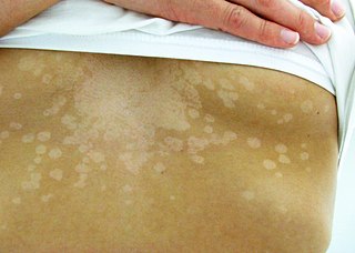
Tinea versicolor is a condition characterized by a skin eruption on the trunk and proximal extremities. The majority of tinea versicolor is caused by the fungus Malassezia globosa, although Malassezia furfur is responsible for a small number of cases. These yeasts are normally found on the human skin and become troublesome only under certain conditions, such as a warm and humid environment, although the exact conditions that cause initiation of the disease process are poorly understood.

A skin condition, also known as cutaneous condition, is any medical condition that affects the integumentary system—the organ system that encloses the body and includes skin, nails, and related muscle and glands. The major function of this system is as a barrier against the external environment.

Lichen planus (LP) is a chronic inflammatory and autoimmune disease that affects the skin, nails, hair, and mucous membranes. It is not an actual lichen, but is named for its appearance. It is characterized by polygonal, flat-topped, violaceous papules and plaques with overlying, reticulated, fine white scale, commonly affecting dorsal hands, flexural wrists and forearms, trunk, anterior lower legs and oral mucosa. The hue may be gray-brown in people with darker skin. Although there is a broad clinical range of LP manifestations, the skin and oral cavity remain as the major sites of involvement. The cause is unknown, but it is thought to be the result of an autoimmune process with an unknown initial trigger. There is no cure, but many different medications and procedures have been used in efforts to control the symptoms.

Hypopigmentation is characterized specifically as an area of skin becoming lighter than the baseline skin color, but not completely devoid of pigment. This is not to be confused with depigmentation, which is characterized as the absence of all pigment. It is caused by melanocyte or melanin depletion, or a decrease in the amino acid tyrosine, which is used by melanocytes to make melanin. Some common genetic causes include mutations in the tyrosinase gene or OCA2 gene. As melanin pigments tend to be in the skin, eye, and hair, these are the commonly affected areas in those with hypopigmentation.

Pityriasis rosea is a type of skin rash. Classically, it begins with a single red and slightly scaly area known as a "herald patch". This is then followed, days to weeks later, by an eruption of many smaller scaly spots; pinkish with a red edge in people with light skin and greyish in darker skin. About 20% of cases show atypical deviations from this pattern. It usually lasts less than three months and goes away without treatment. Sometimes malaise or a fever may occur before the start of the rash or itchiness, but often there are few other symptoms.
Pityriasis commonly refers to flaking of the skin. The word comes from the Greek πίτυρον 'bran'.
Pityriasis lichenoides chronica is an uncommon, idiopathic, acquired dermatosis, characterized by evolving groups of erythematous, scaly papules that may persist for months.
Parapsoriasis refers to one of a group of skin disorders that are characterized primarily by their resemblance to psoriasis, rather than by their underlying cause.
Lichen ruber moniliformis is a rare skin disease named for Fred Wise and Charles R. Rein.

Sweet syndrome (SS), or acute febrile neutrophilic dermatosis, is a skin disease characterized by the sudden onset of fever, an elevated white blood cell count, and tender, red, well-demarcated papules and plaques that show dense infiltrates by neutrophil granulocytes on histologic examination.
Pityriasis alba is a skin condition, a type of dermatitis, commonly seen in children and young adults as dry, fine-scaled, pale patches on the face. It is self-limiting and usually only requires use of moisturizer creams.

Ecthyma gangrenosum is a type of skin lesion characterized by vesicles or blisters which rapidly evolve into pustules and necrotic ulcers with undermined tender erythematous border. "Ecthyma" means a pus forming infection of the skin with an ulcer, "gangrenosum" refers to the accompanying gangrene or necrosis. It is classically associated with Pseudomonas aeruginosa bacteremia, but it is not pathognomonic. Pseudomonas aeruginosa is a gram negative, aerobic bacillus.
Viktor Mucha was a dermatologist from Austria. He was involved in early syphilis research.
Tumid lupus erythematosus is a rare, but distinctive entity in which patients present with edematous erythematous plaques, usually on the trunk.

Annular erythema of infancy(AEI) consists of self-limited eruptions of erythematous, annular to polycyclic patches and plaques. It is an idiopathic figurate erythema. Over several days, a single lesion disappears without leaving behind any scale or hyperpigmentation. Mostly affecting the trunk, face, and extremities, this rash has no symptoms. The diagnosis of AEI is made through a combination of histopathologic and clinical examinations. The disease first manifests in infancy, and if treatment is not received, the periodic eruptions usually stop after the first year of life.
Adrenergic urticaria is a skin condition characterized by an eruption consisting of small (1-5mm) red macules and papules with a pale halo, appearing within 10 to 15 min after emotional upset. There have been 10 cases described in medical literature, and involve a trigger followed by a rise in catecholamine and IgE. Treatment involves propranolol and trigger avoidance.
Doucas and Kapetanakis pigmented purpura is a skin condition characterized by scaly and eczematous patches, which also have petechiae and hemosiderin staining.
Postinflammatory hypopigmentation is a cutaneous condition characterized by decreased pigment in the skin following inflammation of the skin.
