The stomatogastric ganglion (STG) is a much-studied ganglion (collection of neurons) found in arthropods and studied extensively in decapod crustaceans. [1] It is part of the stomatogastric nervous system.

The stomatogastric ganglion (STG) is a much-studied ganglion (collection of neurons) found in arthropods and studied extensively in decapod crustaceans. [1] It is part of the stomatogastric nervous system.
The neurons comprising the stomatogastric ganglion have cell bodies located dorsal to the stomach within the lumen of the opthalmic artery. [2] [3] Most are motor neurons, with neurites that exit through motor nerves and innervate the muscles of the gastric mill and pylorus. [3] [4] [5] These STG motor neurons also form direct synaptic connections with one another. In crabs and lobsters, in which it has been well-studied, the stomatogastric ganglion is a collection of approximately 25-30 neurons. The circuit varies slightly between different crustacean species and between individuals of the same species, but most of the neurons are conserved, and most stomatogastric muscles are innervated by just one motor neuron. [5] The electrical and chemical synaptic connections between all of the STG neurons have been fully mapped and characterized, forming a complete wiring diagram (also called a connectome). [5]
Neural activity in the stomatogastric ganglion produces rhythmic movements of the gastric mill and pyloric region of the digestive system. [6] Neural circuits within the STG are prominent examples of central pattern generators, and their rhythm-generating properties have been studied in detail. The characteristic gastric mill rhythm and pyloric rhythm arise from the intrinsic electrophysiological properties of the neurons and from the strength of synaptic connections between neurons. [7] [8] The stomatogastric ganglion also receives many modulatory inputs. [9] These neuromodulators, such as serotonin, dopamine, proctolin, FLRFamide-like peptides, and red pigment-concentrating hormone (RPCH) can change the speed and form of the rhythmic activity. [9] [10]

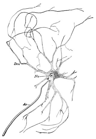
A neuron, neurone, or nerve cell is an excitable cell that fires electric signals called action potentials across a neural network in the nervous system. Neurons communicate with other cells via synapses, which are specialized connections that commonly use minute amounts of chemical neurotransmitters to pass the electric signal from the presynaptic neuron to the target cell through the synaptic gap.

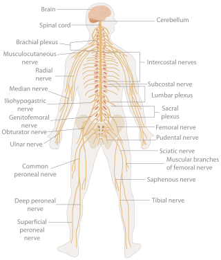
In biology, the nervous system is the highly complex part of an animal that coordinates its actions and sensory information by transmitting signals to and from different parts of its body. The nervous system detects environmental changes that impact the body, then works in tandem with the endocrine system to respond to such events. Nervous tissue first arose in wormlike organisms about 550 to 600 million years ago. In vertebrates, it consists of two main parts, the central nervous system (CNS) and the peripheral nervous system (PNS). The CNS consists of the brain and spinal cord. The PNS consists mainly of nerves, which are enclosed bundles of the long fibers, or axons, that connect the CNS to every other part of the body. Nerves that transmit signals from the brain are called motor nerves (efferent), while those nerves that transmit information from the body to the CNS are called sensory nerves (afferent). The PNS is divided into two separate subsystems, the somatic and autonomic, nervous systems. The autonomic nervous system is further subdivided into the sympathetic, parasympathetic and enteric nervous systems. The sympathetic nervous system is activated in cases of emergencies to mobilize energy, while the parasympathetic nervous system is activated when organisms are in a relaxed state. The enteric nervous system functions to control the gastrointestinal system. Nerves that exit from the brain are called cranial nerves while those exiting from the spinal cord are called spinal nerves.

A motor neuron is a neuron whose cell body is located in the motor cortex, brainstem or the spinal cord, and whose axon (fiber) projects to the spinal cord or outside of the spinal cord to directly or indirectly control effector organs, mainly muscles and glands. There are two types of motor neuron – upper motor neurons and lower motor neurons. Axons from upper motor neurons synapse onto interneurons in the spinal cord and occasionally directly onto lower motor neurons. The axons from the lower motor neurons are efferent nerve fibers that carry signals from the spinal cord to the effectors. Types of lower motor neurons are alpha motor neurons, beta motor neurons, and gamma motor neurons.

The autonomic nervous system (ANS), sometimes called the visceral nervous system and formerly the vegetative nervous system, is a division of the nervous system that operates internal organs, smooth muscle and glands. The autonomic nervous system is a control system that acts largely unconsciously and regulates bodily functions, such as the heart rate, its force of contraction, digestion, respiratory rate, pupillary response, urination, and sexual arousal. This system is the primary mechanism in control of the fight-or-flight response.
The development of the nervous system, or neural development (neurodevelopment), refers to the processes that generate, shape, and reshape the nervous system of animals, from the earliest stages of embryonic development to adulthood. The field of neural development draws on both neuroscience and developmental biology to describe and provide insight into the cellular and molecular mechanisms by which complex nervous systems develop, from nematodes and fruit flies to mammals.

The parasympathetic nervous system is one of the three divisions of the autonomic nervous system, the others being the sympathetic nervous system and the enteric nervous system. The enteric nervous system is sometimes considered part of the autonomic nervous system, and sometimes considered an independent system.

A motor nerve, or efferent nerve, is a nerve that contains exclusively efferent nerve fibers and transmits motor signals from the central nervous system (CNS) to the muscles of the body. This is different from the motor neuron, which includes a cell body and branching of dendrites, while the nerve is made up of a bundle of axons. Motor nerves act as efferent nerves which carry information out from the CNS to muscles, as opposed to afferent nerves, which transfer signals from sensory receptors in the periphery to the CNS. Efferent nerves can also connect to glands or other organs/issues instead of muscles. The vast majority of nerves contain both sensory and motor fibers and are therefore called mixed nerves.

The grey columns are three regions of the somewhat ridge-shaped mass of grey matter in the spinal cord. These regions present as three columns: the anterior grey column, the posterior grey column, and the lateral grey column, all of which are visible in cross-section of the spinal cord.

The Stomatogastric Nervous System (STNS) is a commonly studied neural network composed of several ganglia in arthropods that controls the motion of the gut and foregut. The network of neurons acts as a central pattern generator. It is a model system for motor pattern generation because of the small number of cells, which are comparatively large and can be reliably identified. The system is composed of the stomatogastric ganglion (STG), oesophageal ganglion and the paired commissural ganglia.

Accelerando is a 2005 science fiction novel consisting of a series of interconnected short stories written by British author Charles Stross. As well as normal hardback and paperback editions, it was released as a free e-book under the CC BY-NC-ND license. Accelerando won the Locus Award in 2006, and was nominated for several other awards in 2005 and 2006, including the Hugo, Campbell, Clarke, and British Science Fiction Association Awards.
Central pattern generators (CPGs) are self-organizing biological neural circuits that produce rhythmic outputs in the absence of rhythmic input. They are the source of the tightly-coupled patterns of neural activity that drive rhythmic and stereotyped motor behaviors like walking, swimming, breathing, or chewing. The ability to function without input from higher brain areas still requires modulatory inputs, and their outputs are not fixed. Flexibility in response to sensory input is a fundamental quality of CPG-driven behavior. To be classified as a rhythmic generator, a CPG requires:
Bursting, or burst firing, is an extremely diverse general phenomenon of the activation patterns of neurons in the central nervous system and spinal cord where periods of rapid action potential spiking are followed by quiescent periods much longer than typical inter-spike intervals. Bursting is thought to be important in the operation of robust central pattern generators, the transmission of neural codes, and some neuropathologies such as epilepsy. The study of bursting both directly and in how it takes part in other neural phenomena has been very popular since the beginnings of cellular neuroscience and is closely tied to the fields of neural synchronization, neural coding, plasticity, and attention.

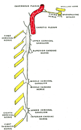
The superior cervical ganglion (SCG) is the upper-most and largest of the cervical sympathetic ganglia of the sympathetic trunk. It probably formed by the union of four sympathetic ganglia of the cervical spinal nerves C1–C4. It is the only ganglion of the sympathetic nervous system that innervates the head and neck. The SCG innervates numerous structures of the head and neck.

Eve Marder is a University Professor and the Victor and Gwendolyn Beinfield Professor of Neuroscience at Brandeis University. At Brandeis, Marder is also a member of the Volen National Center for Complex Systems. Dr. Marder is known for her pioneering work on small neuronal networks which her team has interrogated via a combination of complementary experimental and theoretical techniques.
Models of neural computation are attempts to elucidate, in an abstract and mathematical fashion, the core principles that underlie information processing in biological nervous systems, or functional components thereof. This article aims to provide an overview of the most definitive models of neuro-biological computation as well as the tools commonly used to construct and analyze them.

Retrograde tracing is a research method used in neuroscience to trace neural connections from their point of termination to their source. Retrograde tracing techniques allow for detailed assessment of neuronal connections between a target population of neurons and their inputs throughout the nervous system. These techniques allow the "mapping" of connections between neurons in a particular structure and the target neurons in the brain. The opposite technique is anterograde tracing, which is used to trace neural connections from their source to their point of termination. Both the anterograde and retrograde tracing techniques are based on the visualization of axonal transport.

Non-spiking neurons are neurons that are located in the central and peripheral nervous systems and function as intermediary relays for sensory-motor neurons. They do not exhibit the characteristic spiking behavior of action potential generating neurons.
The evolution of nervous systems dates back to the first development of nervous systems in animals. Neurons developed as specialized electrical signaling cells in multicellular animals, adapting the mechanism of action potentials present in motile single-celled and colonial eukaryotes. Primitive systems, like those found in protists, use chemical signalling for movement and sensitivity; data suggests these were precursors to modern neural cell types and their synapses. When some animals started living a mobile lifestyle and eating larger food particles externally, they developed ciliated epithelia, contractile muscles and coordinating & sensitive neurons for it in their outer layer.

Synaptic plasticity refers to a chemical synapse's ability to undergo changes in strength. Synaptic plasticity is typically input-specific, meaning that the activity in a particular neuron alters the efficacy of a synaptic connection between that neuron and its target. However, in the case of heterosynaptic plasticity, the activity of a particular neuron leads to input unspecific changes in the strength of synaptic connections from other unactivated neurons. A number of distinct forms of heterosynaptic plasticity have been found in a variety of brain regions and organisms. These different forms of heterosynaptic plasticity contribute to a variety of neural processes including associative learning, the development of neural circuits, and homeostasis of synaptic input.

Synthetic Nervous System (SNS) is a computational neuroscience model that may be developed with the Functional Subnetwork Approach (FSA) to create biologically plausible models of circuits in a nervous system. The FSA enables the direct analytical tuning of dynamical networks that perform specific operations within the nervous system without the need for global optimization methods like genetic algorithms and reinforcement learning. The primary use case for a SNS is system control, where the system is most often a simulated biomechanical model or a physical robotic platform. An SNS is a form of a neural network much like artificial neural networks (ANNs), convolutional neural networks (CNN), and recurrent neural networks (RNN). The building blocks for each of these neural networks is a series of nodes and connections denoted as neurons and synapses. More conventional artificial neural networks rely on training phases where they use large data sets to form correlations and thus “learn” to identify a given object or pattern. When done properly this training results in systems that can produce a desired result, sometimes with impressive accuracy. However, the systems themselves are typically “black boxes” meaning there is no readily distinguishable mapping between structure and function of the network. This makes it difficult to alter the function, without simply starting over, or extract biological meaning except in specialized cases. The SNS method differentiates itself by using details of both structure and function of biological nervous systems. The neurons and synapse connections are intentionally designed rather than iteratively changed as part of a learning algorithm.