Strawberry crinkle cytorhabdovirus

Last updated
Strawberry crinkle cytorhabdovirus
Virus classification OOjs UI icon edit-ltr.svg
(unranked): Virus
Realm: Riboviria
Kingdom: Orthornavirae
Phylum: Negarnaviricota
Class: Monjiviricetes
Order: Mononegavirales
Family: Rhabdoviridae
Genus: Cytorhabdovirus
Species:
Strawberry crinkle cytorhabdovirus
Synonyms
  • Strawberry crinkle virus

Strawberry crinkle cytorhabdovirus, commonly called Strawberry crinkle virus (SCV), is a negative sense single stranded RNA virus that threatens strawberry production worldwide. This virus reduces plant rigidity, runner production, fruit size, and production, while causing distortion and crinkling of the leaves. This virus was first described in 1932 in Oregon and California with commercial strawberry varieties, and later became an issue around the world, including North America, South America, Europe, South Africa, New Zealand, Australia, and Japan. Of the family Rhabdoviridae, it is a large family of viruses that affects plants, vertebrates, and invertebrates. Specifically, this virus infects strawberry plants of the genus Fragaria and is transmitted through two aphid vectors that feed on strawberries, Chaetosiphon fragaefolii and C. jacobi . When SCV is combined with other aphid-transmitted strawberry viruses, such as mottle, mild yellow-edge, vein banding, or pallidosis, the damage becomes even more deleterious. Economically, the only significant host of SCV is Fragaria ananassa . [1] [2] [3]

Contents

Baltimore Classification

SCV in its family tree Viruses-11-00982-g003.png
SCV in its family tree

Based on the Baltimore classification, which is a virus classification system the groups viruses into families based on their genome type, we can know the steps the virus must take in order to produce mRNA and how the genome is copied to create more genomes. All viruses want to get to mRNA in order to go through the process of translation; however, only positive sense single stranded RNA virus (+RNA) have the capability to do this. (-)RNA viruses, such as SCV, are not able to be translated by the host because cells do not have the machinery to copy it, indicating that the virus needs to bring their own enzyme, RNA-dependent RNA polymerase, in order to copy the negative strand into mRNA. This allows viruses, such as SCV, to produce (+)RNA, which can therefore be translated by the host.[ citation needed ]

Structure and Genome

SCV is enveloped with a bacilliform morphology. The virus is roughly 163-383 nm long and 74-88 nm in diameter. Glycoproteins are likely said to be the primary surface projections that occur in the cytoplasm, which are either coated or uncoated. Its nucleocapsids is enclosed in a host-derived envelope and are typically helical with a linear genome that is a negative sense single stranded RNA (- RNA). Typically, SCV’s viral genome is roughly 13kb and contains four proteins. The four proteins involved in this virus are glycoprotein (G), nucleocapsid (N), phosphoprotein (P), and the matrix (M) with their respective sizes of 77 kDa, 55 kDa, 45 kDa, and 23 kDa. Like other plant rhabdoviruses there is a fifth protein involved, a large protein (L) that is 230 kDa.[ citation needed ]

The organization of these five proteins in SCV’s genome is highly conserved. The genome’s order is as follows: N, P, M, G, L, while regions in between these proteins exist. It is important to note that the accumulation of M and G proteins are responsible for the bacilliform shaped morphology of SCV. [4] [1]

Replication cycle

Gene Expression

RNA dependent RNA polymerase binds to the SCV genome at the 3’ end where the genes are then transcribed. Once synthesis reaches the L protein near the 5’ end, mRNAs are capped and polyadenylated. [5]

Entry Into Cell

Virus entry into the host cell occurs when viral G glycoproteins attach to host cell surface receptors, initiating endocytosis. Endocytosis is initiated via low pH levels and coated pits that are made up of clathrin. The virus is then able to fuse with the hosts membrane, which occurs in the cytoplasm.[ citation needed ]

Replication

Once the virus fuses to the hosts membrane, replication occurs. The five proteins in SCV are transcribed from their (-)RNA by RNA polymerase, which is also the L protein. Other than this function, the L protein is also enzymatically essential for capping mRNAs and phosphorylating the P protein. The promoter site is located at the 3’-end of the genome where polymerase attaches and it moves along the RNA template towards the 5’-end. This movement from the leader to the trailer end of the mRNA produces the respective N, P, M, G, and L proteins.[ citation needed ]

Genome replication requires newly synthesized N proteins to encase the RNA. This is used to produce more negative sensed genomic RNA.[ citation needed ]

Transcription

Transcription occurs after entry into the cell and is regulated after both the L and P proteins are expressed in replication. This process occurs and accumulates in viroplasms, which are thread like structures that are located in the cytoplasm of infected cells. RNA dependent RNA polymerase or commonly known as Transcriptase, moves from the 3’ end of the genome to the 5’ end and can terminate randomly at any protein sequence, allowing for mRNA to be formed separately from each other. N proteins are usually produced in abundance and accumulate since they are located at the beginning of the genome (3’ end), which is located right after the leader RNA sequence. This is important because the N protein is needed for the virus because it is used to coat the outside of the replicated genomes.[ citation needed ]

During this process, there are start and stop signals which only allows a portion of the polymerase molecule to move past each junction in order for transcription to continue. This interruption results in greater amounts of mRNA that are produced towards the beginning of the genome, resulting in various amounts of mRNA produced as polymerase moves downstream from N, P, M, G, to L. This inconsistency results in greater amounts of nucleocapsid proteins being made than large (L) proteins.[ citation needed ]

After transcription occurs, all of the produced mRNAs are capped at the 5’ end and polyadenylated at the 3’ end. This process produces mRNAs for SCV in order for the translation to occur.[ citation needed ]

Translation

After the mRNAs are formed and are capped and polyadenylated to their respective ends, this structure mimics cellular mRNAs and allows for translation by cellular ribosomes to produce proteins.[ citation needed ]

The Rough Endoplasmic Reticulum is used to translate the G protein, indicating that G proteins have a simple peptide on their mRNA’s start code. Phosphoproteins (P) and glycoproteins (G) then go through post-translational modification. After phosphorylation of the Large (L) protein occurs, trimers of the P protein are made. Then the G protein is glycosylated in the Rough Endoplasmic Reticulum and the Golgi complex.[ citation needed ]

Assembly and Release

The virus exits the host cell through the process of budding and tubule-guided viral movement. Plants are the natural host for SCV and this virus is specifically transmitted by aphid vectors. Principally, Chaetosiphon fragaefolii.

It is important to note that the transmission cycle depends on temperature. Lower temperatures increase the incubation period in the strawberry and the latent period in the vector. [6]

Modulation of Host Processes/Interaction with host

Aphid Vectors during their growth and development stages. Aphid Vectors.jpg
Aphid Vectors during their growth and development stages.

Strawberry aphid vectors, such as Chaetosiphon fragaefolii , is of genus Chaetosiphon, which are the primary causes of  problems seen in strawberry plants worldwide. These vectors infiltrate strawberry plants, and appear to be either translucent yellow-white to pale green-yellow in color. The body length of C. fragaefolii is roughly 0.9–1.8 millimetres (51289128 in) long, while the antennae are 0.9-1.1 times the body length. Damage to plants are caused by aphid vectors sucking on the sap from plants, causing the observed symptoms of SCV. These vectors can be infected with this virus in as little as 24 hours from birth, while a latent period of 10–19 days are observed. Once infected, this vector can transmit SCV for up to 2 weeks. [7] [8]  

Associated Diseases

Signs and Symptoms

The symptoms of Strawberry Crinkle Virus varies between different varieties of plants, and they can also vary in the severity. Common symptoms can include spotting of the plant veins. The spotting can be chlorotic (yellowish spot on the leaf surface) or necrotic with areas of black indicating tissue death in these areas. The petals of the flowers can also be deformed, and thee petals can also have abnormal streaking. The virus can also cause malformed leaflets, crinkled leaves, and also uneven leaf distribution in the plant. Another symptom can be the formation of lesions or areas of damage in the petioles and stolons of the plant. Episnasty can also occur from the virus, which causes increased growth of the upper region of the plant, which in turn makes the plant top-heavy causing it to bend downward. [9]

Geographical Distribution

Tropism

The natural host range of SCV is narrow, including the wild strawberries F. vesca, F. virginiana and F. chiloensis, as well as on the cultivated species, F. ananassa. [6]

In a study, the virus was isolated from strawberry leaf material; however, more experiments are necessary to conclude which strawberry plant tissues are optimal for the virus. [1]

Economic Impact and Solutions

Strawberries are one of the most important berry crops that are grown in the temperate regions. Due to this, the strawberry industry is a multibillion dollar industry across the temperate regions of the world.  Viruses are considered a minor factor of pathogens that cause major arm to crop productions. In order to control SCR, it is advised to control the vectors of this virus, aphids Chaetosiphon fragaefolii and C. jacobi. However, it is important to note that insecticides are not used to regulate aphid vectors due to possible harmful interactions with bumblebees, which are essential for pollinating the plants. Additionally cultivating healthy, virus-free plants can help control the spread of SCR. Plant material that has been infected by the virus should be eradicated. When trading strawberry plants, it is important to verify that the plant material has met the conditions of a virus-free certification scheme; however, apical meristems from diseased plants can be obtained and made virus-free by growing them on culture medium. This is most successful when the parent plants had been treated with heat of 35 °C-41 °C for several months. [6] [10] [7]

Related Research Articles

<i>Rhabdoviridae</i> Family of viruses in the order Mononegavirales

Rhabdoviridae is a family of negative-strand RNA viruses in the order Mononegavirales. Vertebrates, invertebrates, plants, fungi and protozoans serve as natural hosts. Diseases associated with member viruses include rabies encephalitis caused by the rabies virus, and flu-like symptoms in humans caused by vesiculoviruses. The name is derived from Ancient Greek rhabdos, meaning rod, referring to the shape of the viral particles. The family has 40 genera, most assigned to three subfamilies.

<span class="mw-page-title-main">Rabies virus</span> Species of virus

Rabies virus, scientific name Rabies lyssavirus, is a neurotropic virus that causes rabies in animals, including humans. Rabies transmission can occur through the saliva of animals and less commonly through contact with human saliva. Rabies lyssavirus, like many rhabdoviruses, has an extremely wide host range. In the wild it has been found infecting many mammalian species, while in the laboratory it has been found that birds can be infected, as well as cell cultures from mammals, birds, reptiles and insects. Rabies is reported in more than 150 countries and on all continents except Antarctica. The main burden of disease is reported in Asia and Africa, but some cases have been reported also in Europe in the past 10 years, especially in returning travellers.

<i>Bunyavirales</i> Order of RNA viruses

Bunyavirales is an order of segmented negative-strand RNA viruses with mainly tripartite genomes. Member viruses infect arthropods, plants, protozoans, and vertebrates. It is the only order in the class Ellioviricetes. The name Bunyavirales derives from Bunyamwera, where the original type species Bunyamwera orthobunyavirus was first discovered. Ellioviricetes is named in honor of late virologist Richard M. Elliott for his early work on bunyaviruses.

Chandipura vesiculovirus (CHPV) is a member of the Rhabdoviridae family that is associated with an encephalitic illness in humans. It was first identified in 1965 after isolation from the blood of two patients from Chandipura village in Maharashtra state, India and has been associated with a number of otherwise unexplained outbreaks of encephalitic illness in central India. The most recent occurred in Andhra Pradesh and Maharashtra in June–August 2003 with 329 children affected and 183 deaths. Further sporadic cases and deaths in children were observed in Gujarat state in 2004.

<i>Baculoviridae</i> Family of viruses

Baculoviridae is a family of viruses. Arthropods, among the most studied being Lepidoptera, Hymenoptera and Diptera, serve as natural hosts. Currently, 85 species are placed in this family, assigned to four genera.

<i>Alphavirus</i> Genus of viruses

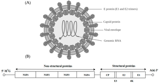
Alphavirus is a genus of RNA viruses, the sole genus in the Togaviridae family. Alphaviruses belong to group IV of the Baltimore classification of viruses, with a positive-sense, single-stranded RNA genome. There are 32 alphaviruses, which infect various vertebrates such as humans, rodents, fish, birds, and larger mammals such as horses, as well as invertebrates. Alphaviruses that could infect both vertebrates and arthropods are referred dual-host alphaviruses, while insect-specific alphaviruses such as Eilat virus and Yada yada virus are restricted to their competent arthropod vector. Transmission between species and individuals occurs mainly via mosquitoes, making the alphaviruses a member of the collection of arboviruses – or arthropod-borne viruses. Alphavirus particles are enveloped, have a 70 nm diameter, tend to be spherical, and have a 40 nm isometric nucleocapsid.

<i>Potyvirus</i> Genus of positive-strand RNA viruses in the family Potyviridae

Potyvirus is a genus of positive-strand RNA viruses in the family Potyviridae. Plants serve as natural hosts. Like begomoviruses, members of this genus may cause significant losses in agricultural, pastoral, horticultural, and ornamental crops. More than 200 species of aphids spread potyviruses, and most are from the subfamily Aphidinae. The genus contains 190 species and potyviruses account for about thirty percent of all currently known plant viruses.

<i>Orthobunyavirus</i> Genus of viruses

Orthobunyavirus is a genus of the Peribunyaviridae family in the order Bunyavirales. There are currently ~170 viruses recognised in this genus. These have been assembled into 103 species and 20 serogroups.

Potato virus Y (PVY) is a plant pathogenic virus of the family Potyviridae, and one of the most important plant viruses affecting potato production.

<i>Strawberry vein banding virus</i> Species of virus

Strawberry vein banding virus (SVBV) is a plant pathogenic virus and a member of the family Caulimoviridae.

Mason-Pfizer monkey virus (M-PMV), formerly Simian retrovirus (SRV), is a species of retroviruses that usually infect and cause a fatal immune deficiency in Asian macaques. The ssRNA virus appears sporadically in mammary carcinoma of captive macaques at breeding facilities which expected as the natural host, but the prevalence of this virus in feral macaques remains unknown. M-PMV was transmitted naturally by virus-containing body fluids, via biting, scratching, grooming, and fighting. Cross contaminated instruments or equipment (fomite) can also spread this virus among animals.

Orchid fleck dichorhavirus, commonly called Orchid fleck virus (OFV), is a non-enveloped, segmented, single-stranded (ss) RNA negative-strand virus, transmitted by the false spider mite, Brevipalpus californicus. OFV causes necrotic and chlorotic lesions on the leaves of many genera in the family Orchidaceae.

Batai orthobunyavirus (BATV) is a RNA virus belonging to order Bunyavirales, genus Orthobunyavirus.

Lily virus X (LVX) is a pathogenic ssRNA(+) plant virus of the family Alphaflexiviridae and the order Tymovirales.

Entebbe bat virus is an infectious disease caused by a Flavivirus that is closely related to yellow fever.

<i>Avian metaavulavirus 2</i> Species of virus

Avian metaavulavirus 2, formerly Avian paramyxovirus 2, is a species of virus belonging to the family Paramyxoviridae and genus Metaavulavirus. The virus is a negative strand RNA virus containing a monopartite genome. Avian metaavulavirus 2 is one of nine species belonging to the genus Metaavulavirus. The most common serotype of Avulavirinae is serotype 1, the cause of Newcastle disease (ND). Avian metaavulavirus 2 has been known to cause disease, specifically mild respiratory infections in domestic poultry, including turkeys and chickens, and has many economic effects on egg production and poultry industries. The virus was first isolated from a strain in Yucaipa, California in 1956. Since then, other isolates of the virus have been isolated worldwide.

West Caucasian bat lyssavirus (WCBL) is a member of genus Lyssavirus, family Rhabdoviridae and order Mononegavirales. This virus was first isolated from Miniopterus schreibersii, in the western Caucasus Mountains of southeastern Europe in 2002. WCBL is the most divergent form of Lyssavirus, and is found in Miniopterus bats (insectivorous), Rousettus aegyptiacus, and Eidolon helvum. The latter two are both fruit bats. The virus is fragile as it can be inactivated by UV light and chemicals, such as ether, chloroform, and bleach. WCBL has not been known to infect humans thus far.

Bat mumps orthorubulavirus, formerly Bat mumps rubulavirus (BMV), is a member of genus Orthorubulavirus, family Paramyxoviridae, and order Mononegavirales. Paramyxoviridae viruses were first isolated from bats using heminested PCR with degenerate primers. This process was then followed by Sanger sequencing. A specific location of this virus is not known because it was isolated from bats worldwide. Although multiple paramyxoviridae viruses have been isolated worldwide, BMV specifically has not been isolated thus far. However, BMV was detected in African fruit bats, but no infectious form has been isolated to date. It is known that BMV is transmitted through saliva in the respiratory system of bats. While the virus was considered its own species for a few years, phylogenetic analysis has since shown that it is a member of Mumps orthorubulavirus.

Rio Negro virus is an alphavirus that was first isolated in Argentina in 1980. The virus was first called Ag80-663 but was renamed to Rio Negro virus in 2005. It is a former member of the Venezuelan equine encephalitis complex (VEEC), which are a group of alphaviruses in the Americas that have the potential to emerge and cause disease. Río Negro virus was recently reclassified as a distinct species. Closely related viruses include Mucambo virus and Everglades virus.

References

  1. 1 2 3 Posthuma, K. I.; Adams, A. N.; Hong, Y.; Kirby, M. J. (2002). "Detection of Strawberry crinkle virus in plants and aphids by RT-PCR using conserved L gene sequences". Plant Pathology. 51 (3): 266–274. doi: 10.1046/j.1365-3059.2002.00725.x . ISSN   1365-3059.
  2. Koloniuk, Igor; Fránová, Jana; Sarkisova, Tatiana; Přibylová, Jaroslava (2018-09-01). "Complete genome sequences of two divergent isolates of strawberry crinkle virus coinfecting a single strawberry plant". Archives of Virology. 163 (9): 2539–2542. doi:10.1007/s00705-018-3860-4. ISSN   1432-8798. PMID   29728910. S2CID   19150995.
  3. Byers, Loren W. (2013-04-15). Second Line of Defense Program, Secondary Screening Operations Questionnaire Datasheet--Users Guide (Report). doi:10.2172/1073755. OSTI   1073755 . Retrieved 14 March 2019.
  4. Posthuma, Karin I.; Adams, Anthony N.; Hong, Yiguo (2000). "Strawberry crinkle virus, a Cytorhabdovirus needing more attention from virologists". Molecular Plant Pathology. 1 (6): 331–336. doi: 10.1046/j.1364-3703.2000.00041.x . ISSN   1364-3703. PMID   20572980.
  5. "Rhabdoviridae ~ ViralZone page". viralzone.expasy.org. Retrieved 2019-03-13.
  6. 1 2 3 "Strawberry crinkle cytorhabdovirus" (PDF). Data Sheets on Quarantine Pests via Prepared by CABI and EPPO for the EU under Contract 90/399003.
  7. 1 2 Cantliffe, Daniel J.; Rondon, Silvia I. (December 2004). "Chaetosiphon Fragaefolii (Homoptera: Aphididae): A Potential New Pest in Florida?". Florida Entomologist. 87 (4): 612–615. doi: 10.1653/0015-4040(2004)087[0612:CFHAAP]2.0.CO;2 . ISSN   0015-4040.
  8. "Chaetosiphon fragaefolii (strawberry aphid) identification, images, ecology, control". influentialpoints.com. Retrieved 2019-03-13.
  9. 1 2 3 Brunt AA, Crabtree K, Dallwitz MJ, Gibbs AJ, Watson L, Zurcher EJ (1996-08-20). "Plant Viruses Online - Strawberry crinkle cytorhabdovirus". srs.im.ac.cn. Retrieved 2020-09-03.
  10. Martin, Robert R.; Tzanetakis, Ioannis E. (October 2013). "High Risk Strawberry Viruses by Region in the United States and Canada: Implications for Certification, Nurseries, and Fruit Production". Plant Disease. 97 (10): 1358–1362. doi: 10.1094/PDIS-09-12-0842-RE . ISSN   0191-2917. PMID   30722134.