Related Research Articles
Working memory is a cognitive system with a limited capacity that can hold information temporarily. Working memory is important for reasoning and the guidance of decision-making and behavior. Working memory is often used synonymously with short-term memory, but some theorists consider the two forms of memory distinct, assuming that working memory allows for the manipulation of stored information, whereas short-term memory only refers to the short-term storage of information. Working memory is a theoretical concept central to cognitive psychology, neuropsychology, and neuroscience.

The precuneus is the portion of the superior parietal lobule on the medial surface of each brain hemisphere. It is located in front of the cuneus. The precuneus is bounded in front by the marginal branch of the cingulate sulcus, at the rear by the parietooccipital sulcus, and underneath by the subparietal sulcus. It is involved with episodic memory, visuospatial processing, reflections upon self, and aspects of consciousness.

Brodmann area 9, or BA9, is part of the frontal cortex in the brain of humans and other primates. It contributes to the dorsolateral and medial prefrontal cortex.
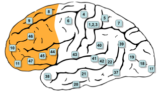
In mammalian brain anatomy, the prefrontal cortex (PFC) is the cerebral cortex which covers the front part of the frontal lobe. The PFC contains the Brodmann areas BA8, BA9, BA10, BA11, BA12, BA13, BA14, BA24, BA25, BA32, BA44, BA45, BA46, and BA47.
Affective neuroscience is the study of the neural mechanisms of emotion. This interdisciplinary field combines neuroscience with the psychological study of personality, emotion, and mood. The putative existence of 'basic emotions' and their defining attributes represents a long lasting and yet unsettled issue in psychology.

The posterior cingulate cortex (PCC) is the caudal part of the cingulate cortex, located posterior to the anterior cingulate cortex. This is the upper part of the "limbic lobe". The cingulate cortex is made up of an area around the midline of the brain. Surrounding areas include the retrosplenial cortex and the precuneus.

The medial dorsal nucleus is a large nucleus in the thalamus.
For the purpose of this article, research on meditation concerns research into the psychological and physiological effects of meditation using the scientific method. In recent years, these studies have increasingly involved the use of modern scientific techniques and instruments, such as fMRI and EEG which are able to directly observe brain physiology and neural activity in living subjects, either during the act of meditation itself, or before and after a meditation effort, thus allowing linkages to be established between meditative practice and changes in brain structure or function.
The ventromedial prefrontal cortex (vmPFC) is a part of the prefrontal cortex in the mammalian brain. The ventral medial prefrontal is located in the frontal lobe at the bottom of the cerebral hemispheres and is implicated in the processing of risk and fear, as it is critical in the regulation of amygdala activity in humans. It also plays a role in the inhibition of emotional responses, and in the process of decision making and self control. It is also involved in the cognitive evaluation of morality.
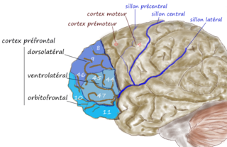
The dorsolateral prefrontal cortex is an area in the prefrontal cortex of the brain of humans and non-human primates. It is one of the most recently derived parts of the human brain. It undergoes a prolonged period of maturation which lasts until adulthood. The DLPFC is not an anatomical structure, but rather a functional one. It lies in the middle frontal gyrus of humans. In macaque monkeys, it is around the principal sulcus. Other sources consider that DLPFC is attributed anatomically to BA 9 and 46 and BA 8, 9 and 10.
In neuroscience, a task-negative (TN) mode, also known as the default mode network, is inversely correlated to the task-positive mode. Its main function is to reorient attention towards salient stimuli. TN is considered to be involved mostly, if not entirely, in involuntary actions. The neural network is right hemisphere lateralized and includes the right temporal-parietal junction and the right ventral frontal cortex. This system shows activity increases upon detection of salient targets, especially when they appear in unexpected locations. Activity increases also are observed in the ventral system after abrupt changes in sensory stimuli, at the onset and offset of task blocks, and at the end of a completed trial.
Scientific studies have found that different brain areas show altered activity in people with major depressive disorder, and this has encouraged advocates of various theories that seek to identify a biochemical origin of the disease, as opposed to theories that emphasize psychological or situational causes. Factors spanning these causative groups include nutritional deficiencies in magnesium, vitamin D, and tryptophan with situational origin but biological impact. Several theories concerning the biologically based cause of depression have been suggested over the years, including theories revolving around monoamine neurotransmitters, neuroplasticity, neurogenesis, inflammation and the circadian rhythm. Physical illnesses, including hypothyroidism and mitochondrial disease, can also trigger depressive symptoms.

In neuroscience, the default mode network (DMN), also default network, or default state network, is a large-scale brain network primarily composed of the medial prefrontal cortex, posterior cingulate cortex/precuneus and angular gyrus. It is best known for being active when a person is not focused on the outside world and the brain is at wakeful rest, such as during daydreaming and mind-wandering. It can also be active during detailed thoughts related to external task performance. Other times that the DMN is active include when the individual is thinking about others, thinking about themselves, remembering the past, and planning for the future.

Resting state fMRI is a method of functional magnetic resonance imaging (fMRI) that is used in brain mapping to evaluate regional interactions that occur in a resting or task-negative state, when an explicit task is not being performed. A number of resting-state conditions are identified in the brain, one of which is the default mode network. These resting brain state conditions are observed through changes in blood flow in the brain which creates what is referred to as a blood-oxygen-level dependent (BOLD) signal that can be measured using fMRI.

The biological basis of personality is the collection of brain systems and mechanisms that underlie human personality. Human neurobiology, especially as it relates to complex traits and behaviors, is not well understood, but research into the neuroanatomical and functional underpinnings of personality are an active field of research. Animal models of behavior, molecular biology, and brain imaging techniques have provided some insight into human personality, especially trait theories.

Fronto-cerebellar dissociation is the disconnection and independent function of frontal and cerebellar regions of the brain. It is characterized by inhibited communication between the two regions, and is notably observed in cases of ADHD, schizophrenia, alcohol abuse, and heroin abuse. The frontal and cerebellar regions make distinctive contributions to cognitive performance, with the left-frontal activations being responsible for selecting a response to a stimulus, while the right-cerebellar activation is responsible for the search for a given response to a stimulus. Left-frontal activation increases when there are many appropriate responses to a stimulus, and right-cerebellar activation increases when there is a single appropriate response to a stimulus. A person with dissociated frontal and cerebellar regions may have difficulties with selecting a response to a stimuli, or difficulties with response initiation. Fronto-cerebellar dissociation can often result in either the frontal lobe or the cerebellum becoming more active in place of the less active region as a compensatory effect.
Large-scale brain networks are collections of widespread brain regions showing functional connectivity by statistical analysis of the fMRI BOLD signal or other recording methods such as EEG, PET and MEG. An emerging paradigm in neuroscience is that cognitive tasks are performed not by individual brain regions working in isolation but by networks consisting of several discrete brain regions that are said to be "functionally connected". Functional connectivity networks may be found using algorithms such as clustering, spatial independent component analysis (ICA), seed based, and others. Synchronized brain regions may also be identified using long-range synchronization of the EEG, MEG, or other dynamic brain signals.

The salience network (SN) is a large scale brain network of the human brain that is primarily composed of the anterior insula (AI) and dorsal anterior cingulate cortex (dACC). It is involved in detecting and filtering salient stimuli, as well as in recruiting relevant functional networks. Together with its interconnected brain networks, the SN contributes to a variety of complex functions, including communication, social behavior, and self-awareness through the integration of sensory, emotional, and cognitive information.
Social cognitive neuroscience is the scientific study of the biological processes underpinning social cognition. Specifically, it uses the tools of neuroscience to study "the mental mechanisms that create, frame, regulate, and respond to our experience of the social world". Social cognitive neuroscience uses the epistemological foundations of cognitive neuroscience, and is closely related to social neuroscience. Social cognitive neuroscience employs human neuroimaging, typically using functional magnetic resonance imaging (fMRI). Human brain stimulation techniques such as transcranial magnetic stimulation and transcranial direct-current stimulation are also used. In nonhuman animals, direct electrophysiological recordings and electrical stimulation of single cells and neuronal populations are utilized for investigating lower-level social cognitive processes.
The frontoparietal network (FPN), generally also known as the central executive network (CEN), is a large-scale brain network primarily composed of the dorsolateral prefrontal cortex and posterior parietal cortex, around the intraparietal sulcus. It is involved in sustained attention, complex problem-solving and working memory.
References
- 1 2 3 4 Fox, M. D.; Snyder, A. Z.; Vincent, J. L.; Corbetta, M.; Van Essen, D. C.; Raichle, M. E. (2005). "From The Cover: The human brain is intrinsically organized into dynamic, anticorrelated functional networks". Proceedings of the National Academy of Sciences. 102 (27): 9673–9678. doi:10.1073/pnas.0504136102. ISSN 0027-8424. PMC 1157105 . PMID 15976020.
- ↑ Somers, David C.; Halko, Mark A.; Levin, Emily J.; Osher, David E.; Tobyne, Sean M.; Brissenden, James A. (2018-11-05). "Topographic Cortico-cerebellar Networks Revealed by Visual Attention and Working Memory". Current Biology. 28 (21): 3364–3372.e5. doi:10.1016/j.cub.2018.08.059. ISSN 0960-9822. PMC 6257946 . PMID 30344119.
- ↑ Somers, David C.; Halko, Mark A.; Osher, David E.; Levin, Emily J.; Brissenden, James A. (2016-06-01). "Functional Evidence for a Cerebellar Node of the Dorsal Attention Network". Journal of Neuroscience. 36 (22): 6083–6096. doi:10.1523/JNEUROSCI.0344-16.2016. ISSN 0270-6474. PMC 4887569 . PMID 27251628.
- ↑ Fransson, P. (2005). "Spontaneous low-frequency BOLD signal fluctuations: an fMRI investigation of the resting-state default mode of brain function hypothesis". Human Brain Mapping. 26 (1): 15–29. doi:10.1002/hbm.20113. PMC 6871700 . PMID 15852468.
- ↑ Lefebvre, Etienne; D’Angiulli, Amedeo (2019). "Imagery-Mediated Verbal Learning Depends on Vividness–Familiarity Interactions: The Possible Role of Dualistic Resting State Network Activity Interference". Brain Sciences. 9 (6): 143. doi:10.3390/brainsci9060143. ISSN 2076-3425. PMC 6627679 . PMID 31216699.
- ↑ Vanhaudenhuyse, Audrey; Demertzi, Athena; Schabus, Manuel; Noirhomme, Quentin; Bredart, Serge; Boly, Melanie; Phillips, Christophe; Soddu, Andrea; Luxen, Andre; Moonen, Gustave; Laureys, Steven (1 March 2011). "Two Distinct Neuronal Networks Mediate the Awareness of Environment and of Self". Journal of Cognitive Neuroscience. 23 (3): 570–578. doi:10.1162/jocn.2010.21488. PMID 20515407.
- ↑ Hamilton, J.Paul (2011). "Default-Mode and Task-Positive Network Activity in Major Depressive Disorder: Implications for Adaptive and Maladaptive Rumination" (PDF). Biological Psychiatry. 70 (4): 327–333. doi:10.1016/j.biopsych.2011.02.003. PMC 3144981 . PMID 21459364 . Retrieved 6 June 2014.