Posterior cingulate cortex

Last updated
Posterior cingulate cortex
MRI posterior cingulate.png
Sagittal MRI slice with highlighting indicating location of the posterior cingulate
Gray727-Brodman.png
Medial surface. (Areas 23 and 31 at center right.)
Details
Part of Cingulate gyrus
Identifiers
Latin cortex cingularis posterior
NeuroNames 162
NeuroLex ID birnlex_950
FMA 61924
Anatomical terms of neuroanatomy

The posterior cingulate cortex (PCC) is the caudal part of the cingulate cortex, located posterior to the anterior cingulate cortex. This is the upper part of the "limbic lobe". The cingulate cortex is made up of an area around the midline of the brain. Surrounding areas include the retrosplenial cortex and the precuneus.

Contents

Cytoarchitectonically the posterior cingulate cortex is associated with Brodmann areas 23 and 31.

The PCC forms a central node in the default mode network of the brain. It has been shown to communicate with various brain networks simultaneously and is involved in diverse functions. [1] Along with the precuneus, the PCC has been implicated as a neural substrate for human awareness in numerous studies of both the anesthetized and vegetative (coma) states. Imaging studies indicate a prominent role for the PCC in pain and episodic memory retrieval. [2] Increased size of the ventral PCC is related to a decline in working memory performance. [3] The PCC has also been strongly implicated as a key part of several intrinsic control networks. [4] [5]

Anatomy

Location and boundaries

The posterior cingulate cortex lies behind the anterior cingulate cortex, forming a part of the posteromedial cortex, along with the retrosplenial cortex (Brodmann areas 29 and 30) and precuneus (located posterior and superior to the PCC). The PCC, together with the retrosplenial cortex, forms the retrosplenial gyrus. The posterior cingulate cortex is bordered by the following brain regions: the marginal ramus of the cingulate sulcus (superiorly), the corpus callosum (inferiorly), the parieto-occipital sulcus (posteriorly), and Brodmann area 24 (anteriorly). [4]

Cytoarchitectural organization

The posterior cingulate cortex is considered a paralimbic cortical structure, consisting of Brodmann areas 23 and 31. As part of the paralimbic cortex, it has fewer than six layers, placing its cell architecture in between the six-layered neocortex and the more primitive allocortex of core limbic structures. It has also been associated with the hippocampocentric subdivision of the paralimbic zone. The cytoarchitecture of the PCC is not entirely uniform, instead it contains distinct anterior and dorsal subregions, which are increasingly understood as distinct in function, as well as cytoarchitectural structure. [4]

Structural connections

Nonhuman structure

In non-human primates the following structural connections of the posterior cingulate cortex are well documented: [4]

As is true in other areas of the posteromedial cortex, the posterior cingulate cortex has no apparent connections to primary sensory or motor areas. Thus, it is unlikely to be involved in low-level sensory or motor processing. [4]

Human structure

While many of the connections in non-human primates may be present in humans, they are less well documented. Studies have shown strong reciprocal connections to medial temporal lobe memory structures, such as the entorhinal cortex and the parahippocampal gyrus, the latter being involved in associative learning and episodic memory. [6] In humans, the PCC is also connected to areas involved in emotion and social behavior, attention (the lateral intraparietal cortex and precuneus), learning and motivation (the anterior and lateral thalamic nucleus, caudate nucleus, orbitofrontal cortex and anterior cingulate cortex). [5] [7]

Function

The posterior cingulate cortex is highly connected and one of the most metabolically active regions in the brain, but there is no consensus as to its cognitive role. [4] [5] Cerebral blood flow and metabolic rate in the PCC are approximately 40% higher than average across the brain. The high functional connectivity of the PCC, signifies extensive intrinsic connectivity networks (networks of brain regions involved in a range of tasks that share common spatio-temporal patterns of activity). [4]

Emotion and memory

The posterior cingulate cortex has been linked by lesion studies to spatial memory, configural learning, and maintenance of discriminative avoidance learning. [6] More recently the PCC was shown to display intense activity when autobiographical memories (such as those concerning friends and family) are recalled successfully. In a study involving autobiographical recollection, the caudal part of the left PCC was the only brain structure highly active in all subjects. [6] Furthermore, the PCC does not show this same activation during attempted but unsuccessful retrieval, implying an important role in successful memory retrieval (see below: Alzheimer's disease). [6]

The posterior cingulate cortex has also been firmly linked to emotional salience. [6] [7] Thus, it has been hypothesized that the emotional importance of autobiographical memories may contribute to the strength and consistency of activity in the PCC upon successful recollection of these memories. [6] The posterior cingulate cortex is significantly bilaterally activated by emotional stimuli, independent of valence (positive or negative). This is in contrast to other structures in the limbic system, such as the amygdala, which are thought to respond disproportionately to negative stimuli, or the left frontal pole, which activated only in response to positive stimuli. These results support the hypothesis that the posterior cingulate cortex mediates interactions between emotion and memory.

Intrinsic control networks

The posterior cingulate cortex exhibits connectivity with a wide range of intrinsic control networks. Its most widely known role is as a central node in the default mode network (DMN). The default mode network (and the PCC) is highly reactive and quickly deactivates during tasks with externally directed, or presently centered, attention (such as working memory or meditation). [4] [8] Conversely, the DMN is active when attention is internally directed (during episodic memory retrieval, planning, and daydreaming). A failure of the DMN to deactivate at proper times is associated with poor cognitive function, thereby indicating its importance in attention. [4]

In addition to the default mode network, the posterior cingulate cortex is also involved in the dorsal attention network (a top-down control of visual attention and eye movement) and the frontoparietal control network (involved in executive motor control). [4] Furthermore, fMRI studies have shown that the posterior cingulate cortex activates during visual tasks when some form of monetary incentive is involved, essentially functioning as a neural interface between motivation-related areas and top-down control of visual attention. [9] [10]

The relationship between these networks within the PCC is not clearly understood. When activity increases in the dorsal attention network and the frontoparietal control network, it must simultaneously decrease in the DMN in a closely correlated way. This anti-correlated pattern is indicative of the various differences and importance of subregions in the posterior cingulate cortex. [4]

Considering the relation of the PCC with the DMN, with suppressed posterior cingulate activity favoring low cognitive introspection and higher external attention and increased activity indicating memory retrieval and planning, it has been hypothesized that this brain region is heavily involved in noticing internal and external changes and in facilitating novel behavior or thought in response. High activity, then, would indicate continued operation with the current cognitive set, while lower activity would indicate exploration, flexibility and renewed learning. [5]

An alternative hypothesis is focused more on the difference between the dorsal and ventral subregions and takes into consideration their functional separation. In this model, the PCC is hypothesized to take a chief regulatory role in focusing internal and external attention. Mounting evidence that the PCC is involved in both integrating memories of experiences and initiating a signal to change behavioral strategies supports this hypothesis. Under this model, the PCC plays a crucial role in controlling state of arousal, the breadth of focus and the internal or external focus of attention. This hypothesis emphasizes the PCC as a dynamic network, rather than a fixed and unchanging structure. [4]

While both of the hypotheses are the result of scientific studies, the role of the PCC is still not well understood and there remains much work to be done to investigate the extent of their veracity. [4] [5]

Meditation

From neuroimaging studies and subjective descriptions, the PCC has been found to be activated during self-related thinking and deactivated during meditation. [11] [12] [13] [14] Using generative topographic mapping, it was further found that undistracted, effortless mind wandering corresponds with PCC deactivation, whereas distracted and controlled awareness corresponds with PCC activation. [11] These results track closely with findings about the role of the PCC in the DMN.

Disorders

Structural and functional abnormalities in the PCC result in a range of neurological and psychiatric disorders. The PCC likely integrates and mediates information in the brain. Therefore, functional abnormalities of the PCC might be an accumulation of remote and widespread damage in the brain. [4]

Alzheimer's disease

The PCC is commonly affected by neurodegenerative disease. [15] In fact, reduced metabolism in the PCC has been identified as an early sign of Alzheimer's disease, and is frequently present before a clinical diagnosis. [4] The reduced metabolism in the PCC is typically one part in a diffuse pattern of metabolic dysfunction in the brain that includes medial temporal lobe structures and the anterior thalamus, abnormalities that may be the result of damage in isolated but connected regions. [4] For instance, Meguro et al. (1999) show that experimental damage of the rhinal cortex results in hypometabolism of the PCC. [16] In Alzheimer's disease, metabolic abnormality is linked to amyloid deposition and brain atrophy with a spatial distribution that resembles the nodes of the default mode network. [4] In early Alzheimer's, functional connectivity within the DMN is reduced, affecting the connection between the PCC and the hippocampus, and these altered patterns can reflect ApoE genetic status (a risk factor associated with the disease). [4] It has been found that neurodegenerative diseases spread 'prion-like' through the brain. [4] For example, when the proteins amyloid-b and TDP-43 are in their abnormal form, they spread across synapses and are associated with neurodegeneration. [4] This transmission of abnormal protein would be constrained by the organization of white matter connections and could potentially explain the spatial distribution of pathology within the DMN, in Alzheimer's . [4] In Alzheimer's disease, the topology of white matter connectivity helps in predicting atrophic patterns, [17] possibly explaining why the PCC is affected in the early stages of the disease. [4]

Autism spectrum disorder

Autism spectrum disorders (ASDs) are associated with metabolic and functional abnormalities of the PCC. Individuals with ASDs show reduction in metabolism, exhibit abnormal functional responses and demonstrate reductions in functional connectivity. [4] One study showed these reductions are prominent in the PCC.[ non-primary source needed ] [18] Studies have shown that the abnormalities in cingulate responses during interpersonal interaction correlate with the severity of symptoms in ASD, and the failure to show task dependent deactivation in the PCC correlates with overall social function. [4] Finally, post-mortem studies show that the PCC in patients with ASD have cytoarchitectonic abnormalities, including reduced levels of GABA A receptors and benzodiazepine binding sites. [4]

Attention deficit hyperactivity disorder

It has been suggested that ADHD is a disorder of the DMN, where neural systems are disrupted by uncontrolled activity that leads to attentional lapses. [19] In a meta-analysis of structural MRI studies, Nakao et al. (2011) found that patients with ADHD exhibit an increased left PCC, [20] suggesting that developmental abnormalities affect the PCC. In fact, PCC function is abnormal in ADHD. [4] Within the DMN, functional connectivity is reduced and resting state activity is used to diagnose ADHD in children. [4] Treatment for ADHD, includes psychostimulant medication that directly affects PCC activity. [4] Other studies addressing medication for PCC abnormalities, report that the PCC may only respond to stimulant treatments and the effectiveness of medication can be dependent on motivation levels. [4] Furthermore, ADHD has been associated with the gene SNAP25. In healthy children, SNAP25 polymorphism is linked to working memory capacity, altered PCC structure, and task-dependent PCC deactivation patterns on working memory task. [21]

Depression

Abnormal PCC functional connectivity has been linked to major depression, with variable results. One study reports increased PCC functional connectivity, [22] while another shows that untreated patients had decreased functional connectivity from the PCC to the caudate. [23] Other studies have looked at interactions between the PCC and the sub-genual cingulate region (Brodmann area 25), a region of the brain that potentially causes depression. [4] The anterior node of the DMN is formed, in part, by the highly connected PCC and Brodmann area 25. These two regions are metabolically overactive in treatment resistant major depression. [24] The link between the activity in the PCC and Brodmann area 25 correlates with rumination, a feature of depression. [25] This link between the two regions could influence medication responses in patients. Already, it has been found that both regions show alterations in metabolism after antidepressant treatment. Furthermore, patients who undergo deep brain stimulation, have increased glucose metabolism and cerebral flow in the PCC, while also showing an altered Brodmann area 25. [4]

Schizophrenia

Abnormal activity in the PCC has been linked to schizophrenia, a mental disorder with common symptoms such as hallucinations, delusions, disorganized thinking, and a lack of emotional intelligence. What is common between symptoms is that they have to do with an inability to distinguish between internal and external events. Two PET studies on patients with schizophrenia showed abnormal metabolism in the PCC. One study reports that glucose metabolism was decreased in people with schizophrenia, [26] while another shows abnormal glucose metabolism that was highly correlated in the pulvinar and the PCC. [27] In the latter study, thalamic interactions with the frontal lobes were reduced, which could mean that schizophrenia affects thalamocortical connections. Further abnormalities in the PCC, abnormal NMDA, cannabinoid, and GABAergic receptor binding have been found with post-mortem autoradiography of people with schizophrenia. [28] Abnormalities in the structure and white matter connections of the PCC have also been recorded in patients with schizophrenia. Those with a poor outcome often have reduced PCC volume. [27] Furthermore, white matter abnormalities in the cingulum bundle, a structure that connects the PCC to other limbic structures, are found in some patients with schizophrenia. [29] In functional MRI studies, abnormal PCC function., has been linked to increases and decreases in functional connectivity. [30] There are also abnormal PCC responses during task performance. [31] These abnormalities may contribute to psychotic symptoms of some persons with schizophrenia. Research on the effect of the psychedelic drug psilocybin shows that the altered state of consciousness induced by this drug can be correlated with abnormal metabolism and functional connectivity of the PCC, as well as a reduction in the strength of anti-correlations between the DMN and the frontoparietal control network (FPCN). [32] Because these networks contribute to internal and external cognition, abnormalities in the PCC might contribute to psychosis in some types of schizophrenia.

Traumatic brain injury

After traumatic brain injury (TBI), abnormalities have been shown in the PCC. Often, head injuries produce widespread axonal injury that disconnect brain regions and lead to cognitive impairment. This is also related to reduced metabolism within the PCC. [33] Studies of performance on simple choice reaction time tasks after TBI [34] show, in particular, that the pattern of functional connectivity from the PCC to the rest of the DMN can predict TBI impairments. They also found that greater damage to the cingulum bundle, that connects the PCC to the anterior DMN, was correlated with sustained attention impairment. In a subsequent study, it was found that TBIs are related to a difficulty in switching from automatic to controlled responses. [35] Within selected tasks, patients with TBI showed impaired motor inhibition that was associated with failure to rapidly reactive the PCC. Collectively, this suggests that the failure to control the PCC/DMN activity can lead to attentional lapses in TBI patients.

Anxiety disorders

There is accumulating evidence for PCC dysfunction underlying many childhood/adolescent-onset mental disorders. [36] Further, anxiety disorder patients show an association between increased extinction–related PCC activity and greater symptom severity. [37] PCC dysfunction may also play a role in anxiety disorders during adolescence. [38]

See also

Related Research Articles

<span class="mw-page-title-main">Cingulate cortex</span> Part of the brain within the cerebral cortex

The cingulate cortex is a part of the brain situated in the medial aspect of the cerebral cortex. The cingulate cortex includes the entire cingulate gyrus, which lies immediately above the corpus callosum, and the continuation of this in the cingulate sulcus. The cingulate cortex is usually considered part of the limbic lobe.

<span class="mw-page-title-main">Anterior cingulate cortex</span> Brain region

In the human brain, the anterior cingulate cortex (ACC) is the frontal part of the cingulate cortex that resembles a "collar" surrounding the frontal part of the corpus callosum. It consists of Brodmann areas 24, 32, and 33.

<span class="mw-page-title-main">Precuneus</span> Region of the parietal lobe of the brain

In neuroanatomy, the precuneus is the portion of the superior parietal lobule on the medial surface of each brain hemisphere. It is located in front of the cuneus. The precuneus is bounded in front by the marginal branch of the cingulate sulcus, at the rear by the parieto-occipital sulcus, and underneath by the subparietal sulcus. It is involved with episodic memory, visuospatial processing, reflections upon self, and aspects of consciousness.

<span class="mw-page-title-main">Brodmann area 9</span> Part of the frontal cortex in the brain of humans and other primates

Brodmann area 9, or BA9, refers to a cytoarchitecturally defined portion of the frontal cortex in the brain of humans and other primates. It contributes to the dorsolateral and medial prefrontal cortex.

<span class="mw-page-title-main">Claustrum</span> Structure in the brain

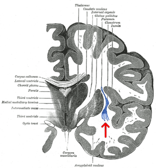
The claustrum is a thin sheet of neurons and supporting glial cells, that connects to the cerebral cortex and subcortical regions including the amygdala, hippocampus and thalamus of the brain. It is located between the insular cortex laterally and the putamen medially, encased by the extreme and external capsules respectively. Blood to the claustrum is supplied by the middle cerebral artery. It is considered to be the most densely connected structure in the brain, and thus hypothesized to allow for the integration of various cortical inputs such as vision, sound and touch, into one experience. Other hypotheses suggest that the claustrum plays a role in salience processing, to direct attention towards the most behaviorally relevant stimuli amongst the background noise. The claustrum is difficult to study given the limited number of individuals with claustral lesions and the poor resolution of neuroimaging.

<span class="mw-page-title-main">Brodmann area 22</span>

Brodmann area 22 is a Brodmann's area that is cytoarchitecturally located in the posterior superior temporal gyrus of the brain. In the left cerebral hemisphere, it is one portion of Wernicke's area. The left hemisphere BA22 helps with generation and understanding of individual words. On the right side of the brain, BA22 helps to discriminate pitch and sound intensity, both of which are necessary to perceive melody and prosody. Wernicke's area is active in processing language and consists of the left Brodmann area 22 and Brodmann area 40, the supramarginal gyrus.

<span class="mw-page-title-main">Brodmann area 25</span> Part of the brain

Brodmann area 25 (BA25) is the subgenual area, area subgenualis or subgenual cingulate area in the cerebral cortex of the brain and delineated based on its cytoarchitectonic characteristics.

<span class="mw-page-title-main">Orbitofrontal cortex</span> Region of the prefrontal cortex of the brain

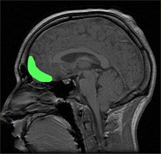
The orbitofrontal cortex (OFC) is a prefrontal cortex region in the frontal lobes of the brain which is involved in the cognitive process of decision-making. In non-human primates it consists of the association cortex areas Brodmann area 11, 12 and 13; in humans it consists of Brodmann area 10, 11 and 47.

<span class="mw-page-title-main">Superior longitudinal fasciculus</span> Association fiber tract of the brain

The superior longitudinal fasciculus (SLF) is an association tract in the brain that is composed of three separate components. It is present in both hemispheres and can be found lateral to the centrum semiovale and connects the frontal, occipital, parietal, and temporal lobes. This bundle of tracts (fasciculus) passes from the frontal lobe through the operculum to the posterior end of the lateral sulcus where they either radiate to and synapse on neurons in the occipital lobe, or turn downward and forward around the putamen and then radiate to and synapse on neurons in anterior portions of the temporal lobe.

<span class="mw-page-title-main">Dorsolateral prefrontal cortex</span> Area of the prefrontal cortex of primates

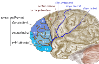
The dorsolateral prefrontal cortex is an area in the prefrontal cortex of the primate brain. It is one of the most recently derived parts of the human brain. It undergoes a prolonged period of maturation which lasts into adulthood. The DLPFC is not an anatomical structure, but rather a functional one. It lies in the middle frontal gyrus of humans. In macaque monkeys, it is around the principal sulcus. Other sources consider that DLPFC is attributed anatomically to BA 9 and 46 and BA 8, 9 and 10.

<span class="mw-page-title-main">Temporoparietal junction</span> Area of the brain where the temporal and parietal lobes meet

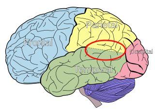
The temporoparietal junction (TPJ) is an area of the brain where the temporal and parietal lobes meet, at the posterior end of the lateral sulcus. The TPJ incorporates information from the thalamus and the limbic system as well as from the visual, auditory, and somatosensory systems. The TPJ also integrates information from both the external environment as well as from within the body. The TPJ is responsible for collecting all of this information and then processing it.

<span class="mw-page-title-main">Default mode network</span> Large-scale brain network active when not focusing on an external task

In neuroscience, the default mode network (DMN), also known as the default network, default state network, or anatomically the medial frontoparietal network (M-FPN), is a large-scale brain network primarily composed of the dorsal medial prefrontal cortex, posterior cingulate cortex, precuneus and angular gyrus. It is best known for being active when a person is not focused on the outside world and the brain is at wakeful rest, such as during daydreaming and mind-wandering. It can also be active during detailed thoughts related to external task performance. Other times that the DMN is active include when the individual is thinking about others, thinking about themselves, remembering the past, and planning for the future.

<span class="mw-page-title-main">Resting state fMRI</span> Type of functional magnetic resonance imaging

Resting state fMRI is a method of functional magnetic resonance imaging (fMRI) that is used in brain mapping to evaluate regional interactions that occur in a resting or task-negative state, when an explicit task is not being performed. A number of resting-state brain networks have been identified, one of which is the default mode network. These brain networks are observed through changes in blood flow in the brain which creates what is referred to as a blood-oxygen-level dependent (BOLD) signal that can be measured using fMRI.

The dorsal nexus is an area within the dorsal medial prefrontal cortex that serves as an intersection point for multiple brain networks. Research suggests it plays a role in the maintenance and manipulation of information, as well as supporting the control of cognitive functions such as behavior, memory, and conflict resolution. Abnormally increased connectivity between these networks through the dorsal nexus has been associated with certain types of depression. The activity generated by this abnormally high level of connectivity during a depressive state can be identified through magnetic resonance imaging (MRI) and positron emission tomography (PET).

Amplitude of Low Frequency Fluctuations (ALFF) and fractional Amplitude of Low Frequency Fluctuations (f/ALFF) are neuroimaging methods used to measure spontaneous fluctuations in BOLD-fMRI signal intensity for a given region in the resting brain. Electrophysiological studies suggest that low-frequency oscillations arise from spontaneous neuronal activity. Though ALFFs have been researched extensively in fMRI based theoretical models of brain function, their actual significance is still unknown.

<span class="mw-page-title-main">Salience network</span> Large-scale brain network involved in detecting and attending to relevant stimuli

The salience network (SN), also known anatomically as the midcingulo-insular network (M-CIN) or ventral attention network, is a large scale network of the human brain that is primarily composed of the anterior insula (AI) and dorsal anterior cingulate cortex (dACC). It is involved in detecting and filtering salient stimuli, as well as in recruiting relevant functional networks. Together with its interconnected brain networks, the SN contributes to a variety of complex functions, including communication, social behavior, and self-awareness through the integration of sensory, emotional, and cognitive information.

Social cognitive neuroscience is the scientific study of the biological processes underpinning social cognition. Specifically, it uses the tools of neuroscience to study "the mental mechanisms that create, frame, regulate, and respond to our experience of the social world". Social cognitive neuroscience uses the epistemological foundations of cognitive neuroscience, and is closely related to social neuroscience. Social cognitive neuroscience employs human neuroimaging, typically using functional magnetic resonance imaging (fMRI). Human brain stimulation techniques such as transcranial magnetic stimulation and transcranial direct-current stimulation are also used. In nonhuman animals, direct electrophysiological recordings and electrical stimulation of single cells and neuronal populations are utilized for investigating lower-level social cognitive processes.

Network neuroscience is an approach to understanding the structure and function of the human brain through an approach of network science, through the paradigm of graph theory. A network is a connection of many brain regions that interact with each other to give rise to a particular function. Network Neuroscience is a broad field that studies the brain in an integrative way by recording, analyzing, and mapping the brain in various ways. The field studies the brain at multiple scales of analysis to ultimately explain brain systems, behavior, and dysfunction of behavior in psychiatric and neurological diseases. Network neuroscience provides an important theoretical base for understanding neurobiological systems at multiple scales of analysis.

<span class="mw-page-title-main">Frontoparietal network</span> Large-scale brain network involved in sustained attention and complex cognition

The frontoparietal network (FPN), generally also known as the central executive network (CEN) or, more specifically, the lateral frontoparietal network (L-FPN), is a large-scale brain network primarily composed of the dorsolateral prefrontal cortex and posterior parietal cortex, around the intraparietal sulcus. It is involved in sustained attention, complex problem-solving and working memory.

Functional MRI imaging methods have allowed researchers to combine neurocognitive testing with structural neuroanatomical measures, take into consideration both cognitive and affective paradigms, and subsequently create computer-aided diagnosis techniques and algorithms. Functional MRI has several benefits, such as its non-invasive quality, relatively high spatial resolution, and decent temporal resolution. One particular method used in recent research is resting-state functional magnetic resonance imaging, rs-fMRI. fMRI imaging has been applied to numerous behavioral studies for schizophrenia, the findings of which have hinted toward potential brain regions that govern key characteristics in cognition and affect.

References

  1. R Leech; R Braga; DJ Sharp (2012). "Echoes of the brain within the posterior cingulate cortex". The Journal of Neuroscience. 32 (1): 215–222. doi:10.1523/JNEUROSCI.3689-11.2012. PMC   6621313 . PMID   22219283.
  2. Nielsen FA, Balslev D, Hansen LK (2005). "Mining the posterior cingulate: segregation between memory and pain components" (PDF). NeuroImage. 27 (3): 520–532. doi:10.1016/j.neuroimage.2005.04.034. PMID   15946864. S2CID   18509039.
  3. Kozlovskiy SA, Vartanov AV, Nikonova EY, Pyasik MM, Velichkovsky BM (2012). "The Cingulate Cortex and Human Memory Processes". Psychology in Russia: State of the Art. 5: 231–243. doi: 10.11621/pir.2012.0014 .
  4. 1 2 3 4 5 6 7 8 9 10 11 12 13 14 15 16 17 18 19 20 21 22 23 24 25 26 27 28 29 30 31 Leech R, Sharp DJ (July 2013). "The role of the posterior cingulate cortex in cognition and disease". Brain. 137 (Pt 1): 12–32. doi:10.1093/brain/awt162. PMC   3891440 . PMID   23869106.
  5. 1 2 3 4 5 Pearson, John M.; Heilbronner, Sarah R.; Barack, David L.; Hayden, Benjamin Y.; Platt, Michael L. (April 2011). "Posterior cingulate cortex: adapting behavior to a changing world". Trends in Cognitive Sciences. 15 (4): 143–151. doi:10.1016/j.tics.2011.02.002. PMC   3070780 . PMID   21420893.
  6. 1 2 3 4 5 6 Maddock, R. J.; A. S. Garrett; M. H. Buonocore (2001). "Remembering Familiar People: The Posterior Cingulate Cortex and Autobiographical Memory Retrieval". Neuroscience. 104 (3): 667–676. CiteSeerX   10.1.1.397.7614 . doi:10.1016/s0306-4522(01)00108-7. PMID   11440800. S2CID   15412482.
  7. 1 2 Maddock, Richard J.; Garrett, Amy S.; Buonocore, Michael H. (January 2003). "Posterior cingulate cortex activation by emotional words: fMRI evidence from a valence decision task". Human Brain Mapping. 18 (1): 30–41. CiteSeerX   10.1.1.529.1671 . doi:10.1002/hbm.10075. PMC   6871991 . PMID   12454910.
  8. Brewer, Judson A.; Garrison, Kathleen A.; Whitfield-Gabrieli, Susan (2013). "What about the "Self" is Processed in the Posterior Cingulate Cortex?". Frontiers in Human Neuroscience. 7: 647. doi: 10.3389/fnhum.2013.00647 . PMC   3788347 . PMID   24106472.
  9. Small, Dana A.; Gitelman, Darren; Simmons, Katharine; Bloise, Suzanne; Parrish, Todd; Mesulam, Marsel M. (2005). "Monetary Incentives Enhance Processing in Brain Regions Mediating Top-down Control of Attention". Cerebral Cortex. 15 (12): 1855–1865. doi: 10.1093/cercor/bhi063 . PMID   15746002.
  10. Engelmann, Jan B.; Damaraju, Eswar; Padmala, Srikanth; Pessoa, Luiz (2009). "Combined Effects of Attention and Motivation on Visual Task Performance: Transient and Sustained Motivational Effects". Frontiers in Human Neuroscience. 3: 4. doi: 10.3389/neuro.09.004.2009 . PMC   2679199 . PMID   19434242.
  11. 1 2 Garrison KA, Santoyo JF, Davis JH, Thornhill TA, Kerr CE, Brewer JA (2013). "Effortless awareness: using real time neurofeedback to investigate correlates of posterior cingulate cortex activity in meditators' self-report". Front Hum Neurosci. 7: 440. doi: 10.3389/fnhum.2013.00440 . PMC   3734786 . PMID   23964222.
  12. Brewer, Judson A.; Worhunsky, Patrick D.; Gray, Jeremy R.; Tang, Yi-Yuan; Weber, Jochen; Kober, Hedy (2011-12-13). "Meditation experience is associated with differences in default mode network activity and connectivity". Proceedings of the National Academy of Sciences of the United States of America. 108 (50): 20254–20259. Bibcode:2011PNAS..10820254B. doi: 10.1073/pnas.1112029108 . ISSN   1091-6490. PMC   3250176 . PMID   22114193.
  13. Garrison, Kathleen A.; Scheinost, Dustin; Worhunsky, Patrick D.; Elwafi, Hani M.; Thornhill, Thomas A.; Thompson, Evan; Saron, Clifford; Desbordes, Gaëlle; Kober, Hedy (2013-11-01). "Real-time fMRI links subjective experience with brain activity during focused attention". NeuroImage. 81: 110–118. doi:10.1016/j.neuroimage.2013.05.030. ISSN   1095-9572. PMC   3729617 . PMID   23684866.
  14. Garrison, Kathleen A.; Zeffiro, Thomas A.; Scheinost, Dustin; Constable, R. Todd; Brewer, Judson A. (September 2015). "Meditation leads to reduced default mode network activity beyond an active task". Cognitive, Affective, & Behavioral Neuroscience. 15 (3): 712–720. doi:10.3758/s13415-015-0358-3. ISSN   1531-135X. PMC   4529365 . PMID   25904238.
  15. Buckner, RL; Andrews-Hanna JR; Schacter DL (2008). "The brain's default network: anatomy, function, and relevance to disease". Ann N Y Acad Sci. 1124 (1): 1–38. Bibcode:2008NYASA1124....1B. CiteSeerX   10.1.1.689.6903 . doi:10.1196/annals.1440.011. PMID   18400922. S2CID   3167595.
  16. Meguro, K. (1999). "Neocortical and hippocampal glucose hypometabolism following neurotoxic lesions of the entorhinal and perirhinal cortices in the non-human primate as shown by PET: Implications for Alzheimer's disease". Brain. 122 (8): 1519–1531. doi: 10.1093/brain/122.8.1519 . ISSN   1460-2156. PMID   10430835.
  17. Raj, A; Kuceyeski A; Weiner M (2012). "A network diffusion model of disease progression in dementia". Neuron. 73 (6): 1204–15. doi:10.1016/j.neuron.2011.12.040. PMC   3623298 . PMID   22445347.
  18. Cherkassky, VL; Kana, RK; Keller, TA; Just, MA (Nov 6, 2006). "Functional connectivity in a baseline resting-state network in autism". NeuroReport (Submitted manuscript). 17 (16): 1687–90. doi:10.1097/01.wnr.0000239956.45448.4c. PMID   17047454. S2CID   568233.
  19. Sonuga-Barke, EJ; Castellanos, FX (2007). "Spontaneous attentional fluctuations in impaired states and pathological conditions: a neurobiological hypothesis". Neuroscience and Biobehavioral Reviews. 31 (7): 977–86. doi:10.1016/j.neubiorev.2007.02.005. PMID   17445893. S2CID   16831759.
  20. Nakao, T; Radua, J; Rubia, K; Mataix-Cols, D (November 2011). "Gray matter volume abnormalities in ADHD: voxel-based meta-analysis exploring the effects of age and stimulant medication". The American Journal of Psychiatry. 168 (11): 1154–63. doi:10.1176/appi.ajp.2011.11020281. PMID   21865529.
  21. Latasch, L; Christ, R (March 1988). "[Problems in anesthesia of drug addicts]". Der Anaesthesist. 37 (3): 123–39. PMID   3289412.
  22. Zhou, Y; Yu, C; Zheng, H; Liu, Y; Song, M; Qin, W; Li, K; Jiang, T (March 2010). "Increased neural resources recruitment in the intrinsic organization in major depression". Journal of Affective Disorders. 121 (3): 220–30. doi:10.1016/j.jad.2009.05.029. PMID   19541369.
  23. Bluhm, R; Williamson, P; Lanius, R; Théberge, J; Densmore, M; Bartha, R; Neufeld, R; Osuch, E (December 2009). "Resting state default-mode network connectivity in early depression using a seed region-of-interest analysis: decreased connectivity with caudate nucleus". Psychiatry and Clinical Neurosciences. 63 (6): 754–61. doi: 10.1111/j.1440-1819.2009.02030.x . PMID   20021629. S2CID   35725401.
  24. Mayberg, HS; Liotti, M; Brannan, SK; McGinnis, S; Mahurin, RK; Jerabek, PA; Silva, JA; Tekell, JL; Martin, CC; Lancaster, JL; Fox, PT (May 1999). "Reciprocal limbic-cortical function and negative mood: converging PET findings in depression and normal sadness". The American Journal of Psychiatry. 156 (5): 675–82. doi:10.1176/ajp.156.5.675. PMID   10327898. S2CID   16258492.
  25. Berman, MG; Peltier, S; Nee, DE; Kross, E; Deldin, PJ; Jonides, J (October 2011). "Depression, rumination and the default network". Social Cognitive and Affective Neuroscience. 6 (5): 548–55. doi:10.1093/scan/nsq080. PMC   3190207 . PMID   20855296.
  26. Haznedar, MM; Buchsbaum, MS; Hazlett, EA; Shihabuddin, L; New, A; Siever, LJ (Dec 1, 2004). "Cingulate gyrus volume and metabolism in the schizophrenia spectrum". Schizophrenia Research. 71 (2–3): 249–62. doi:10.1016/j.schres.2004.02.025. PMID   15474896. S2CID   28889346.
  27. 1 2 Mitelman, SA; Byne, W; Kemether, EM; Hazlett, EA; Buchsbaum, MS (September 2005). "Metabolic disconnection between the mediodorsal nucleus of the thalamus and cortical Brodmann's areas of the left hemisphere in schizophrenia". The American Journal of Psychiatry. 162 (9): 1733–5. doi:10.1176/appi.ajp.162.9.1733. PMID   16135634.
  28. Newell, KA; Zavitsanou, K; Huang, XF (Aug 22, 2005). "Ionotropic glutamate receptor binding in the posterior cingulate cortex in schizophrenia patients". NeuroReport. 16 (12): 1363–7. doi:10.1097/01.wnr.0000174056.11403.71. PMID   16056140. S2CID   29764510.
  29. Kubicki, M; McCarley, RW; Nestor, PG; Huh, T; Kikinis, R; Shenton, ME; Wible, CG (December 2003). "An fMRI study of semantic processing in men with schizophrenia". NeuroImage. 20 (4): 1923–33. doi:10.1016/s1053-8119(03)00383-5. PMC   2806220 . PMID   14683698.
  30. Liang, M; Zhou, Y; Jiang, T; Liu, Z; Tian, L; Liu, H; Hao, Y (Feb 6, 2006). "Widespread functional disconnectivity in schizophrenia with resting-state functional magnetic resonance imaging". NeuroReport. 17 (2): 209–13. doi:10.1097/01.wnr.0000198434.06518.b8. PMID   16407773. S2CID   10743973.
  31. Whitfield-Gabrieli, S; Thermenos, HW; Milanovic, S; Tsuang, MT; Faraone, SV; McCarley, RW; Shenton, ME; Green, AI; Nieto-Castanon, A; LaViolette, P; Wojcik, J; Gabrieli, JD; Seidman, LJ (Jan 27, 2009). "Hyperactivity and hyperconnectivity of the default network in schizophrenia and in first-degree relatives of persons with schizophrenia". Proceedings of the National Academy of Sciences of the United States of America. 106 (4): 1279–84. Bibcode:2009PNAS..106.1279W. doi: 10.1073/pnas.0809141106 . PMC   2633557 . PMID   19164577.
  32. Carhart-Harris RL, Erritzoe D, Williams T, Stone JM, Reed LJ, Colasanti A, Tyacke RJ, Leech R, Malizia AL, Murphy K, Hobden P, Evans J, Feilding A, Wise RG, Nutt DJ (2012). "Neural correlates of the psychedelic state as determined by fMRI studies with psilocybin". Proceedings of the National Academy of Sciences of the United States of America. 109 (6): 2138–43. doi: 10.1073/pnas.1119598109 . PMC   3277566 . PMID   22308440.
  33. Nakashima, T; Nakayama, N; Miwa, K; Okumura, A; Soeda, A; Iwama, T (February 2007). "Focal brain glucose hypometabolism in patients with neuropsychologic deficits after diffuse axonal injury". AJNR. American Journal of Neuroradiology. 28 (2): 236–42. PMC   7977405 . PMID   17296986.
  34. Bonnelle V, Leech R, Kinnunen KM, Ham TE, Beckmann CF, De Boissezon X, Greenwood RJ, Sharp DJ (2011). "Default mode network connectivity predicts sustained attention deficits after traumatic brain injury". J. Neurosci. 31 (38): 13442–51. doi:10.1523/JNEUROSCI.1163-11.2011. PMC   6623308 . PMID   21940437.
  35. Bonnelle, V; Ham, TE; Leech, R; Kinnunen, KM; Mehta, MA; Greenwood, RJ; Sharp, DJ (Mar 20, 2012). "Salience network integrity predicts default mode network function after traumatic brain injury". Proceedings of the National Academy of Sciences of the United States of America. 109 (12): 4690–5. Bibcode:2012PNAS..109.4690B. doi: 10.1073/pnas.1113455109 . PMC   3311356 . PMID   22393019.
  36. Leech, Robert; Sharp, David J. (2014-01-01). "The role of the posterior cingulate cortex in cognition and disease". Brain. 137 (1): 12–32. doi:10.1093/brain/awt162. ISSN   0006-8950. PMC   3891440 . PMID   23869106.
  37. Milad, Mohammed R.; Furtak, Sharon C.; Greenberg, Jennifer L.; Keshaviah, Aparna; Im, Jooyeon J.; Falkenstein, Martha J.; Jenike, Michael; Rauch, Scott L.; Wilhelm, Sabine (2013-06-01). "Deficits in Conditioned Fear Extinction in Obsessive-Compulsive Disorder and Neurobiological Changes in the Fear Circuit". JAMA Psychiatry. 70 (6): 608–618. doi: 10.1001/jamapsychiatry.2013.914 . ISSN   2168-622X. PMID   23740049.
  38. Ganella, Despina E.; Drummond, Katherine D.; Ganella, Eleni P.; Whittle, Sarah; Kim, Jee Hyun (2018). "Extinction of Conditioned Fear in Adolescents and Adults: A Human fMRI Study". Frontiers in Human Neuroscience. 11: 647. doi: 10.3389/fnhum.2017.00647 . ISSN   1662-5161. PMC   5766664 . PMID   29358913.

For details regarding MRI definitions of the cingulate cortex based on the Desikan-Killiany Brain atlas, see: