Terminal amine isotopic labeling of substrates

Last updated

Terminal amine isotopic labeling of substrates (TAILS) is a method in quantitative proteomics that identifies the protein content of samples based on N-terminal fragments of each protein (N-terminal peptides) and detects differences in protein abundance among samples.

Contents

Like other methods based on N-terminal peptides, this assay uses trypsin to break proteins into fragments and separates the N-terminal peptides (the fragments containing the N-termini of the original proteins) from the other fragments (internal tryptic peptides). TAILS isolates the N-terminal peptides by identifying and removing the internal tryptic peptides. This negative selection allows the TAILS method to detect all N-termini in the given samples. Alternative methods that rely on the free amino group of the N-terminus to identify the N-terminal peptides cannot detect some N-termini because they are "naturally blocked" (i.e. the natural protein does not have a free amino group).

The TAILS method has a number of applications including the identification of new substrates and proteases (including those that have an unknown and broad specificity) [1] and as a way to define the termini of proteins that enables protein annotation. TAILS can also be used to link proteases with a variety of defined biological pathways in diseases such as cancer, in order to gain a clearer understanding of the substrates and proteases involved in the disease state. [2]

Method

TAILS is a 2D or 3D proteomics based assay for the labeling and isolation of N-terminal peptides, developed by a group at the University of British Columbia. [1] The TAILS method is designed for comparison of multiple protease treated cells and control proteome cells. [2] Samples can be derived from a variety of sources including tissue, fibroblasts, cancer cells and from fluid effusions.

This assay isolates the N-terminal peptides by removing the internal tryptic peptides via ultrafiltration leaving the labeled mature N-terminal and neo-N-Terminal peptides to be analyzed by tandem mass spectrometry (MS/MS). This negative selection allows the TAILS method to detect all N-termini in the given samples. Alternative methods that rely on the free amino group of the N-terminus to isolate the N-terminal peptides cannot detect naturally blocked N-termini because they do not have a free amino group.

TAILS requires only small sample of peptide for experimentation (100–300 ug), can be used with proteases which have unknown or broad specificity and supports a variety of methods for sample labeling. However, it identifies ~ 50% of proteins by two or more different and unique peptides (one of the original mature N-terminus and/or one or more neo-N-terminal peptide via cleavage site) that do not represent independent biological events thus cannot be averaged for quantification[ clarification needed ]. It also has difficulty validating results for single peptide-base N-terminome analysis[ clarification needed ]. [1]

TAILS overview picture.JPG
1
2, 3, 4
5, 6
7
8
9
10
TAILS overview picture.JPG
TAILS Procedure Overview. Numbers in red identify steps in the text.

The following steps are for the dimethylation-TAILS assay, comparing a control sample (exhibiting normal proteolytic activity) and a treated sample (which in this example exhibits an additional proteolytic activity).

  1. Proteome-wide proteolysis occurring in both the treated and control samples with additional proteolytic activity in the treated sample.
  2. Inactivation of the proteases and protein denaturation and reduction.
  3. Labelling with stable isotopes. This allows peptides that originated in the control sample to be distinguished from those that originated in the treated sample so their relative abundance can be compared. In this example, the labelling is applied by reductive dimethylation of the primary amines using either heavy (d(2)C13)-formaldehyde for the treated samples or light (d(O)C12)-formaldehyde for the controls. This reaction is catalyzed by sodium cyanoborohydride and attaches the labeled methyl groups to lysine-amines and the free (∝)- amino groups at the N-termini of the proteins and protease cleavage products.
  4. Blocking of reactive amino groups. This allows the internal tryptic peptides to be identified later in the process because they will be the only peptides with reactive amino groups. In this example the labelling reaction (reductive dimethylation) also blocks the reactive amino groups.
  5. Pooling. The two labeled proteomes are now mixed. This ensures that the samples are treated identically at all subsequent steps allowing the relative quantities of the proteins in the two samples to be more accurately measured.
  6. Trypsinization. This breaks each protein into fragments. The labeled N-termini of the original proteins remain blocked, while the new internal tryptic peptides have a free N-terminus.
  7. Negative selection. A hyperbranched polyglycerol and aldehyde (HPG) polymer specific for tryptic peptide binding is added to the sample and reacts with the newly generated tryptic peptides through their free N-termini. As in step 3 above, this reaction is catalyzed by sodium cyanoborohydride. The dimethylated lysine's acetylated and isotopically labeled protein peptides and neo(new)-N-terminal peptides are unreactive and remain unbound and can be separated from the poly-internal tryptic peptide complexes using ultrafiltration.
  8. The eluted unbound proteins are highly concentrated with the N-terminal peptides and neo-N-terminal peptides.
  9. This eluted sample is then quantified and analysis completed by MS/MS.
  10. The final step in TAILS involves bioinformatics. Using a hierarchical substrate winnowing process that discriminates from background proteolysis products and non-cleaved proteins by a peptide isotope quantification and certain bioinformatic search criteria. [1] [2]

Types

The types of TAILS differ in the methods used to block and label the amino groups of the proteins and protease cleavage products. These amino groups include lysine-amines and the free (∝)-amino groups of the N-termini of the proteins.

Dimethylation-TAILS procedure is a chemical labeling based procedure that is performed in one step using amine-reactive isotopic reagents. The labeling of two samples uses either 12CH2-formaldehyde (light) or 13CD2-formaldehyde (heavy) and uses sodium cyanoborohydride as the catalyst. [1] The advantage of this method is that it is robust, efficient and cost effective. The labeling procedure for the controls and protease treated samples must be carried out separately before they can be pooled, and it is limited to two samples per experiment, which may be a disadvantage if multiple samples need to be studied simultaneously. [1]

Stable isotope labeling with amino acids in cell culture ( SILAC ) is a procedure that can be done in vivo . This procedure can be used in all cell culture laboratories and is a routinely used labeling technique. This metabolic labeling enables inhibition of a given protease in biological samples and analysis of ex vivo processing. [1] An advantage of using this metabolic labeling method over chemical labeling is that it allows for reliable, fast and efficient discrimination between the real cellular derived proteins that are being investigated from contaminants such as serum proteins. SILAC TAILS can be used for analysis of up to five multiplex samples. SILAC is not suitable for clinically relevant human samples that are not able to be metabolically labeled. SILAC is an expensive method and may not be a feasible option for most laboratories. [1]

The isobaric tag for relative and absolute quantification (iTRAQ) method or iTRAQ-TAILS enables the quantification of multiple samples simultaneously. This method has the ability to simultaneously analyze from 4-8 samples in multiplex experiments using four- and eight- plex iTRAQ reagents. This method provides high accuracy identification and quantification of samples and allows for more reproducible analysis of sample replicates. [1] Like other iTRAQ methods, iTRAQ-TAILS requires a MALDI mass spectrometer and costly iTRAQ reagents.

Alternative methods

There are several alternative approaches to studying N termini and proteolysis products.

Acetylation of amines followed by tryptic digestion and biotinylation of free N-terminal peptides uses chemical (acetylation) to label free lysines and N-termini. The blocked N-termini is then negatively selected. However, the naturally free internal N-termini and blocked N-termini cannot be distinguished after acetylation. This method does not use isotopic labeling, thus it is difficult to quantify the findings. Also, it is hard distinguish between experimental and background proteolysis products. [3]

Lysine guanidination followed by biotinylation of N-termini uses a chemical to block lysine residues and tag free N-termini. The tagged free N-termini is then selected. The down side to this method is that the findings cannot be applied to a statistical model using non-cleaved peptides due to not being able to capture naturally blocked N-termini. Since it does not involve isotopic labeling, the results cannot be quantified. The cleavage site also has to be already known to do labeling. [4]

Subtiligase biotinylation of N-termini uses enzymatic labeling of N-terminal peptides, but does not use lysine blocking chemicals. Without lysine blocking, many of the cleaved N-terminal peptide will be too short for identification. The results can be highly dependent on the properties of subtiligase thus may be biased. This method does not capture naturally blocked N-termini and also does not use isotopic labeling thus it would be difficult to quantify findings. [5]

ITRAQ-labeling of N-termini uses iTRAQ to label the N-termini. Neo-N-termini peptides are selected through in silico. The down side to this technique is that MALDI mass spectrometer is needed and the iTRAQ reagents required are costly. This method does not capture naturally blocked N-termini. The whole process will require 50-100mg of peptide samples. [6]

Combined fractional diagonal chromatography (COFRADIC) allows different labeling for naturally blocked N-termini and protease generated neo-N-termini. All blocked N-termini are negatively selected. However the process requires many chemical processing, chromatography and mass spectrometry. The best separation results are dependent on the amino acid modification such as methionine oxidation not occurring during handling. This method requires 150 MS/MS analyses per sample but samples can be pooled for mass spectrometry (and number of analyses can be reduced). This technique is suitable to be used for proteases with unknown or broad specificity. [7]

See also

Related Research Articles

Protein primary structure Linear sequence of amino acids in a peptide or protein

Protein primary structure is the linear sequence of amino acids in a peptide or protein. By convention, the primary structure of a protein is reported starting from the amino-terminal (N) end to the carboxyl-terminal (C) end. Protein biosynthesis is most commonly performed by ribosomes in cells. Peptides can also be synthesized in the laboratory. Protein primary structures can be directly sequenced, or inferred from DNA sequences.

Proteolysis the breakdown of proteins into smaller polypeptides or amino acids

Proteolysis is the breakdown of proteins into smaller polypeptides or amino acids. Uncatalysed, the hydrolysis of peptide bonds is extremely slow, taking hundreds of years. Proteolysis is typically catalysed by cellular enzymes called proteases, but may also occur by intra-molecular digestion.

Trypsin

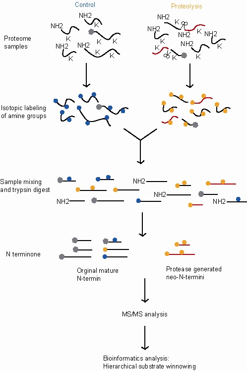
Trypsin is a serine protease from the PA clan superfamily, found in the digestive system of many vertebrates, where it hydrolyzes proteins. Trypsin is formed in the small intestine when its proenzyme form, the trypsinogen produced by the pancreas, is activated. Trypsin cuts peptide chains mainly at the carboxyl side of the amino acids lysine or arginine. It is used for numerous biotechnological processes. The process is commonly referred to as trypsin proteolysis or trypsinization, and proteins that have been digested/treated with trypsin are said to have been trypsinized. Trypsin was discovered in 1876 by Wilhelm Kühne and was named from the Ancient Greek word for rubbing since it was first isolated by rubbing the pancreas with glycerin.

Protease Enzyme that cleaves other proteins into smaller peptides

A protease is an enzyme that catalyzes proteolysis, the breakdown of proteins into smaller polypeptides or single amino acids. They do this by cleaving the peptide bonds within proteins by hydrolysis, a reaction where water breaks bonds. Proteases are involved in many biological functions, including digestion of ingested proteins, protein catabolism, and cell signaling.

The C-terminus is the end of an amino acid chain, terminated by a free carboxyl group (-COOH). When the protein is translated from messenger RNA, it is created from N-terminus to C-terminus. The convention for writing peptide sequences is to put the C-terminal end on the right and write the sequence from N- to C-terminus.

The N-terminus (also known as the amino-terminus, NH2-terminus, N-terminal end or amine-terminus) is the start of a protein or polypeptide referring to the free amine group (-NH2) located at the end of a polypeptide. Within a peptide, the amine group is bonded to another carboxylic group in a protein to make it a chain, but since the end amino acid of a protein is only connected at the carboxy- end, the remaining free amine group is called the N-terminus. By convention, peptide sequences are written N-terminus to C-terminus, left to right (in LTR writing systems). This correlates the translation direction to the text direction (because when a protein is translated from messenger RNA, it is created from N-terminus to C-terminus - amino acids are added to the carboxyl end).

Protein sequencing

Protein sequencing is the practical process of determining the amino acid sequence of all or part of a protein or peptide. This may serve to identify the protein or characterize its post-translational modifications. Typically, partial sequencing of a protein provides sufficient information to identify it with reference to databases of protein sequences derived from the conceptual translation of genes.

Stable isotope labeling by amino acids in cell culture

Stable Isotope Labeling by/with Amino acids in Cell culture (SILAC) is a technique based on mass spectrometry that detects differences in protein abundance among samples using non-radioactive isotopic labeling. It is a popular method for quantitative proteomics.

PEAKS is a proteomics software program for tandem mass spectrometry designed for peptide sequencing, protein identification and quantification.

Protein mass spectrometry

Protein mass spectrometry refers to the application of mass spectrometry to the study of proteins. Mass spectrometry is an important method for the accurate mass determination and characterization of proteins, and a variety of methods and instrumentations have been developed for its many uses. Its applications include the identification of proteins and their post-translational modifications, the elucidation of protein complexes, their subunits and functional interactions, as well as the global measurement of proteins in proteomics. It can also be used to localize proteins to the various organelles, and determine the interactions between different proteins as well as with membrane lipids.

Quantitative proteomics

Quantitative proteomics is an analytical chemistry technique for determining the amount of proteins in a sample. The methods for protein identification are identical to those used in general proteomics, but include quantification as an additional dimension. Rather than just providing lists of proteins identified in a certain sample, quantitative proteomics yields information about the physiological differences between two biological samples. For example, this approach can be used to compare samples from healthy and diseased patients. Quantitative proteomics is mainly performed by two-dimensional gel electrophoresis (2-DE) or mass spectrometry (MS). However, a recent developed method of quantitative dot blot (QDB) analysis is able to measure both the absolute and relative quantity of an individual proteins in the sample in high throughput format, thus open a new direction for proteomic research. In contrast to 2-DE, which requires MS for the downstream protein identification, MS technology can identify and quantify the changes.

Isobaric tag for relative and absolute quantitation

Isobaric tags for relative and absolute quantitation (iTRAQ) is an isobaric labeling method used in quantitative proteomics by tandem mass spectrometry to determine the amount of proteins from different sources in a single experiment. It uses stable isotope labeled molecules that can be covalent bonded to the N-terminus and side chain amines of proteins.

The in-gel digestion step is a part of the sample preparation for the mass spectrometric identification of proteins in course of proteomic analysis. The method was introduced in 1992 by Rosenfeld. Innumerable modifications and improvements in the basic elements of the procedure remain.

Label-free quantification is a method in mass spectrometry that aims to determine the relative amount of proteins in two or more biological samples. Unlike other methods for protein quantification, label-free quantification does not use a stable isotope containing compound to chemically bind to and thus label the protein.

An Isotope-coded affinity tag (ICAT) is an in-vitro isotopic labeling method used for quantitative proteomics by mass spectrometry that uses chemical labeling reagents. These chemical probes consist of three elements: a reactive group for labeling an amino acid side chain, an isotopically coded linker, and a tag for the affinity isolation of labeled proteins/peptides. The samples are combined and then separated through chromatography, then sent through a mass spectrometer to determine the mass-to-charge ratio between the proteins. Only cysteine containing peptides can be analysed. Since only cysteine containing peptides are analysed, often the post translational modification is lost.

Isobaric labeling

Isobaric labeling is a mass spectrometry strategy used in quantitative proteomics. Peptides or proteins are labeled with various chemical groups that are identical masses (isobaric), but vary in terms of distribution of heavy isotopes around their structure. These tags, commonly referred to as tandem mass tags, are designed so that the mass tag is cleaved at a specific linker region upon high-energy CID (HCD) during tandem mass spectrometry yielding reporter ions of different masses. The most common isobaric tags are amine-reactive tags. However, tags that react with cysteine residues and carbonyl groups have also been described. These amine-reactive groups go through N-hydroxysuccinimide (NHS) reactions, which are based around three types of functional groups. Isobaric labeling methods include tandem mass tags (TMT), isobaric tags for relative and absolute quantification (iTRAQ), mass differential tags for absolute and relative quantification, and dimethyl labeling. TMTs and iTRAQ methods are most common and developed of these methods. Tandem mass tags have a mass reporter region, a cleavable linker region, a mass normalization region, and a protein reactive group and have the same total mass.

TopFIND is the Termini oriented protein Function Inferred Database (TopFIND) is an integrated knowledgebase focused on protein termini, their formation by proteases and functional implications. It contains information about the processing and the processing state of proteins and functional implications thereof derived from research literature, contributions by the scientific community and biological databases.

Degradomics Sub-discipline of biology

Degradomics is a sub-discipline of biology encompassing all the genomic and proteomic approaches devoted to the study of proteases, their inhibitors, and their substrates on a system-wide scale. This includes the analysis of the protease and protease-substrate repertoires, also called "protease degradomes". The scope of these degradomes can range from cell, tissue, and organism-wide scales.

Stable isotope standards and capture by anti-peptide antibodies (SISCAPA) is a mass spectrometry method for measuring the amount of a protein in a biological sample.

Translatomics

Translatomics is the study of all open reading frames (ORFs) that are being actively translated in a cell or organism. This collection of ORFs is called the translatome. Characterizing a cell's translatome can give insight into the array of biological pathways that are active in the cell. According to the central dogma of molecular biology, the DNA in a cell is transcribed to produce RNA, which is then translated to produce a protein. Thousands of proteins are encoded in an organism's genome, and the proteins present in a cell cooperatively carry out many functions to support the life of the cell. Under various conditions, such as during stress or specific timepoints in development, the cell may require different biological pathways to be active, and therefore require a different collection of proteins. Depending on intrinsic and environmental conditions, the collection of proteins being made at one time varies. Translatomic techniques can be used to take a "snapshot" of this collection of actively translating ORFs, which can give information about which biological pathways the cell is activating under the present conditions.

References

  1. 1 2 3 4 5 6 7 8 9 Kleifeld, Oded; Doucet, Alain; Prudova, Anna; Auf Dem Keller, Ulrich; Gioia, Magda; Kizhakkedathu, Jayachandran N; Overall, Christopher M (2011). "Identifying and quantifying proteolytic events and the natural N terminome by terminal amine isotopic labeling of substrates". Nature Protocols. 6 (10): 1578–611. doi:10.1038/nprot.2011.382. PMID   21959240.
  2. 1 2 3 Kleifeld, Oded; Doucet, Alain; Auf Dem Keller, Ulrich; Prudova, Anna; Schilling, Oliver; Kainthan, Rajesh K; Starr, Amanda E; Foster, Leonard J; et al. (2010). "Isotopic labeling of terminal amines in complex samples identifies protein N-termini and protease cleavage products". Nature Biotechnology. 28 (3): 281–8. doi:10.1038/nbt.1611. PMID   20208520.
  3. McDonald, Lucy; Robertson, Duncan H L; Hurst, Jane L; Beynon, Robert J (2005). "Positional proteomics: Selective recovery and analysis of N-terminal proteolytic peptides". Nature Methods. 2 (12): 955–7. doi:10.1038/nmeth811. PMID   16299481.
  4. Timmer, John C.; Enoksson, Mari; Wildfang, Eric; Zhu, Wenhong; Igarashi, Yoshinobu; Denault, Jean-Benard; Ma, Yuliang; Dummitt, Benjamin; et al. (2007). "Profiling constitutive proteolytic events in vivo". Biochemical Journal. 407 (1): 41–8. doi:10.1042/BJ20070775. PMC   2267409 . PMID   17650073.
  5. Mahrus, Sami; Trinidad, Jonathan C.; Barkan, David T.; Sali, Andrej; Burlingame, Alma L.; Wells, James A. (2008). "Global Sequencing of Proteolytic Cleavage Sites in Apoptosis by Specific Labeling of Protein N Termini". Cell. 134 (5): 866–76. doi:10.1016/j.cell.2008.08.012. PMC   2566540 . PMID   18722006.
  6. Enoksson, Mari; Li, Jingwei; Ivancic, Melanie M.; Timmer, John C.; Wildfang, Eric; Eroshkin, Alexey; Salvesen, Guy S.; Tao, W. Andy (2007). "Identification of Proteolytic Cleavage Sites by Quantitative Proteomics". Journal of Proteome Research. 6 (7): 2850–8. doi:10.1021/pr0701052. PMID   17547438.
  7. Gevaert, Kris; Goethals, Marc; Martens, Lennart; Van Damme, Jozef; Staes, An; Thomas, Grégoire R.; Vandekerckhove, Joël (2003). "Exploring proteomes and analyzing protein processing by mass spectrometric identification of sorted N-terminal peptides". Nature Biotechnology. 21 (5): 566–9. doi:10.1038/nbt810. PMID   12665801.