A transverse ligament is a ligament on a transverse plane, orthogonal to the anteroposterior or oral-aboral axiscan of the body.
A ligament is the fibrous connective tissue that connects bones to other bones. It is also known as articular ligament, articular larua, fibrous ligament, or true ligament. Other ligaments in the body include the:
The transverse plane or axial plane is an imaginary plane that divides the body into superior and inferior parts. It is perpendicular to the coronal plane and sagittal plane.
In human anatomy, examples are:
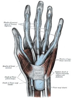
The flexor retinaculum is a fibrous band on the palmar side of the hand near the wrist. It arches over the carpal bones of the hands, covering them and forming the carpal tunnel.
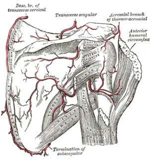
The inferior transverse ligament is a weak membranous band, situated behind the neck of the scapula and stretching from the lateral border of the spine to the margin of the glenoid cavity.
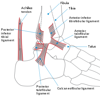
The inferior transverse ligament of the tibiofibular syndesmosis is a connective tissue structure in the lower leg that lies in front of the posterior ligament. It is a strong, thick band, of yellowish fibers which passes transversely across the back of the ankle joint, from the lateral malleolus to the posterior border of the articular surface of the tibia, almost as far as its malleolar process.
This article includes a list of related items that share the same name (or similar names). If an internal link incorrectly led you here, you may wish to change the link to point directly to the intended article. |
In anatomy, the atlas (C1) is the most superior (first) cervical vertebra of the spine.
The trapezius is a large paired surface muscle that extends longitudinally from the occipital bone to the lower thoracic vertebrae of the spine and laterally to the spine of the scapula. It moves the scapula and supports the arm.
In anatomy, the scapula, also known as shoulder bone, shoulder blade, wing bone or blade bone, is the bone that connects the humerus with the clavicle. Like their connected bones the scapulae are paired, with the scapula on either side of the body being roughly a mirror image of the other. The name derives from early Roman times when it was thought that the bone resembled a trowel or small shovel.
The dorsal scapular nerve arises from the brachial plexus, usually from the plexus root of the cervical nerve C5. Once the nerve leaves C5 it commonly pierces the middle scalene muscle, and continues deep to levator scapulae and the rhomboids.
The ankle, or the talocrural region, is the region where the foot and the leg meet. The ankle includes three joints: the ankle joint proper or talocrural joint, the subtalar joint, and the inferior tibiofibular joint. The movements produced at this joint are dorsiflexion and plantarflexion of the foot. In common usage, the term ankle refers exclusively to the ankle region. In medical terminology, "ankle" can refer broadly to the region or specifically to the talocrural joint.
The levator scapulae is a skeletal muscle situated at the back and side of the neck. As the Latin name suggests, its main function is to lift the scapula.
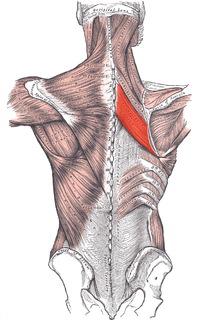
The rhomboid major is a skeletal muscle on the back that connects the scapula with the vertebrae of the spinal column. In human anatomy, it acts together with the rhomboid minor to keep the scapula pressed against thoracic wall and to retract the scapula toward the vertebral column.
In human anatomy, the rhomboid minor is a small skeletal muscle on the back that connects the scapula with the vertebrae of the spinal column.
The omohyoid muscle is a muscle that depresses the hyoid. It is located in the front of the neck and consists of two bellies separated by an intermediate tendon. Its superior belly serves as the most lateral member of the infrahyoid muscles, located lateral to both the sternothyroid and thyrohyoid muscles. Its name derives from the Greek "omos" meaning shoulder, giving one of its attachments, and "hyoid", giving the other attachment - the hyoid bone.
The suprascapular nerve is a nerve that arises from the brachial plexus. It is responsible for the innervation of some of the muscles that attach on the scapula, namely the supraspinatus and infraspinatus muscles.
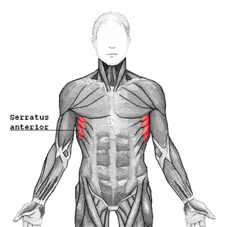
The serratus anterior is a muscle that originates on the surface of the 1st to 8th ribs at the side of the chest and inserts along the entire anterior length of the medial border of the scapula. The serratus anterior acts to pull the scapula forward around the thorax. The muscle is named from Latin: serrare = to saw, referring to the shape, anterior = on the front side of the body.
The circumflex scapular artery is a branch of the subscapular artery and part of the scapular anastomoses.
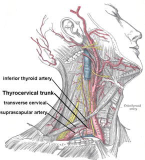
The transverse cervical artery is an artery in the neck and a branch of the thyrocervical trunk, running at a higher level than the suprascapular artery.
The suprascapular artery is a branch of the thyrocervical trunk on the neck.
The supraspinatous fossa of the posterior aspect of the scapula is smaller than the infraspinatous fossa, concave, smooth, and broader at its vertebral than at its humeral end. Its medial two-thirds give origin to the Supraspinatus.
The Investing layer of deep cervical fascia is the most superficial part of the deep cervical fascia, and it encloses the whole neck.
The following outline is provided as an overview of and topical guide to human anatomy:
Transverse ligament of scapula may refer to:
Transverse scapular ligament may refer to: