Related Research Articles

In anatomy, the atlas (C1) is the most superior (first) cervical vertebra of the spine and is located in the neck.

The sacrum, in human anatomy, is a large, triangular bone at the base of the spine that forms by the fusing of the sacral vertebrae (S1–S5) between ages 18 and 30.

The tibia, also known as the shinbone or shankbone, is the larger, stronger, and anterior (frontal) of the two bones in the leg below the knee in vertebrates ; it connects the knee with the ankle. The tibia is found on the medial side of the leg next to the fibula and closer to the median plane. The tibia is connected to the fibula by the interosseous membrane of leg, forming a type of fibrous joint called a syndesmosis with very little movement. The tibia is named for the flute tibia. It is the second largest bone in the human body, after the femur. The leg bones are the strongest long bones as they support the rest of the body.

The olecranon, is a large, thick, curved bony eminence of the ulna, a long bone in the forearm that projects behind the elbow. It forms the most pointed portion of the elbow and is opposite to the cubital fossa or elbow pit. The olecranon serves as a lever for the extensor muscles that straighten the elbow joint.

The sacrotuberous ligament is situated at the lower and back part of the pelvis. It is flat, and triangular in form; narrower in the middle than at the ends.

The erector spinae or spinal erectors is a set of muscles that straighten and rotate the back. The spinal erectors work together with the glutes to maintain stable posture standing or sitting.

The lateral parts of the occipital bone are situated at the sides of the foramen magnum; on their under surfaces are the condyles for articulation with the superior facets of the atlas.

The transversalis fascia is the fascial lining of the anterolateral abdominal wall situated between the inner surface of the transverse abdominal muscle, and the preperitoneal fascia. It is directly continuous with the iliac fascia, the internal spermatic fascia, and pelvic fascia.
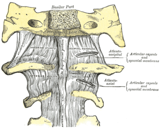
The atlanto-axial joint is a joint in the upper part of the neck between the atlas bone and the axis bone, which are the first and second cervical vertebrae. It is a pivot joint.

The sacrococcygeal symphysis is an amphiarthrodial joint, formed between the oval surface at the apex of the sacrum, and the base of the coccyx.

In anatomy, the transverse ligament of the atlas is a ligament which arches across the ring of the atlas, and keeps the odontoid process in contact with the atlas.

The posterior longitudinal ligament is a ligament connecting the posterior surfaces of the vertebral bodies of all of the vertebrae of humans. It weakly prevents hyperflexion of the vertebral column. It also prevents posterior spinal disc herniation, although problems with the ligament can cause it.

The tectorial membrane of atlanto-axial joint is situated within the vertebral canal. It represents the superior-ward prolongation of the posterior longitudinal ligament. It attaches to the body of the axis before terminating superiorly by attaching onto the basilar part of occipital bone superior to the foramen magnum.
The posterior sacrococcygeal ligament or dorsal sacrococcygeal ligament is a ligament which stretches from the sacrum to the coccyx and thus dorsally across the sacrococcygeal symphysis shared by these two bones.

The deep transverse metacarpal ligament is a narrow fibrous band which runs across the palmar surfaces of the heads of the second, third, fourth and fifth metacarpal bones, connecting them together.
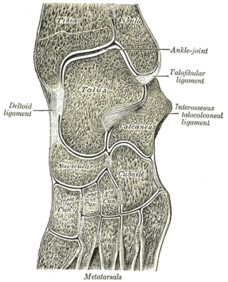
The intercuneiform joints are the joints the cuneiform bones.

The deep fascia of leg, or crural fascia forms a complete investment to the muscles, and is fused with the periosteum over the subcutaneous surfaces of the bones.
The lumbar fascia is the lumbar portion of the thoracolumbar fascia. It consists of three fascial layers - posterior, middle, and anterior - that enclose two muscular compartments. The anterior and middle layers occur only in the lumbar region, whereas the posterior layer extends superiorly to the inferior part of the neck, and the inferiorly to the dorsal surface of the sacrum. The quadratus lumborum is contained in the anterior muscular compartment, and the erector spinae in the posterior compartment. Psoas major lies anterior to the anterior layer. Various superficial muscles of the posterior thorax and abdomen arise from the posterior layer - namely the latissimus dorsi, and serratus posterior inferior.

The following outline is provided as an overview of and topical guide to human anatomy:

The transverse or [anterior] meniscomeniscal ligament is a ligament in the knee joint that connects the anterior convex margin of the lateral meniscus to the anterior end of the medial meniscus.
References
![]() This article incorporates text in the public domain from page 349 of the 20th edition of Gray's Anatomy (1918)
This article incorporates text in the public domain from page 349 of the 20th edition of Gray's Anatomy (1918)
- ↑ A. M. R. Agur; Arthur F. Dalley (2009). Grant's atlas of anatomy. Lippincott Williams & Wilkins. pp. 442–. ISBN 978-0-7817-7055-2 . Retrieved 29 October 2010.
| | This ligament-related article is a stub. You can help Wikipedia by expanding it. |