Wrist arthroscopy

Last updated
Wrist arthroscopy
Wrist arthroscopy.png
Wrist Arthroscopy

Wrist arthroscopy can be used to look inside the joint of the wrist. It is a minimally invasive technique which can be utilized for diagnostic purposes as well as for therapeutic interventions. Wrist arthroscopy has been used for diagnostic purposes since it was first introduced in 1979. However, it only became accepted as diagnostic tool around the mid-1980s. At that time, arthroscopy of the wrist was an innovative technique to determine whether a problem could be found in the wrist. A few years later, wrist arthroscopy could also be used as a therapeutic tool. [1]

Contents

Medical uses

There are several therapeutic wrist arthroscopy indications, in this article the focus will be on the TFCC-lesion, the SL-lesion, the dorsal ganglion resection and the distal radius fracture.

TFCC lesion

The Triangular Fibrocartilage Complex (TFCC) is a fibrous structure covering both the radiocarpal and distal radioulnar joint. Tears in this ligament occur commonly after a person falls or secondary to a wrist fracture. Abnormalities in the TFCC are classified with the Palmer Classification, which divides the tears in a traumatic or degenerative stage. [2] Both stages of TFCC tears are treatable with an arthroscopic intervention, although the degenerative stage is operated according to the "Arthroscopic wafer procedure". [3] In this procedure, the surgeon debrides the TFCC and a limited part of the ulnar head. If the patient has a Class 1 TFCC tear, a different arthroscopic technique is used. Damaged tissue will be debrided until fresh edges are available for arthroscopic repair. [4] Good to excellent results have been reported on TFCC Class 1B repair for 85-90% of the patients. And 90% of the results for the Arthroscopic wafer procedure were good to excellent. [5]

Scapholunate and lunotriquetral instability

The initial treatment of patients with a suspected tear of the scapholunate and lunotriquetral interosseous ligament is a splint of the wrist. If the pain and instability persists, one could undergo an open surgery to reconstruct the scapholunate ligament. The lunotriquetral shear test may also be used. Arthroscopy is until today in an experimental stage but research suggest that in the near future it will be a reasonable alternative for open surgery due to faster recovery time. [6] For a tear in the lunotriquetral ligament, arthroscopic debridement is the prime treatment with a loss or reduction of symptoms of 78-100%. [7]

Dorsal ganglion resection

28 to 58% of the dorsal ganglia resolve spontaneously, still some patients choose to undergo cosmetic intervention for resection of the ganglion when non-operative treatment failed. Some examples of this non-operative treatment are immobilization through a splint or aspiration of the ganglion with or without injection of a steroid. In some cases the ganglia are associated with serious loss of wrist function or weakness in the affected finger, which makes a surgical intervention highly indicated. [8]

Resection of dorsal ganglion are still widely treated with open surgery. However, recent studies show that treatment with an arthroscopic intervention is a reasonable alternative. Arthroscopic intervention has the advantages of smaller incisions, faster recovery of wrist function and less pain postoperative. Overall outcome of arthroscopy is not superior to the outcome in open surgery. Recurrence rates and final wrist function are similar. [9]

Distal radius fracture

Distal radius fractures might occur when a person falls on an outstretched hand (FOOSH). Immediate pain, swelling and loss of wrist function are the most common symptoms. These fractures have wide variety of classification systems, but for arthroscopic intervention a difference is made between extra- or intra-articular fractures. Arthroscopy can be used to treat an intra-articular distal radius fracture and at the same time examine the Scapholunate ligament, Lunotriquetral ligament and articular disk. [5] The shifted bone segment is relocated, in necessary a bone autograft can be used to fill up the fracture. At the end the fracture is stabilized using percutane wires. Arthroscopy has good results as treatment for distal radius fracture, with over 85% of the patiënts showing good to excellent postoperative results. [10]

Diagnostic uses

Persistent pain in the wrist after conservative treatment is the major indication for a diagnostic wrist arthroscopy. Conservative treatment consists of wrist immobilization, oral NSAIDs and/or injection with corticoids. [11] Diagnostic wrist arthroscopy may also be indicated when other imaging techniques, such as MRI and ultrasonography, need confirmation on observed uncertainties or when other imaging techniques failed to establish a clear diagnosis. This makes arthroscopy an important diagnostic tool for some common anomalies around the wrist. [12] However, relying on arthroscopic findings in the setting of an unclear preoperative diagnosis yields limited diagnostic benefit. Therefore, its use should be limited to the cases with other objective clinical findings. [13]

Diagnostic IndicationsExplanation
GanglionA cavity in a joint capsule filled with thick fluid
SL-lesionTear of the Scapholunate ligament
LT-lesionTear of the Lunotriquetral ligament
TFCC-lesionTear in the TFCC. Classification according Palmer Classification in traumatic or degenerative lesion
Synovitis Inflammation of the synovial membrane
Cartilage decreaseA short of cartilage can cause arthritis

Procedure

Before surgery can be started, the patient has to be positioned in such a way that the wrist is stabilized. In order to do this, the patient has to be placed on the operating table with their face upward. The wrist of the arm on which will be operated on, has to be placed on a separate operating table on the side of the other operating table. To properly stabilize the wrist, the patient's elbow is flexed and the forearm is immobilized by using a traction apparatus. By using a traction apparatus, it will also become easier to insert the instruments. [5]

Traction Tower Traction Towe.JPG
Traction Tower

In general, 4.5-6.8 kilograms of traction is implemented with the aid of finger traps. The surgeon can either opt for nylon finger traps or for wire traps. In most cases, nylon finger traps will be preferred to wire traps, as nylon is a more resilient material and thus distributes the traction force to the skin more evenly. Usually, traction is applied to two fingers, namely the index finger and middle finger. Sometimes it may be necessary to more evenly distribute the traction force on the skin, as the patient's skin may be too fragile or atrophic. The traction brought about by the traction apparatus is then applied to three or even four fingers. [5] The surgeon can either opt for dry arthroscopy or for wet arthroscopy. When performing a dry procedure the wrist can still, if necessary, be approached using open surgery. This may, for example, be required when a TFCC lesion is diagnosed. The latter is not possible when a wet procedure is performed, because the fluid that is used causes expansion of the tissue. To improve visualization within the wrist, an inflow irrigation system is used during surgery. Currently, several systems are available, such as gravity flow, a hand pump, large syringes, or a mechanical infusion pump. For irrigation, lactated Ringer’s solution is generally used. The characteristics of lactated Ringer’s solution are that it is physiological and rapidly absorbed. Moreover, the right equipment must be available to finish a complete diagnostic arthroscopy, as well as to perform various operative procedures. Complications of using an irrigation system is that fluid extravasation into the soft tissues of the forearm can result in a compartment syndrome. [5]

Materials

To perform a wrist arthroscopy, the surgeon can use a standard arthroscopy set. This set contains a variety of tools for both a diagnostic and therapeutic arthroscopic intervention. The sets currently available contain: scopes; probes; punches; graspers; joint shavers and electrocautery. [14] To get a clear view of the wrist a high quality camera with a small diameter between 2 and 3 mm is needed. This camera is attached to a fiberoptic light source and shows the image of the inside of the joint. Probes are used for examination of internal structures by palpating, pulling of moving tissue. They are essential for diagnostic investigation of the joint. All probes bear markings at 5 mm intervals to indicate the scale, since the cameras used in arthroscopy have different magnifications. [14] Punches or basket forceps have sharp edges to cut away redundant and damaged tissue. Suction punches are punches which can be connected to a suction pump to immediately remove cut tissue from the joint. Graspers or grasping forceps are useful to grab tissue floating loosely through the joint. A surgeon might want to remove this particular piece of tissue for tactile feedback. Every different arthroscopic intervention has its own set of tools, depending on difficulty, type of tissue and available working space. [14]

Techniques

In recent years, wrist arthroscopy is becoming a more familiar technique for the diagnosis and treatment of various wrist abnormalities. Individual surgeons will have their own preferences, concerning the variety of available techniques. In this subsection, not all techniques will be described. A description of the most commonly used techniques will be given. [15]

Before surgery

To prevent postoperative infections, the amount of pathogens in the operating-room has to be minimized. In order to achieve this, the surgeon, assistants, equipment and the operation area have to be sterile during the operation. Furthermore, the doors in the operating-room should not be opened, or this should at least be limited. Before the patient can undergo wrist arthroscopy, they must be positioned in a specific way. This operation procedure has already been thoroughly described earlier in subsection ‘’Procedure’’. The patient will only be operated on when every requirement for surgery is implemented.[ citation needed ]

Entrance

To perform wrist arthroscopy, the instruments will have to be inserted in the wrist. The surgeon will start by making a small incision for creating a so-called portal. The different portals will be further discussed in the next section. [5] After the incision has been made, the portal is used to insert a scope. This scope will provide the visualization. In that way, the surgeon will be able to orient within the joint of the wrist. Once a clear view of the wrist is obtained, a possible problem can be identified. This problem, for example a TFCC lesion, may then immediately be solved by operative treatment.[ citation needed ]

Portals

A patient may suffer from various illnesses regarding the wrist. Examples include TFCC lesion, scapholunate and lunotriquetral instability, ganglions, and synovitis. These illnesses concern many different tissues and structures. By using just one portal, one will never be able to visualize all these structures and tissues. For that reason, several portals exist, each having its own function (Table 1). [16] The traditional portals are at the back of the hand (dorsal side), because many neurovascular structures are at risk at the palmar side of the hand.

Table 1. Different portals that can be used during Wrist Arthroscopy Portals wrist arthroscopy.jpg
Table 1. Different portals that can be used during Wrist Arthroscopy

Dorsal Radial portals The portals lay between the dorsal extensor compartments. [17]

1-2 portal:

3-4 portal:

4-5 portal:

6R portal:

6U portal:

Volar radiocarpal(VR) portal

Midcarpal portals:

Midcarpal Radial portal (MCR)

Location of the Portals used for Wrist Arthroscopy Portals hand.jpg
Location of the Portals used for Wrist Arthroscopy

Midcarpal Ulnar portal (MCU)

Dorsal Distal Radioulnar joint portal (DDRUJ)

Volar Distal Radioulnar joint portal (VDRUJ)

Related Research Articles

<span class="mw-page-title-main">Carpal tunnel syndrome</span> Medical condition

Carpal tunnel syndrome (CTS) is the collection of symptoms and signs associated with median neuropathy at the carpal tunnel. Most CTS is related to idiopathic compression of the median nerve as it travels through the wrist at the carpal tunnel (IMNCT). Idiopathic means that there is no other disease process contributing to pressure on the nerve. As with most structural issues, it occurs in both hands, and the strongest risk factor is genetics.

<span class="mw-page-title-main">Arthroscopy</span> Medical procedure

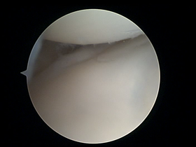
Arthroscopy is a minimally invasive surgical procedure on a joint in which an examination and sometimes treatment of damage is performed using an arthroscope, an endoscope that is inserted into the joint through a small incision. Arthroscopic procedures can be performed during ACL reconstruction.

<span class="mw-page-title-main">Wrist</span> Part of the arm between the lower arm and the hand

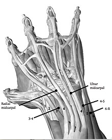
In human anatomy, the wrist is variously defined as (1) the carpus or carpal bones, the complex of eight bones forming the proximal skeletal segment of the hand; (2) the wrist joint or radiocarpal joint, the joint between the radius and the carpus and; (3) the anatomical region surrounding the carpus including the distal parts of the bones of the forearm and the proximal parts of the metacarpus or five metacarpal bones and the series of joints between these bones, thus referred to as wrist joints. This region also includes the carpal tunnel, the anatomical snuff box, bracelet lines, the flexor retinaculum, and the extensor retinaculum.

<span class="mw-page-title-main">Ganglion cyst</span> Medical condition

A ganglion cyst is a fluid-filled bump associated with a joint or tendon sheath. It most often occurs at the back of the wrist, followed by the front of the wrist.

<span class="mw-page-title-main">Orthopedic surgery</span> Branch of surgery concerned with the musculoskeletal and bones system

Orthopedic surgery or orthopedics is the branch of surgery concerned with conditions involving the musculoskeletal system. Orthopedic surgeons use both surgical and nonsurgical means to treat musculoskeletal trauma, spine diseases, sports injuries, degenerative diseases, infections, tumors, and congenital disorders.

<span class="mw-page-title-main">Maisonneuve fracture</span> Medical condition

The Maisonneuve fracture is a spiral fracture of the proximal third of the fibula associated with a tear of the distal tibiofibular syndesmosis and the interosseous membrane. There is an associated fracture of the medial malleolus or rupture of the deep deltoid ligament of the ankle. This type of injury can be difficult to detect.

<span class="mw-page-title-main">Distal radius fracture</span> Fracture of the radius bone near the wrist

A distal radius fracture, also known as wrist fracture, is a break of the part of the radius bone which is close to the wrist. Symptoms include pain, bruising, and rapid-onset swelling. The ulna bone may also be broken.

<span class="mw-page-title-main">Galeazzi fracture</span> Medical condition

The Galeazzi fracture is a fracture of the distal third of the radius with dislocation of the distal radioulnar joint. It classically involves an isolated fracture of the junction of the distal third and middle third of the radius with associated subluxation or dislocation of the distal radio-ulnar joint; the injury disrupts the forearm axis joint.

<span class="mw-page-title-main">Madelung's deformity</span> Medical condition

Madelung's deformity is usually characterized by malformed wrists and wrist bones and is often associated with Léri-Weill dyschondrosteosis. It can be bilateral or just in the one wrist. It has only been recognized within the past hundred years. Named after Otto Wilhelm Madelung (1846–1926), a German surgeon, who described it in detail, it was noted by others. Guillaume Dupuytren mentioned it in 1834, Auguste Nélaton in 1847, and Joseph-François Malgaigne in 1855.

<span class="mw-page-title-main">Triangular fibrocartilage</span> Anatomical feature in the wrist

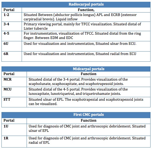
The triangular fibrocartilage complex (TFCC) is formed by the triangular fibrocartilage discus (TFC), the radioulnar ligaments (RULs) and the ulnocarpal ligaments (UCLs).

Shoulder surgery is a means of treating injured shoulders. Many surgeries have been developed to repair the muscles, connective tissue, or damaged joints that can arise from traumatic or overuse injuries to the shoulder.

<span class="mw-page-title-main">Microfracture surgery</span> Cartilage repair technique

Microfracture surgery is an articular cartilage repair surgical technique that works by creating tiny fractures in the underlying bone. This causes new cartilage to develop from a so-called super-clot.

<span class="mw-page-title-main">Jammed finger</span> Medical condition

Jammed finger is a colloquialism referring to a variety of injuries to the joints of the fingers, resulting from axial loading beyond that which the ligaments can withstand. Common parts of the finger susceptible to this type of injury are ligaments, joints, and bones. The severity of the damage to the finger increases with the magnitude of the force exerted by the external object on the fingertip. Toes may become jammed as well, with similar results.

<span class="mw-page-title-main">Radial tunnel syndrome</span> Medical condition

Radial tunnel syndrome (RTS) is caused by increased pressure on the radial nerve as it travels from the upper arm to the hand and wrist.

<span class="mw-page-title-main">Scapholunate ligament</span>

The scapholunate ligament is a ligament of the wrist.

<span class="mw-page-title-main">Hip arthroscopy</span>

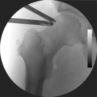
Hip arthroscopy refers to the viewing of the interior of the acetabulofemoral (hip) joint through an arthroscope and the treatment of hip pathology through a minimally invasive approach. This technique is sometimes used to help in the treatment of various joint disorders and has gained popularity because of the small incisions used and shorter recovery times when compared with conventional surgical techniques. Hip arthroscopy was not feasible until recently, new technology in both the tools used and the ability to distract the hip joint has led to a recent surge in the ability to do hip arthroscopy and the popularity of it.

Posterolateral corner injuries of the knee are injuries to a complex area formed by the interaction of multiple structures. Injuries to the posterolateral corner can be debilitating to the person and require recognition and treatment to avoid long term consequences. Injuries to the PLC often occur in combination with other ligamentous injuries to the knee; most commonly the anterior cruciate ligament (ACL) and posterior cruciate ligament (PCL). As with any injury, an understanding of the anatomy and functional interactions of the posterolateral corner is important to diagnosing and treating the injury.

<span class="mw-page-title-main">Wrist osteoarthritis</span> Medical condition

Wrist osteoarthritis is gradual loss of articular cartilage and hypertrophic bone changes (osteophytes). While in many joints this is part of normal aging (senescence), in the wrist osteoarthritis usually occurs over years to decades after scapholunate interosseous ligament rupture or an unhealed fracture of the scaphoid. Characteristic symptoms including pain, deformity and stiffness. Pain intensity and incapability are notably variable and do not correspond with arthritis severity on radiographs.

<span class="mw-page-title-main">Lister's tubercle</span>

Lister's tubercle or dorsal tubercle of radius is a bony prominence located at the distal end of the radius. It is palpable on the dorsum of the wrist.

<span class="mw-page-title-main">Labral reconstruction</span> Medical procedure

Labral reconstruction is a type of hip arthroscopy in which the patient's native labrum is partially or completely removed and reconstructed using either autograft or allograft tissue. Originally described in 2009 using the ligamentum teres capitis, arthroscopic labral reconstruction using a variety of graft tissue has demonstrated promising short and mid-term clinical outcomes. Most importantly, labral reconstruction has demonstrated utility when the patient's native labral tissue is far too damaged for debridement or repair.

References

  1. Geissler, William (2005). Wrist Arthroscopy. New York: Springler-Verlag. ISBN   978-0387208978.
  2. Verheyden, James (February 17, 2012). "Triangular Fibrocartilage Complex Injuries". Medscape. Retrieved 29 October 2013.
  3. Fontès, D. (2006). "Arthroscopic management of recent or chronic lesions of triangular fibrocartilage complex of the wrist". Chirurgie de la Main. 25 (Suppl 1): S178–S186. doi:10.1016/j.main.2006.07.012. PMID   17361887.
  4. Wysocki, R. W.; Richard, M. J.; Crowe, M. M.; Leversedge, F. J.; Ruch, D. S. (2012). "Arthroscopic Treatment of Peripheral Triangular Fibrocartilage Complex Tears with the Deep Fibers Intact". The Journal of Hand Surgery. 37 (3): 509–516. doi:10.1016/j.jhsa.2011.12.023. PMID   22305741.
  5. 1 2 3 4 5 6 Elkowitz, S. J.; Posner, M. A. (2006). "Wrist arthroscopy". Bulletin of the NYU Hospital for Joint Diseases. 64 (3–4): 156–165. PMID   17155924.
  6. Stuffmann, E. S.; McAdams, T. R.; Shah, R. P.; Yao, J. (2010). "Arthroscopic Repair of the Scapholunate Interosseous Ligament". Techniques in Hand & Upper Extremity Surgery. 14 (4): 204–208. doi:10.1097/BTH.0b013e3181df0a93. PMID   21107214. S2CID   7819090.
  7. Sachar, K. (2012). "Ulnar-sided Wrist Pain: Evaluation and Treatment of Triangular Fibrocartilage Complex Tears, Ulnocarpal Impaction Syndrome, and Lunotriquetral Ligament Tears". The Journal of Hand Surgery. 37 (7): 1489–1500. doi:10.1016/j.jhsa.2012.04.036. PMID   22721461.
  8. Chow, James (2000). Advanced Arthroscopy. New York: Springer-Verlag. ISBN   978-0387988085.
  9. Kang, L.; Akelman, E.; Weiss, A. P. C. (2008). "Arthroscopic Versus Open Dorsal Ganglion Excision: A Prospective, Randomized Comparison of Rates of Recurrence and of Residual Pain". The Journal of Hand Surgery. 33 (4): 471–475. doi:10.1016/j.jhsa.2008.01.009. PMID   18406949.
  10. Khanchandani, P.; Badia, A. (2013). "Functional outcome of arthroscopic assisted fixation of distal radius fractures". Indian Journal of Orthopaedics. 47 (3): 288–294. doi: 10.4103/0019-5413.109872 . PMC   3687907 . PMID   23798761.
  11. Chung, Kevin C. (2012). Operative Techniques: Hand and Wrist Surgery. Philadelphia: Elsevier. p. 600. ISBN   978-1-4557-4024-6.
  12. Farr, S.; Grill, F.; Ganger, R.; Graf, A.; Girsch, W. (2012). "Pathomorphologic Findings of Wrist Arthroscopy in Children and Adolescents with Chronic Wrist Pain". Arthroscopy. 28 (11): 1634–1643. doi:10.1016/j.arthro.2012.04.152. PMID   22951372.
  13. Mohamadi, Amin; Claessen, Femke M. A. P.; Ozkan, Sezai; Kolovich, Gregory P.; Ring, David; Chen, Neal C. (March 2017). "Diagnostic Wrist Arthroscopy for Nonspecific Wrist Pain". Hand. 12 (2): 193–196. doi:10.1177/1558944716661993. ISSN   1558-9455. PMC   5349414 . PMID   28344533.
  14. 1 2 3 Strobel, Mickael (2012). Manual of Arthroscopy. New York: Springler-Verlag. ISBN   978-3540857372.
  15. "Wrist Arthroscopy". American Society for Surgery of the Hand. Retrieved 29 October 2013.
  16. 1 2 Frank, Rachel (June 10, 2013). "Wrist Arthroscopy". Medscape. Retrieved 29 October 2013.
  17. Slutsky, D. J. (2012). "Current Innovations in Wrist Arthroscopy". The Journal of Hand Surgery. 37 (9): 1932–1941. doi:10.1016/j.jhsa.2012.06.028. PMID   22916867.