
An injury is any physiological damage to living tissue caused by immediate physical stress. Injuries to humans can occur intentionally or unintentionally and may be caused by blunt trauma, penetrating trauma, burning, toxic exposure, asphyxiation, or overexertion. Injuries can occur in any part of the body, and different symptoms are associated with different injuries.

A bone fracture is a medical condition in which there is a partial or complete break in the continuity of any bone in the body. In more severe cases, the bone may be broken into several fragments, known as a comminuted fracture. A bone fracture may be the result of high force impact or stress, or a minimal trauma injury as a result of certain medical conditions that weaken the bones, such as osteoporosis, osteopenia, bone cancer, or osteogenesis imperfecta, where the fracture is then properly termed a pathologic fracture.

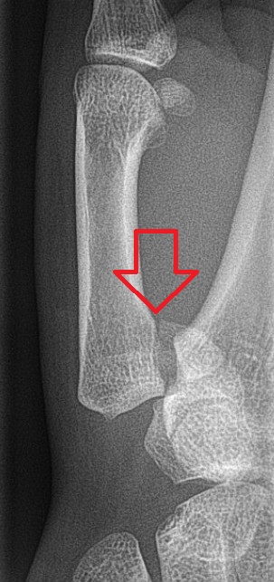
A distal radius fracture, also known as wrist fracture, is a break of the part of the radius bone which is close to the wrist. Symptoms include pain, bruising, and rapid-onset swelling. The ulna bone may also be broken.

In medicine, a joint injection is a procedure used in the treatment of inflammatory joint conditions, such as rheumatoid arthritis, psoriatic arthritis, gout, tendinitis, bursitis, Carpal Tunnel Syndrome, and occasionally osteoarthritis. A hypodermic needle is injected into the affected joint where it delivers a dose of any one of many anti-inflammatory agents, the most common of which are corticosteroids. Hyaluronic acid, because of its high viscosity, is sometimes used to replace bursa fluids. The technique may be used to also withdraw excess fluid from the joint.
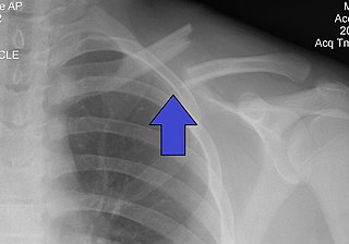
A clavicle fracture, also known as a broken collarbone, is a bone fracture of the clavicle. Symptoms typically include pain at the site of the break and a decreased ability to move the affected arm. Complications can include a collection of air in the pleural space surrounding the lung (pneumothorax), injury to the nerves or blood vessels in the area, and an unpleasant appearance.

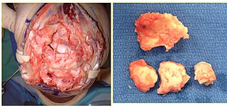
Synovial osteochondromatosis (SOC) (synonyms include synovial chondromatosis, primary synovial chondromatosis, synovial chondrometaplasia) is a rare disease that creates a benign change or proliferation in the synovium or joint-lining tissue, which changes to form bone-forming cartilage. In most occurrences, there is only one joint affected, either the knee, the hip, or the elbow. Rarely involves the TMJ.

The calcaneocuboid joint is the joint between the calcaneus and the cuboid bone.

A calcaneal fracture is a break of the calcaneus. Symptoms may include pain, bruising, trouble walking, and deformity of the heel. It may be associated with breaks of the hip or back.

In human anatomy, the adductor hiatus also known as hiatus magnus is a hiatus (gap) between the adductor magnus muscle and the femur that allows the passage of the femoral vessels from the anterior thigh to the posterior thigh and then the popliteal fossa. It is the termination of the adductor canal and lies about 8–13.5 cm (3.1–5.3 in) superior to the adductor tubercle.
Bennett fracture is a type of partial broken finger involving the base of the thumb, and extends into the carpometacarpal (CMC) joint.
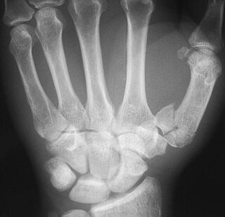
The Rolando fracture is a type of broken finger involving the base of the thumb.
Crutch paralysis is a form of paralysis which can occur when either the radial nerve or part of the brachial plexus, containing various nerves that innervate sense and motor function to the arm and hand, is under constant pressure, such as by the use of a crutch. This can lead to paralysis of the muscles innervated by the compressed nerve. Generally, crutches that are not adjusted to the correct height can cause the radial nerve to be constantly pushed against the humerus. This can cause any muscle that is innervated by the radial nerve to become partially or fully paralyzed. An example of this is wrist drop, in which the fingers, hand, or wrist is chronically in a flexed position because the radial nerve cannot innervate the extensor muscles due to paralysis. This condition, like other injuries from compressed nerves, normally improves quickly through therapy.
Gene therapy for osteoarthritis is the application of gene therapy to treat osteoarthritis (OA). Unlike pharmacological treatments which are administered locally or systemically as a series of interventions, gene therapy aims to establish sustained therapeutic effect after a single, local injection.
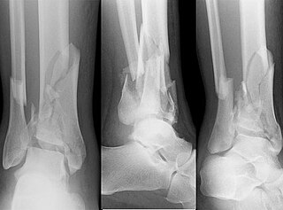
A pilon fracture, is a fracture of the distal part of the tibia, involving its articular surface at the ankle joint. Pilon fractures are caused by rotational or axial forces, mostly as a result of falls from a height or motor vehicle accidents. Pilon fractures are rare, comprising 3 to 10 percent of all fractures of the tibia and 1 percent of all lower extremity fractures, but they involve a large part of the weight-bearing surface of the tibia in the ankle joint. Because of this, they may be difficult to fixate and are historically associated with high rates of complications and poor outcome.
Post-traumatic arthritis (PTA) is a form of osteoarthritis following an injury to a joint.

Raju Vaishya is an Indian researcher with contributions in the field of orthopaedics. He is former President and founder member of Indian Cartilage Society (2018–19) and Founder President of Arthritis Care Foundation. He has established a center for Autologous Chondrocyte Implantation (ACI) at Indraprastha Apollo Hospitals, New Delhi, India. Instrumental (PSI) in starting the first cartilage club in Delhi, to enhance the awareness about the cartilage science and regenerative treatments used in Orthopaedics. He has the credit of doing the first preplan patient specific instruments (PSI) total knee arthroplasty, in Northern India in May 2013.
A disease-modifying osteoarthritis drug (DMOAD) is a disease-modifying drug that would inhibit or even reverse the progression of osteoarthritis. Since the main hallmark of osteoarthritis is cartilage loss, a typical DMOAD would prevent the loss of cartilage and potentially regenerate it. Other DMOADs may attempt to help repair adjacent tissues by reducing inflammation. A successful DMOAD would be expected to show an improvement in patient pain and function with an improvement of the health of the joint tissues.
Hans-Christoph Pape is a German surgeon and trauma surgeon and was appointed full professor of traumatology at the Medical Faculty of the University of Zurich on 31 October 2016, effective 1 February 2017. He heads the Department of Traumatology at the University Hospital Zurich. In particular, his research on polytrauma, pelvic fractures and severe joint injuries (articular) helped him to achieve a high international profile. From 2005 to 2009, he was head of the trauma surgery department at the University of Pittsburgh Medical Center (UPMC), Pittsburgh, USA. From 2009 to 2016, he was head of the Department of Trauma and Reconstructive Surgery at the University Hospital RWTH Aachen. Since March 2018, he has also once again been an adjunct professor at the University of Pittsburgh Medical Center (UPMC), Pittsburgh.
Elbow pain generally refers to discomfort in the joint (elbow) between the upper arm and forearm. Elbow pain is a common complaint in both the emergency department and in primary care offices. The CDC estimated that 1.15 million people visited an emergency room for elbow or forearm-related injuries in 2020. There are many possible causes of elbow discomfort but the most common are trauma, infection, and inflammation. Pain may be acute, chronic or associated with a number of other symptoms. Treatments range from conservative measures, such as ice and rest, to surgical interventions, depending on the underlying cause and severity.
Radiosynoviorthesis (RSO) is a minimally invasive therapeutic procedure for managing joint inflammation, particularly synovitis associated with osteoarthritis. Radiosynoviorthesis involves the intra-articular injection of radioactive isotopes to specifically treat the inflamed synovial membrane. Synovitis, a hallmark of various joint disorders, including osteoarthritis, manifests as inflammation within the synovial membrane lining the joints. RSO aims to suppress overactive macrophage and synovial cells responsible for the inflammatory response, providing relief from pain and improving joint functionality.
