
In humans and other primates, the knee joins the thigh with the leg and consists of two joints: one between the femur and tibia, and one between the femur and patella. It is the largest joint in the human body. The knee is a modified hinge joint, which permits flexion and extension as well as slight internal and external rotation. The knee is vulnerable to injury and to the development of osteoarthritis.

Coxa vara is a deformity of the hip, whereby the angle between the head and the shaft of the femur is reduced to less than 120 degrees. This results in the leg being shortened and the development of a limp. It may be congenital and is commonly caused by injury, such as a fracture. It can also occur when the bone tissue in the neck of the femur is softer than normal, causing it to bend under the weight of the body. This may either be congenital or the result of a bone disorder. The most common cause of coxa vara is either congenital or developmental. Other common causes include metabolic bone diseases, post-Perthes deformity, osteomyelitis, and post traumatic. Shepherd's Crook deformity is a severe form of coxa vara where the proximal femur is severely deformed with a reduction in the neck shaft angle beyond 90 degrees. It is most commonly a sequela of osteogenesis imperfecta, Pagets disease, osteomyelitis, tumour and tumour-like conditions.

Hip replacement is a surgical procedure in which the hip joint is replaced by a prosthetic implant, that is, a hip prosthesis. Hip replacement surgery can be performed as a total replacement or a hemi (half) replacement. Such joint replacement orthopaedic surgery is generally conducted to relieve arthritis pain or in some hip fractures. A total hip replacement consists of replacing both the acetabulum and the femoral head while hemiarthroplasty generally only replaces the femoral head. Hip replacement is one of the most common orthopaedic operations, though patient satisfaction varies widely. Approximately 58% of total hip replacements are estimated to last 25 years. The average cost of a total hip replacement in 2012 was $40,364 in the United States, and about $7,700 to $12,000 in most European countries.
A hip fracture is a break that occurs in the upper part of the femur. Symptoms may include pain around the hip, particularly with movement, and shortening of the leg. Usually the person cannot walk.

In vertebrate anatomy, hip refers to either an anatomical region or a joint.
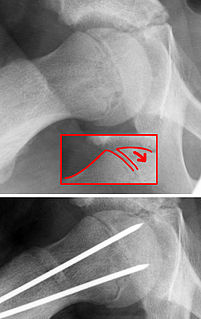
Slipped capital femoral epiphysis is a medical term referring to a fracture through the growth plate (physis), which results in slippage of the overlying end of the femur (metaphysis).

A hip dislocation is when the thighbone (femur) separates from the hip bone (pelvis). Specifically it is when the ball–shaped head of the femur separates from its cup–shaped socket in the hip bone, known as the acetabulum. The joint of the femur and pelvis is very stable, secured by both bony and soft-tissue constraints. With that, dislocation would require significant force which typically results from significant trauma such as from a motor vehicle collision or from a fall from elevation. Hip dislocations can also occur following a hip replacement or from a developmental abnormality known as hip dysplasia.

The medial circumflex femoral artery is an artery in the upper thigh that helps supply blood to the neck of the femur. Damage to the artery following a femoral neck fracture may lead to avascular necrosis (ischemic) of the femoral neck/head.

The lateral condyle is one of the two projections on the lower extremity of the femur. The other one is the medial condyle. The lateral condyle is the more prominent and is broader both in its front-to-back and transverse diameters.
A femoral head ostectomy is a surgical operation to remove the head and neck from the femur. It is performed to alleviate pain, and is a salvage procedure, reserved for condition where pain can not be alleviated in any other way. It is common in veterinary surgery. Other names are excision arthroplasty of the femoral head and neck, Girdlestone's operation, Girdlestone procedure, and femoral head and neck ostectomy.

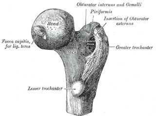
The femoral neck is a flattened pyramidal process of bone, connecting the femoral head with the femoral shaft, and forming with the latter a wide angle opening medialward.

In human anatomy, the adductor hiatus also known as hiatus magnus is a hiatus (gap) between the adductor magnus muscle and the femur that allows the passage of the femoral vessels from the anterior thigh to the posterior thigh and then the popliteal fossa. It is the termination of the adductor canal and lies about 8–13.5 cm. superior to the adductor tubercle.

Dynamic hip screw (DHS) or Sliding Screw Fixation is a type of orthopaedic implant designed for fixation of certain types of hip fractures which allows controlled dynamic sliding of the femoral head component along the construct. It is the most commonly used implant for extracapsular fractures of the hip, which are common in older osteoporotic patients. There are 3 components of a dynamic hip screw, including a lag screw, a sideplate and several cortical screws. The idea behind the dynamic compression is that the femoral head component is allowed to move along one plane; since bone responds to dynamic stresses, the native femur may undergo primary healing: cells join along boundaries, resulting in a robust joint requiring no remodeling.
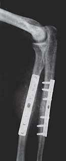
Internal fixation is an operation in orthopedics that involves the surgical implementation of implants for the purpose of repairing a bone, a concept that dates to the mid-nineteenth century and was made applicable for routine treatment in the mid-twentieth century. An internal fixator may be made of stainless steel, titanium alloy, or cobalt-chrome alloy. or plastics.
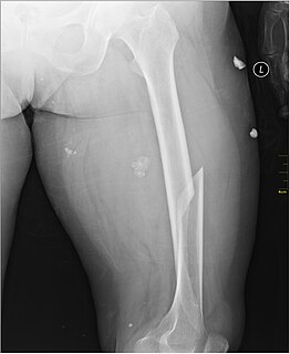
A femoral fracture is a bone fracture that involves the femur. They are typically sustained in high-impact trauma, such as car crashes, due to the large amount of force needed to break the bone. Fractures of the diaphysis, or middle of the femur, are managed differently from those at the head, neck, and trochanter

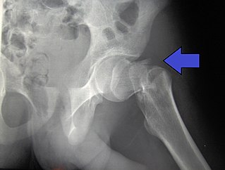
The Garden classification is a system of categorizing intracapsular hip fractures of the femoral neck. This fracture often disrupt the blood supply to the femoral head.
The Pipkin classification is a system of categorizing femoral head hip fractures based on the fracture pattern.

An intraarticular fracture is a bone fracture in which the break crosses into the surface of a joint. This always results in damage to the cartilage. Compared to extraarticular fractures, intraarticular have a higher risk for developing long-term complications, such as posttraumatic osteoarthritis. Treatment considerations include restoring joint surface congruity and maintaining joint alignment and stability.
An extracapsular fracture is a bone fracture near a joint but still located outside the joint capsule.