Femoral neck

Last updated
Femoral neck
Gray243.png
Upper extremity of right femur viewed from behind and above.
Gray244.png
Right femur. Anterior surface.
Details
Identifiers
Latin collum femoris
MeSH D005272
TA98 A02.5.04.004
TA2 1363
FMA 42385
Anatomical terms of bone

The femoral neck (femur neck or neck of the femur) is a flattened pyramidal process of bone, connecting the femoral head with the femoral shaft, and forming with the latter a wide angle opening medialward.

Contents

Structure

The neck is flattened from before backward, contracted in the middle, and broader laterally than medially.

The vertical diameter of the lateral half is increased by the obliquity of the lower edge, which slopes downward to join the body at the level of the lesser trochanter, so that it measures one-third more than the antero-posterior diameter.

The medial half is smaller and of a more circular shape.

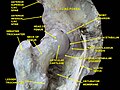
The anterior surface of the neck is perforated by numerous vascular foramina.

Along the upper part of the line of junction of the anterior surface with the head is a shallow groove, best marked in elderly subjects; this groove lodges the orbicular fibers of the capsule of the hip joint.

The posterior surface is smooth, and is broader and more concave than the anterior: the posterior part of the capsule of the hip-joint is attached to it about 1 cm above the intertrochanteric crest.

The superior border is short and thick, and ends laterally at the greater trochanter; its surface is perforated by large foramina.

The inferior border, long and narrow, curves a little backward, to end at the lesser trochanter.

Angle of inclination

The angle is widest in infancy, and becomes lessened during growth, so that at puberty it forms a gentle curve from the axis of the body of the bone. In the adult, the neck forms an angle of about 125° with the body, but this varies in inverse proportion to the development of the pelvis and the stature. The angle decreases during the period of growth, but after full growth has been attained it does not usually undergo any change, even in old age; it varies considerably in different persons of the same age. Coxa vara is a deformity of the hip, whereby the angle between the head and the shaft of the femur is reduced to less than 120 degrees. Its opposite is coxa valga.

Designations of abnormal femur angles. FemurAngles.jpg
Designations of abnormal femur angles.

In the female, in consequence of the increased width of the pelvis, the neck of the femur forms more nearly a right angle with the body than it does in the male.

It is smaller in short than in long bones, and when the pelvis is wide.

In addition to projecting upward and medialward from the body of the femur, the neck also projects somewhat forward; the amount of this forward projection is extremely variable, but on an average is from 12° to 14°.

Fracture

Hip fracture classification. "Neck" is labeled near top. Hip fracture classification.png
Hip fracture classification. "Neck" is labeled near top.

A fracture of the femoral neck is classified as a type of hip fracture. It is often due to osteoporosis; in the vast majority of cases, a hip fracture is a fragility fracture due to a fall or minor trauma in someone with weakened osteoporotic bone. Most hip fractures in people with normal bone are the result of high-energy trauma such as car accidents, falling from heights, or sports injuries.

Garden classification of femoral neck fractures.
TypesDescription
1Incomplete stable fracture with impaction in valgus
2Complete but non displaced with two group of trabeculle in line
3completely displaced with varus with all three trabeculle disturb.
4Completely displaced with no contact between the fracture fragments

For low-grade fractures (Garden types 1 and 2), standard treatment is fixation of the fracture in situ with screws or a sliding screw/plate device. In elderly patients with displaced or intracapsular fractures many surgeons prefer to undertake a hemiarthroplasty, replacing the broken part of the bone with a metal implant. In elderly patients who are medically well and still active, a total hip replacement may be indicated.

Additional images

Related Research Articles

<span class="mw-page-title-main">Human leg</span> Lower extremity or limb of the human body (foot, lower leg, thigh and hip)

The leg is the entire lower limb of the human body, including the foot, thigh or sometimes even the hip or buttock region. The major bones of the leg are the femur, tibia, and adjacent fibula. The thigh is between the hip and knee, while the calf (rear) and shin (front) are between the knee and foot.

<span class="mw-page-title-main">Femur</span> Thigh bone

The femur, or thigh bone is the only bone in the thigh. The thigh is the region of the lower limb between the hip and the knee. In many four-legged animals the femur is the upper bone of the hindleg.

<span class="mw-page-title-main">Knee</span> Leg joint in primates

In humans and other primates, the knee joins the thigh with the leg and consists of two joints: one between the femur and tibia, and one between the femur and patella. It is the largest joint in the human body. The knee is a modified hinge joint, which permits flexion and extension as well as slight internal and external rotation. The knee is vulnerable to injury and to the development of osteoarthritis.

<span class="mw-page-title-main">Humerus</span> Long bone of the upper arm

The humerus is a long bone in the arm that runs from the shoulder to the elbow. It connects the scapula and the two bones of the lower arm, the radius and ulna, and consists of three sections. The humeral upper extremity consists of a rounded head, a narrow neck, and two short processes. The body is cylindrical in its upper portion, and more prismatic below. The lower extremity consists of 2 epicondyles, 2 processes, and 3 fossae. As well as its true anatomical neck, the constriction below the greater and lesser tubercles of the humerus is referred to as its surgical neck due to its tendency to fracture, thus often becoming the focus of surgeons.

<span class="mw-page-title-main">Sacrum</span> Bone of the spine

The sacrum, in human anatomy, is a large, triangular bone at the base of the spine that forms by the fusing of the sacral vertebrae (S1–S5) between ages 18 and 30.

<span class="mw-page-title-main">Tibia</span> Leg bone in vertebrates

The tibia, also known as the shinbone or shankbone, is the larger, stronger, and anterior (frontal) of the two bones in the leg below the knee in vertebrates ; it connects the knee with the ankle. The tibia is found on the medial side of the leg next to the fibula and closer to the median plane. The tibia is connected to the fibula by the interosseous membrane of leg, forming a type of fibrous joint called a syndesmosis with very little movement. The tibia is named for the flute tibia. It is the second largest bone in the human body, after the femur. The leg bones are the strongest long bones as they support the rest of the body.

<span class="mw-page-title-main">Fibula</span> Leg bone in vertebrates

The fibula or calf bone is a leg bone on the lateral side of the tibia, to which it is connected above and below. It is the smaller of the two bones and, in proportion to its length, the most slender of all the long bones. Its upper extremity is small, placed toward the back of the head of the tibia, below the knee joint and excluded from the formation of this joint. Its lower extremity inclines a little forward, so as to be on a plane anterior to that of the upper end; it projects below the tibia and forms the lateral part of the ankle joint.

<span class="mw-page-title-main">Coxa vara</span> Deformity of the hip

Coxa vara is a deformity of the hip, whereby the angle between the head and the shaft of the femur is reduced to less than 120 degrees. This results in the leg being shortened and the development of a limp. It may be congenital and is commonly caused by injury, such as a fracture. It can also occur when the bone tissue in the neck of the femur is softer than normal, causing it to bend under the weight of the body. This may either be congenital or the result of a bone disorder. The most common cause of coxa vara is either congenital or developmental. Other common causes include metabolic bone diseases, post-Perthes deformity, osteomyelitis, and post traumatic. Shepherd's Crook deformity is a severe form of coxa vara where the proximal femur is severely deformed with a reduction in the neck shaft angle beyond 90 degrees. It is most commonly a sequela of osteogenesis imperfecta, Paget's disease, osteomyelitis, tumour and tumour-like conditions.

<span class="mw-page-title-main">Greater trochanter</span> Quadrilateral part of the thigh bone

The greater trochanter of the femur is a large, irregular, quadrilateral eminence and a part of the skeletal system.

<span class="mw-page-title-main">Hip</span> Anatomical region between the torso and the legs, holding the buttocks and genital region

In vertebrate anatomy, the hip, or coxa in medical terminology, refers to either an anatomical region or a joint on the outer (lateral) side of the pelvis.

<span class="mw-page-title-main">Hip dislocation</span> Orthopedic injury

A hip dislocation is when the thighbone (femur) separates from the hip bone (pelvis). Specifically it is when the ball–shaped head of the femur separates from its cup–shaped socket in the hip bone, known as the acetabulum. The joint of the femur and pelvis is very stable, secured by both bony and soft-tissue constraints. With that, dislocation would require significant force which typically results from significant trauma such as from a motor vehicle collision or from a fall from elevation. Hip dislocations can also occur following a hip replacement or from a developmental abnormality known as hip dysplasia.

<span class="mw-page-title-main">Lesser trochanter</span> Conical projection from the shaft of the femur

In human anatomy, the lesser trochanter is a conical, posteromedial, bony projection from the shaft of the femur. It serves as the principal insertion site of the iliopsoas muscle.

<span class="mw-page-title-main">Obturator artery</span>

The obturator artery is a branch of the internal iliac artery that passes antero-inferiorly on the lateral wall of the pelvis, to the upper part of the obturator foramen, and, escaping from the pelvic cavity through the obturator canal, it divides into an anterior branch and a posterior branch.

<span class="mw-page-title-main">Upper extremity of femur</span>

The upper extremity, proximal extremity or superior epiphysis of the femur is the part of the femur closest to the pelvic bone and the trunk. It contains the following structures:

In human anatomy, the body of femur is the almost cylindrical, long part of the femur. It is a little broader above than in the center, broadest and somewhat flattened from before backward below. It is slightly arched, so as to be convex in front, and concave behind, where it is strengthened by a prominent longitudinal ridge, the linea aspera.

<span class="mw-page-title-main">Intertrochanteric line</span>

The intertrochanteric line is a line upon the anterior aspect of the proximal end of the femur, extending between the lesser trochanter and the greater trochanter. It is a rough, variable ridge.

<span class="mw-page-title-main">Femoral head</span>

The femoral head is the highest part of the thigh bone (femur). It is supported by the femoral neck.

<span class="mw-page-title-main">Hip bone</span> Bone of the pelvis

The hip bone is a large flat bone, constricted in the center and expanded above and below. In some vertebrates it is composed of three parts: the ilium, ischium, and the pubis.

<span class="mw-page-title-main">Pelvis</span> Lower torso of the human body

The pelvis is the lower part of the trunk, between the abdomen and the thighs, together with its embedded skeleton.

X-rays of hip dysplasia are one of the two main methods of medical imaging to diagnose hip dysplasia, the other one being medical ultrasonography. Ultrasound imaging yields better results defining the anatomy until the cartilage is ossified. When the infant is around 3 months old a clear roentgenographic image can be achieved. Unfortunately the time the joint gives a good x-ray image is also the point at which nonsurgical treatment methods cease to give good results.

References

    • "Prevention and Management of Hip Fracture on Older People. Section 7: Surgical management". Scottish Intercollegiate Guidelines Network. Archived from the original on 2017-04-24. Retrieved 2017-04-23. Last modified 15/7/02
    • Area of trochanteric fractures: Ernst Raaymakers, Inger Schipper, Rogier Simmermacher, Chris van der Werken. "Proximal femur". AO Foundation . Retrieved 2017-04-23.{{cite web}}: CS1 maint: multiple names: authors list (link)
    • Area of subtrochanteric fractures: Mark A Lee. "Subtrochanteric Hip Fractures" . Retrieved 2017-04-25. Updated: Jun 22, 2016
    • Area of femoral neck fractures: Page 333 in: Paul Tornetta, III, Sam W. Wiesel (2010). Operative Techniques in Orthopaedic Trauma Surgery. Lippincott Williams & Wilkins. ISBN   9781451102604.{{cite book}}: CS1 maint: multiple names: authors list (link)

PD-icon.svgThis article incorporates text in the public domain from page 243 of the 20th edition of Gray's Anatomy (1918)