9-Hydroxyoctadecadienoic acid

Last updated
9-Hydroxyoctadecadienoic acid
9-HODE.svg
Names
Preferred IUPAC name
(9S,10E,12Z)-9-Hydroxyoctadeca-10,12-dienoic acid
Other names
  • α-Dimorphecolic acid
  • 9-hydroxy-10(E),12(Z)-octadecadienoic acid
Identifiers
3D model (JSmol)
ChemSpider
ECHA InfoCard 100.230.886 OOjs UI icon edit-ltr-progressive.svg
PubChem CID
UNII
  • InChI=1S/C18H32O3/c1-2-3-4-5-6-8-11-14-17(19)15-12-9-7-10-13-16-18(20)21/h6,8,11,14,17,19H,2-5,7,9-10,12-13,15-16H2,1H3,(H,20,21)/b8-6-,14-11+/t17-/m1/s1 COPY
    Key: NPDSHTNEKLQQIJ-UINYOVNOSA-N
  • CCCCC/C=C\C=C\[C@H](CCCCCCCC(=O)O)O
Properties
C18H32O3
Molar mass 296.451 g·mol−1
Except where otherwise noted, data are given for materials in their standard state (at 25 °C [77 °F], 100 kPa).

9-Hydroxyoctadecadienoic acid (or 9-HODE) has been used in the literature to designate either or both of two stereoisomer metabolites of the essential fatty acid, linoleic acid: 9(S)-hydroxy-10(E),12(Z)-octadecadienoic acid (9(S)-HODE) and 9(R)-hydroxy-10(E),12(Z)-octadecadienoic acid (9(R)-HODE); these two metabolites differ in having their hydroxy residues in the S or R configurations, respectively. The accompanying figure gives the structure for 9(S)-HETE. Two other 9-hydroxy linoleic acid derivatives occur in nature, the 10E,12E isomers of 9(S)-HODE and 9(R)-HODE viz., 9(S)-hydroxy-10E,12E-octadecadienoic acid (9(S)-EE-HODE) and 9(R)-hydroxy-10E,12E-octadecadienoic acid (13(R)-EE-HODE); these two derivatives have their double bond at carbon 12 in the E or trans configuration as opposed to the Z or cis configuration. The four 9-HODE isomers, particularly under conditions of oxidative stress, may form together in cells and tissues; they have overlapping but not identical biological activities and significances. Because many studies have not distinguished between the S and R stereoisomers and, particularly in identifying tissue levels, the two EE isomers, 9-HODE is used here when the isomer studied is unclear.

Contents

A similar set of 13-Hydroxyoctadecadienoic acid (13-HODE) metabolites (13(S)-HODE), 13(R)-HODE, 13(S)-EE-HODE), and 13(R)-EE-HODE) also occurs naturally and, again particularly under conditions of oxidative stress, may form concurrently with 9-HODEs; these 13-HODEs also have overlapping and complementary but not identical activities with the 9-HODEs. Some recent studies measuring HODE levels in tissue have lumped the four 9-HODEs and four 13-HODEs together to report only on total HODEs (tHODEs): tHODEs have been proposed to be markers for certain human disease. Other recent studies have lumped together the 9-(S), 9(R), 13 (S)-, and 13(R)-HODE along with the two ketone metabolites of these HODEs, 9-oxoODE (9-oxo-10(E),12(Z)-octadecadienoic acid) and 13-oxoODE, reporting only on total OXLAMs (oxidized linoleic acid metabolites); the OXLAMs have been implicated in working together to signal for pain perception.

Pathways making 9-HODEs

Cyclooxygenases 1 and 2

The enzymes cyclooxygenase 1 (COX-1) and cyclooxygenase 2 (COX-2), which are best known for metabolizing arachidonic acid to prostaglandins, are also able to metabolize linoleic acid predominantly to 9(R)-hydroperoxy-10(E),12(Z)-octadecadienoic acid (i.e. 9(R)-HpODE)-HODE) and lesser amounts of 9(S)-hydroperoxy-10(E),12(Z)-octadecadienoic acid (i.e. 9(S)-HpODE); in cells and tissues, the two hydroperoxy metabolites are rapidly reduce to 9(R)-HODE and 9(S)-HODE, respectively. [1] [2] [3] COX-2 exhibits a greater preference for linoleic acid than does Cox-1 and is therefore credited with producing most of these products in cells expressing both COX enzymes. [2] The COXs also metabolize linoleic acid to 13(S)-hydroperoxy-octadecadionoic acid (13(S)-HpODE and lesser amounts of 13(R)-hydroperoxy-octadecadienoic acid (13(R)-HpODE, which are then rapidly reduced to 13(S)-HODE) and 13(R)-HODE; the two enzymes therefore metabolize linoleic acid predominantly to the R stereoisomer of 9-HODE and (S) stereoisomer of 13-HODE with the 13-HODE products predominating over the 9-HODE products. [1] [2] [4]

Cytochrome P450

Cytochrome P450 microsomal enzymes metabolize linoleic acid to a mixture of 9(S)-HpODE and 9(R)-HpODE which are subsequently reduced to their corresponding hydroxy products; these reactions produce racemic mixtures in which the R stereoisomer predominates, for instance by a R/S ratio of 80%/20% in human liver microsomes. [5] [6] [7] In cells and tissues, the cytochrome enzymes concurrently metabolize linoleic acid to 13(S)-HpODE and 13(R)-HpODE which are reduced to 13(S)-HODE and 13(R)-HODE in an R/S ratio similar to than of the 9-HODES, i.e. 80%/20%. [6]

Free-radical and singlet-oxygen oxidations

Oxidative stress in cells and tissues produces Free-radical-induced and singlet-oxygen-induced oxidations of linoleic acid to generate the various racemic mixtures of 9-HpODE and 9-HODE in non-enzymatic reactions that produce, or are suspected but not proven to produce, approximately equal amounts of their S and R stereoisomers. [8] [9] [10] These oxidations are credited with being the major contributors to 9-HODE and 13-HODE isomer production in tissues undergoing oxidative stress such as occurs in any tissue suffering inadequate blood flow, inflammation, or other serious insult, in liver steatohepatitis, in the atheroma plaques of cardiovascular disease, in nerve tissues of neurodegenerative diseases, and in the various tissues compromised by diabetes (see oxidative stress). [11] [12] Free-radical oxidation of linoleic acid produces racemic mixtures of 9-HODE and 9-EE-HODE; singlet-oxygen attack on linoleic acid produces (presumably) racemic mixtures of 9-HODE, 10-hydroxy-8E,12Z-octadecadienoic acid, and 12-hydroxy-9Z-13-E-octadecadienoic acid. [13] [12] Since free-radical-induced and singlet-oxygen-induced oxidations of linoleic acid produce a similar set of 13-HODE metabolites (see 13-Hydroxyoctadecadienoic acid), since both free radicals and singlet oxygen attack not only free linoleic acid but also linoleic acid bound to phospholipids, glycerides, cholesterol, and other lipids, and since free-radical and singlet-oxygen reactions may occur together, oxygen-stressed tissues often contain an array of free and lipid-bound 9-HODE and 13-HODE products. For example, laboratory studies find that 9-HODE and 9-EE-HODE (along with their 13-HODE counterparts) are found in the phospholipid and cholesterol components of low-density lipoproteins that have been oxidized by human monocytes; the reaction appears due to the in situ free-radical- and/or superoxide-induced oxidation of the lipoproteins. [14]

Mouse 8(S)-lipoxygenase

The murine homolog of human 15(S)-lipoxygenase-2 (ALOX15B), 8(S)-lipoxygenase, while preferring arachidonic acid over linoleic acid, metabolizes linoleic acid predominantly to (9(S)-HpODE, which in tissues and cells is rapidly reduced to 9(S)-HODE. [15] [16] However, ALOX15B, similar to human 15-lipoxygenase-1 (ALOX15), metabolizes linoleic acid to 13(S)-HODE but not to 9(S)-HODEs. [17] [18]

Metabolism

Like most unsaturated fatty acids, the 9-HODEs formed in cells are incorporated into cellular phospholipids principally at the sn-2 position of the phospholipid (see Phospholipase A2); [19] [20] since, however, the linoleic acid bound to cellular phospholipids is susceptible to non-enzymatic peroxidation and free-radical attack, [21] [22] [23] the 9-HODEs in cellular phospholipids may also derive more directly from in-situ oxidation. 9-HODE esterified to the sn-2 position of phosphatidylserine is subject to be released as free 9-HODE by the action of cytosol (see phospholipase A2 section on cPLA2) and therefore may serve as a storage pool that is mobilized by cell stimulation. [23]

9-HODE may be further metabolized to 9-oxo-10(E),12(Z)-octadecadienoic acid (9-oxoODE or 9-oxo-ODE), possibly by the same hydroxy-fatty-acid dehydrogenase which metabolizes other hydroxy fatty acids, such as 13-HODE, to their oxo derivatives. [24]

Direct actions

9-HODE, 9-oxoODE, and 9-EE-HODE (along with their 13-HODE counterparts) directly activate peroxisome proliferator-activated receptor gamma (PPARγ). [25] [26] [27] This activation appears responsible for the ability of 13-HODE (and 9-HODE) to induce the transcription of PPARγ-inducible genes in human monocytes as well as to stimulate the maturation of these cells to macrophages. [25] 13(S)-HODE (and 9(S)-HODE) also stimulate the activation of peroxisome proliferator-activated receptor beta (PPARβ) in a model cell system; 13-HODE (and 9-HODE) are also proposed to contribute to the ability of oxidized low-density lipoprotein (LDL) to activate PPARβl: LDL containing phospholipid-bound 13-HODE (and 9-HODE) is taken up by the cell and then acted on by phospholipases to release the HODEs which in turn directly activate PPARβl. [28]

13(S)-HODE, 13(R)-HODE and 13-oxoODE, along with their 9-HODE counterparts, also act on cells through TRPV1. TRPV1 is the transient receptor potential cation channel subfamily V member 1 receptor (also termed capsaicin receptor or vanilloid receptor 1). These 6 HODEs, dubbed, oxidized linoleic acid metabolites (OXLAMs), individually but also and possibly to a greater extent when acting together, stimulate TRPV1-dependent responses in rodent neurons, rodent and human bronchial epithelial cells, and in model cells made to express rodent or human TRPV1. This stimulation appears due to a direct interaction of these agents on TRPV1 although reports disagree on the potencies of the (OXLAMs) with, for example, the most potent OXLAM, 9(S)-HODE, requiring at least 10 micromoles/liter [29] or a more physiological concentration of 10 nanomoles/liter [30] to activate TRPV1 in rodent neurons. The OXLAM-TRPV1 interaction is credited with mediating pain sensation in rodents (see below).

9(S)-HODE and with progressively lesser potencies 9(S)-HpODE, a racemic mixture of 9-HODE, 13(S)-HpODE, and 13(S)-HODE directly activate human (but not mouse) GPR132 (i.e. G protein coupled receptor 132 or G2A) in Chinese hamster ovary cells made to express these receptors; 9(S)-HODE was also a more potent stimulator of human G2A than a series of mono-hydroxy arachidonic acid metabolites. [31] [32] GPR132 was initially described as a pH sensing receptor; the role(s) of 9-HODEs as well as other linoleic and arachidonic acid metabolites in activating GPR132 under the physiological and pathological conditions in which it is implicated to be involved(see (see GPR132 for a listing of these conditions) have not yet been determined. This determination, as it might apply to humans, is made difficult by the inability of these HODEs to activate rodent GPR132 and therefore to be analyzed in rodent models.

Biological and clinical relevancy

As markers of disease involving oxidative stress

Various measurements of tissue and blood levels of reactive oxygen species have been used as markers of diseases in which these species are generated and may contribute to tissue injury and systemic disturbances; examples of such diseases include a wide range of neurological, cardiovascular, infectious, autoimmune, and genetic diseases (see oxidative stress). HODEs measurements have been evaluated as markers for many of these oxygen-stress-related diseases. These measurements commonly use saponification methods to release HODEs bound by acylation to other molecules; they therefore measure not only free HODEs but also HODEs acylated to phospholipids, glycerides, cholesterol, and other lipids.

Studies find that 1) 9(S)-HODE (and 13(S)-HODE) levels are elevated in the plasma of older patients with early-stage cataracts compared to non-cataract subjects; 2) 9-HODE (and 13-HODE) are increased in the low density lipoproteins of patients with rheumatoid arthritis compared to healthy subjects as well as in the destructive but not normal bone tissue of the rheumatoid arthritic patients; 3) total HODEs (includes 9-HODE and 13-HODE stereoisomers) are higher in the plasma and liver of patients with hepatitis C and hepatitis B chronic viral infections as well as in the plasma and red blood cells of patients with Alzheimer's disease compared to healthy subjects; 4) 9-HODE and 9-oxoODE (as well as 13-HODE and 13-oxo-ODE) levels were elevated in the serum and/or pancreatic secretions of patients with pancreatitis compared to control subjects; 5) levels of the hydroperoxy precursors to 9-HODE and 13-HODE are elevated in the plasma and/or red blood cells of patients with Alzheimer's disease, atherosclerosis, diabetes, diabetic nephritis, non-alcoholic steatohepatitis, and alcoholic steatohepatitis compared to healthy subjects. [33] [34] [35] [36] [37] [38] [39] These studies suggest that high levels of the HODEs may be useful to indicate the presence and progression of the cited diseases. Since, however, the absolute values of HODEs found in different studies vary greatly, since HODE levels vary with dietary linoleic acid intake, since HODEs may form during the processing of tissues, and since abnormal HODE levels are not linked to a specific disease, the use of these metabolites as markers has not attained clinical usefulness. [11] [37] [40] [12] HODE markers may find usefulness as markers of specific disease, type of disease, and/or progression of disease when combined with other disease markers. [12] [41]

Some of the studies cited above have suggested that 9-HODEs, 13-HODEs, their hydroperoxy counterparts, and/or their oxo counterparts contribute mechanistically to these oxidative-stress-related diseases. That is, the free radical oxidation of linoleic acid makes these products which then proceed to contribute to the tissue injury, DNA damage, and/or systemic dysfunctions that characterize the diseases. [42] [43] [44] [45] [46] Furthermore, certain of these HODE-related products may serve as signals to activate pathways that combat the reactive oxygen species and in this and other ways the oxidative stress. It remains unclear whether or not the HODEs and their counterparts promote, dampen, or merely reflect oxidative-stress-related diseases.

As mediators of pain perception

9(S)-HODE, 9(R)-HODE, and 9-oxoODE, along with the other OXLAMs, appear to act through the TRPV1 receptor (see above section on Direct actions) mediate the perception of acute and chronic pain induced by heat, UV light, and inflammation in the skin of rodents. [30] [47] [48] [49] [50] These studies propose that the OXLAM-TRPV1 circuit (with 9(S)-HODE being the most potent TRPV1-activating OXLAM) similarly contributes to the perception of pain in humans.

As contributors to atherosclerosis

9-HODEs, 13-HODEs, and low density lipoprotein which has been oxidized so that it contains HODEs stimulate the expression of interleukin 1β mRNA in and its extracellular release from human peripheral blood monocyte-derived macrophages; interleukin 1β is implicated in the proliferation of smooth muscle cells that occurs in atherosclerosis and contributes to blood vessel narrowing. [51]

Related Research Articles

<span class="mw-page-title-main">Eicosanoid</span> Class of compounds

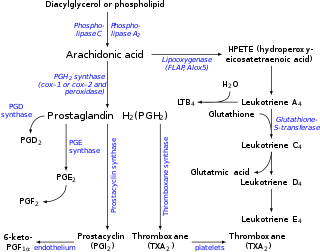
Eicosanoids are signaling molecules made by the enzymatic or non-enzymatic oxidation of arachidonic acid or other polyunsaturated fatty acids (PUFAs) that are, similar to arachidonic acid, around 20 carbon units in length. Eicosanoids are a sub-category of oxylipins, i.e. oxidized fatty acids of diverse carbon units in length, and are distinguished from other oxylipins by their overwhelming importance as cell signaling molecules. Eicosanoids function in diverse physiological systems and pathological processes such as: mounting or inhibiting inflammation, allergy, fever and other immune responses; regulating the abortion of pregnancy and normal childbirth; contributing to the perception of pain; regulating cell growth; controlling blood pressure; and modulating the regional flow of blood to tissues. In performing these roles, eicosanoids most often act as autocrine signaling agents to impact their cells of origin or as paracrine signaling agents to impact cells in the proximity of their cells of origin. Eicosanoids may also act as endocrine agents to control the function of distant cells.

<span class="mw-page-title-main">Resolvin</span> Class of chemical compounds

Resolvins are specialized pro-resolving mediators (SPMs) derived from omega-3 fatty acids, primarily eicosapentaenoic acid (EPA) and docosahexaenoic acid (DHA), as well as from two isomers of docosapentaenoic acid (DPA), one omega-3 and one omega-6 fatty acid. As autacoids similar to hormones acting on local tissues, resolvins are under preliminary research for their involvement in promoting restoration of normal cellular function following the inflammation that occurs after tissue injury. Resolvins belong to a class of polyunsaturated fatty acid (PUFA) metabolites termed specialized proresolving mediators (SPMs).

<span class="mw-page-title-main">Oxidative stress</span> Free radical toxicity

Oxidative stress reflects an imbalance between the systemic manifestation of reactive oxygen species and a biological system's ability to readily detoxify the reactive intermediates or to repair the resulting damage. Disturbances in the normal redox state of cells can cause toxic effects through the production of peroxides and free radicals that damage all components of the cell, including proteins, lipids, and DNA. Oxidative stress from oxidative metabolism causes base damage, as well as strand breaks in DNA. Base damage is mostly indirect and caused by the reactive oxygen species generated, e.g., O2 (superoxide radical), OH (hydroxyl radical) and H2O2 (hydrogen peroxide). Further, some reactive oxidative species act as cellular messengers in redox signaling. Thus, oxidative stress can cause disruptions in normal mechanisms of cellular signaling.

<span class="mw-page-title-main">Lipoxygenase</span>

Lipoxygenases (LOX) are a family of (non-heme) iron-containing enzymes most of which catalyze the dioxygenation of polyunsaturated fatty acids in lipids containing a cis,cis-1,4-pentadiene into cell signaling agents that serve diverse roles as autocrine signals that regulate the function of their parent cells, paracrine signals that regulate the function of nearby cells, and endocrine signals that regulate the function of distant cells.

<span class="mw-page-title-main">Hepoxilin</span> Chemical compound

Hepoxilins (Hx) are a set of epoxyalcohol metabolites of polyunsaturated fatty acids (PUFA), i.e. they possess both an epoxide and an alcohol residue. HxA3, HxB3, and their non-enzymatically formed isomers are nonclassic eicosanoid derived from acid the (PUFA), arachidonic acid. A second group of less well studied hepoxilins, HxA4, HxB4, and their non-enzymatically formed isomers are nonclassical eicosanoids derived from the PUFA, eicosapentaenoic acid. Recently, 14,15-HxA3 and 14,15-HxB3 have been defined as arachidonic acid derivatives that are produced by a different metabolic pathway than HxA3, HxB3, HxA4, or HxB4 and differ from the aforementioned hepoxilins in the positions of their hydroxyl and epoxide residues. Finally, hepoxilin-like products of two other PUFAs, docosahexaenoic acid and linoleic acid, have been described. All of these epoxyalcohol metabolites are at least somewhat unstable and are readily enzymatically or non-enzymatically to their corresponding trihydroxy counterparts, the trioxilins (TrX). HxA3 and HxB3, in particular, are being rapidly metabolized to TrXA3, TrXB3, and TrXC3. Hepoxilins have various biological activities in animal models and/or cultured mammalian tissues and cells. The TrX metabolites of HxA3 and HxB3 have less or no activity in most of the systems studied but in some systems retain the activity of their precursor hepoxilins. Based on these studies, it has been proposed that the hepoxilins and trioxilins function in human physiology and pathology by, for example, promoting inflammation responses and dilating arteries to regulate regional blood flow and blood pressure.

<span class="mw-page-title-main">TRPV1</span> Human protein for regulating body temperature

The transient receptor potential cation channel subfamily V member 1 (TRPV1), also known as the capsaicin receptor and the vanilloid receptor 1, is a protein that, in humans, is encoded by the TRPV1 gene. It was the first isolated member of the transient receptor potential vanilloid receptor proteins that in turn are a sub-family of the transient receptor potential protein group. This protein is a member of the TRPV group of transient receptor potential family of ion channels. Fatty acid metabolites with affinity for this receptor are produced by cyanobacteria, which diverged from eukaryotes at least 2000 million years ago (MYA). The function of TRPV1 is detection and regulation of body temperature. In addition, TRPV1 provides a sensation of scalding heat and pain (nociception). In primary afferent sensory neurons, it cooperates with TRPA1 to mediate the detection of noxious environmental stimuli.

In biochemistry, docosanoids are signaling molecules made by the metabolism of twenty-two-carbon fatty acids (EFAs), especially the omega-3 fatty acid, Docosahexaenoic acid (DHA) by lipoxygenase, cyclooxygenase, and cytochrome P450 enzymes. Other docosanoids are metabolites of n-3 docosapentaenoic acid, n-6 DHA (i.e. 4Z,7Z,10Z,13Z,16Z-docosahexaenoic acid, and docosatetraenoic acid. Prominent docosanoid metabolites of DHA and n-3 DHA are members of the specialized proresolving mediator class of polyunsaturated fatty acid metabolites that possess potent anti-inflammation, tissue healing, and other activities.

<span class="mw-page-title-main">ALOX15</span> Lipoxygenase found in humans

ALOX15 is, like other lipoxygenases, a seminal enzyme in the metabolism of polyunsaturated fatty acids to a wide range of physiologically and pathologically important products. ▼ Gene Function

<span class="mw-page-title-main">ALOX12</span> Protein-coding gene in the species Homo sapiens

ALOX12, also known as arachidonate 12-lipoxygenase, 12-lipoxygenase, 12S-Lipoxygenase, 12-LOX, and 12S-LOX is a lipoxygenase-type enzyme that in humans is encoded by the ALOX12 gene which is located along with other lipoyxgenases on chromosome 17p13.3. ALOX12 is 75 kilodalton protein composed of 663 amino acids.

<span class="mw-page-title-main">5-Hydroxyeicosatetraenoic acid</span> Chemical compound

5-Hydroxyeicosatetraenoic acid (5-HETE, 5(S)-HETE, or 5S-HETE) is an eicosanoid, i.e. a metabolite of arachidonic acid. It is produced by diverse cell types in humans and other animal species. These cells may then metabolize the formed 5(S)-HETE to 5-oxo-eicosatetraenoic acid (5-oxo-ETE), 5(S),15(S)-dihydroxyeicosatetraenoic acid (5(S),15(S)-diHETE), or 5-oxo-15-hydroxyeicosatetraenoic acid (5-oxo-15(S)-HETE).

<span class="mw-page-title-main">ALOX12B</span> Protein-coding gene in the species Homo sapiens

Arachidonate 12-lipoxygenase, 12R type, also known as ALOX12B, 12R-LOX, and arachidonate lipoxygenase 3, is a lipoxygenase-type enzyme composed of 701 amino acids and encoded by the ALOX12B gene. The gene is located on chromosome 17 at position 13.1 where it forms a cluster with two other lipoxygenases, ALOXE3 and ALOX15B. Among the human lipoxygenases, ALOX12B is most closely related in amino acid sequence to ALOXE3

<span class="mw-page-title-main">ALOXE3</span> Protein-coding gene in the species Homo sapiens

Epidermis-type lipoxygenase 3 is a member of the lipoxygenase family of enzymes; in humans, it is encoded by the ALOXE3 gene. This gene is located on chromosome 17 at position 13.1 where it forms a cluster with two other lipoxygenases, ALOX12B and ALOX15B. Among the human lipoxygenases, ALOXE3 is most closely related in amino acid sequence to ALOX12B. ALOXE3, ALOX12B, and ALOX15B are often classified as epidermal lipoxygenases, in distinction to the other three human lipoxygenases, because they were initially defined as being highly or even exclusively expressed and functioning in skin. The epidermis-type lipoxygenases are now regarded as a distinct subclass within the multigene family of mammalian lipoxygenases with mouse Aloxe3 being the ortholog to human ALOXE3, mouse Alox12b being the ortholog to human ALOX12B, and mouse Alox8 being the ortholog to human ALOX15B [supplied by OMIM]. ALOX12B and ALOXE3 in humans, Alox12b and Aloxe3 in mice, and comparable orthologs in other in other species are proposed to act sequentially in a multistep metabolic pathway that forms products that are structurally critical for creating and maintaining the skin's water barrier function.

<span class="mw-page-title-main">Oxylipin</span> Class of lipids

Oxylipins constitute a family of oxygenated natural products which are formed from fatty acids by pathways involving at least one step of dioxygen-dependent oxidation. Oxylipins are derived from polyunsaturated fatty acids (PUFAs) by COX enzymes (cyclooxygenases), by LOX enzymes (lipoxygenases), or by cytochrome P450 epoxygenase.

<span class="mw-page-title-main">12-Hydroxyeicosatetraenoic acid</span> Chemical compound

12-Hydroxyeicosatetraenoic acid (12-HETE) is a derivative of the 20 carbon polyunsaturated fatty acid, arachidonic acid, containing a hydroxyl residue at carbon 12 and a 5Z,8Z,10E,14Z Cis–trans isomerism configuration (Z=cis, E=trans) in its four double bonds. It was first found as a product of arachidonic acid metabolism made by human and bovine platelets through their 12S-lipoxygenase (i.e. ALOX12) enzyme(s). However, the term 12-HETE is ambiguous in that it has been used to indicate not only the initially detected "S" stereoisomer, 12S-hydroxy-5Z,8Z,10E,14Z-eicosatetraenoic acid (12(S)-HETE or 12S-HETE), made by platelets, but also the later detected "R" stereoisomer, 12(R)-hydroxy-5Z,8Z,10E,14Z-eicosatetraenoic acid (also termed 12(R)-HETE or 12R-HETE) made by other tissues through their 12R-lipoxygenase enzyme, ALOX12B. The two isomers, either directly or after being further metabolized, have been suggested to be involved in a variety of human physiological and pathological reactions. Unlike hormones which are secreted by cells, travel in the circulation to alter the behavior of distant cells, and thereby act as Endocrine signalling agents, these arachidonic acid metabolites act locally as Autocrine signalling and/or Paracrine signaling agents to regulate the behavior of their cells of origin or of nearby cells, respectively. In these roles, they may amplify or dampen, expand or contract cellular and tissue responses to disturbances.

<span class="mw-page-title-main">15-Hydroxyeicosatetraenoic acid</span> Chemical compound

15-Hydroxyeicosatetraenoic acid (also termed 15-HETE, 15(S)-HETE, and 15S-HETE) is an eicosanoid, i.e. a metabolite of arachidonic acid. Various cell types metabolize arachidonic acid to 15(S)-hydroperoxyeicosatetraenoic acid (15(S)-HpETE). This initial hydroperoxide product is extremely short-lived in cells: if not otherwise metabolized, it is rapidly reduced to 15(S)-HETE. Both of these metabolites, depending on the cell type which forms them, can be further metabolized to 15-oxo-eicosatetraenoic acid (15-oxo-ETE), 5(S),15(S)-dihydroxy-eicosatetraenoic acid (5(S),15(S)-diHETE), 5-oxo-15(S)-hydroxyeicosatetraenoic acid (5-oxo-15(S)-HETE), a subset of specialized pro-resolving mediators viz., the lipoxins, a class of pro-inflammatory mediators, the eoxins, and other products that have less well-defined activities and functions. Thus, 15(S)-HETE and 15(S)-HpETE, in addition to having intrinsic biological activities, are key precursors to numerous biologically active derivatives.

<span class="mw-page-title-main">13-Hydroxyoctadecadienoic acid</span> Chemical compound

13-Hydroxyoctadecadienoic acid (13-HODE) is the commonly used term for 13(S)-hydroxy-9Z,11E-octadecadienoic acid (13(S)-HODE). The production of 13(S)-HODE is often accompanied by the production of its stereoisomer, 13(R)-hydroxy-9Z,11E-octadecadienoic acid (13(R)-HODE). The adjacent figure gives the structure for the (S) stereoisomer of 13-HODE. Two other naturally occurring 13-HODEs that may accompany the production of 13(S)-HODE are its cis-trans (i.e., 9E,11E) isomers viz., 13(S)-hydroxy-9E,11E-octadecadienoic acid (13(S)-EE-HODE) and 13(R)-hydroxy-9E,11E-octadecadienoic acid (13(R)-EE-HODE). Studies credit 13(S)-HODE with a range of clinically relevant bioactivities; recent studies have assigned activities to 13(R)-HODE that differ from those of 13(S)-HODE; and other studies have proposed that one or more of these HODEs mediate physiological and pathological responses, are markers of various human diseases, and/or contribute to the progression of certain diseases in humans. Since, however, many studies on the identification, quantification, and actions of 13(S)-HODE in cells and tissues have employed methods that did not distinguish between these isomers, 13-HODE is used here when the actual isomer studied is unclear.

<span class="mw-page-title-main">5-Oxo-eicosatetraenoic acid</span> Chemical compound

5-Oxo-eicosatetraenoic acid is a Nonclassic eicosanoid metabolite of arachidonic acid and the most potent naturally occurring member of the 5-HETE family of cell signaling agents. Like other cell signaling agents, 5-oxo-ETE is made by a cell and then feeds back to stimulate its parent cell and/or exits this cell to stimulate nearby cells. 5-Oxo-ETE can stimulate various cell types particularly human leukocytes but possesses its highest potency and power in stimulating the human eosinophil type of leukocyte. It is therefore suggested to be formed during and to be an important contributor to the formation and progression of eosinophil-based allergic reactions; it is also suggested that 5-oxo-ETE contributes to the development of inflammation, cancer cell growth, and other pathological and physiological events.

5-Hydroxyeicosanoid dehydrogenase (5-HEDH) or more formally, nicotinamide adenine dinucleotide phosphate (NADP+)-dependent dehydrogenase, is an enzyme that metabolizes an eicosanoid product of arachidonate 5-lipoxygenase (5-LOX), 5(S)-hydroxy-6S,8Z,11Z,14Z-eicosatetraenoic acid (i.e. 5-(S)-HETE; see 5-HETE) to its 5-keto analog, 5-oxo-eicosatetraenoic acid (i.e. 5-oxo-6S,8Z,11Z,14Z-eicosatetraenoic acid or 5-oxo-ETE). It also acts in the reverse direction, metabolizing 5-oxo-ETE to 5(S)-HETE. Since 5-oxo-ETE is 30-100-fold more potent than 5(S)-HETE in stimulating various cell types, 5-HEDH is regarded as a regulator and promoter of 5(S)HETE's and thereby 5-LOX's influences on cell function. Although 5-HEDH has been evaluated in a wide range of intact cells and in crude microsome preparations, it has not yet been evaluated for its structure, for its gene, of in pure form; furthermore, most studies on it have been conducted in human tissues.

Cytochrome P450 omega hydroxylases, also termed cytochrome P450 ω-hydroxylases, CYP450 omega hydroxylases, CYP450 ω-hydroxylases, CYP omega hydroxylase, CYP ω-hydroxylases, fatty acid omega hydroxylases, cytochrome P450 monooxygenases, and fatty acid monooxygenases, are a set of cytochrome P450-containing enzymes that catalyze the addition of a hydroxyl residue to a fatty acid substrate. The CYP omega hydroxylases are often referred to as monoxygenases; however, the monooxygenases are CYP450 enzymes that add a hydroxyl group to a wide range of xenobiotic and naturally occurring endobiotic substrates, most of which are not fatty acids. The CYP450 omega hydroxylases are accordingly better viewed as a subset of monooxygenases that have the ability to hydroxylate fatty acids. While once regarded as functioning mainly in the catabolism of dietary fatty acids, the omega oxygenases are now considered critical in the production or break-down of fatty acid-derived mediators which are made by cells and act within their cells of origin as autocrine signaling agents or on nearby cells as paracrine signaling agents to regulate various functions such as blood pressure control and inflammation.

Specialized pro-resolving mediators are a large and growing class of cell signaling molecules formed in cells by the metabolism of polyunsaturated fatty acids (PUFA) by one or a combination of lipoxygenase, cyclooxygenase, and cytochrome P450 monooxygenase enzymes. Pre-clinical studies, primarily in animal models and human tissues, implicate SPM in orchestrating the resolution of inflammation. Prominent members include the resolvins and protectins.

References

  1. 1 2 J Biol Chem. 1995 Aug 18;270(33):19330-6
  2. 1 2 3 J Invest Dermatol. 1996 Nov;107(5):726-32
  3. rch Biochem Biophys. 1998 Jan 15;349(2):376-80
  4. Prostaglandins. 1989 Aug;38(2):203-14
  5. Arch Biochem Biophys. 1984 Aug 15;233(1):80-7
  6. 1 2 Biochim Biophys Acta. 1993 Feb 24;1166(2-3):258-63
  7. Ruparel, Shivani; Green, Dustin; Chen, Paul; Hargreaves, Kenneth M. (2012). "The Cytochrome P450 Inhibitor, Ketoconazole, Inhibits Oxidized Linoleic Acid Metabolite-Mediated Peripheral Inflammatory Pain". Molecular Pain. 8: 1744–8069–8–73. doi: 10.1186/1744-8069-8-73 . PMC   3488501 . PMID   23006841.
  8. Prog Lipid Res. 1984;23(4):197-221
  9. Biochim Biophys Acta. 1998 May 20;1392(1):23-40
  10. Chem Res Toxicol. 2005 Feb;18(2):349-56
  11. 1 2 Ramsden, Christopher E.; Ringel, Amit; Feldstein, Ariel E.; Taha, Ameer Y.; MacIntosh, Beth A.; Hibbeln, Joseph R.; Majchrzak-Hong, Sharon F.; Faurot, Keturah R.; Rapoport, Stanley I.; Cheon, Yewon; Chung, Yoon-Mi; Berk, Michael; Douglas Mann, J. (2012). "Lowering dietary linoleic acid reduces bioactive oxidized linoleic acid metabolites in humans". Prostaglandins, Leukotrienes and Essential Fatty Acids. 87 (4–5): 135–141. doi:10.1016/j.plefa.2012.08.004. PMC   3467319 . PMID   22959954.
  12. 1 2 3 4 Yoshida, Yasukazu; Umeno, Aya; Akazawa, Yoko; Shichiri, Mototada; Murotomi, Kazutoshi; Horie, Masanori (2015). "Chemistry of Lipid Peroxidation Products and Their Use as Biomarkers in Early Detection of Diseases". Journal of Oleo Science. 64 (4): 347–356. doi: 10.5650/jos.ess14281 . PMID   25766928.
  13. Akazawa-Ogawa, Yoko; Shichiri, Mototada; Nishio, Keiko; Yoshida, Yasukazu; Niki, Etsuo; Hagihara, Yoshihisa (2015). "Singlet-oxygen-derived products from linoleate activate Nrf2 signaling in skin cells". Free Radical Biology and Medicine. 79: 164–175. doi:10.1016/j.freeradbiomed.2014.12.004. PMID   25499849.
  14. J Lipid Res. 1994 Sep;35(9):1570-82
  15. Mol Carcinog. 1999 Feb;24(2):108-17
  16. Oncogene. 2005 Feb 10;24(7):1174-87
  17. Eur. J. Biochem. 1999 Nov;266(1):83-93
  18. Proc Natl Acad Sci U S A. 1997 Jun 10;94(12):6148-52
  19. Exp Dermatol. 1993 Feb;2(1):38-4
  20. J Lipid Res. 1993 Sep;34(9):1473-82
  21. Free Radic Biol Med. 1995 Jun;18(6):1003-12
  22. Biochim Biophys Acta. 1999 May 18;1438(2):204-12
  23. 1 2 Tyurin, Vladimir A.; Yanamala, Naveena; Tyurina, Yulia Y.; Klein-Seetharaman, Judith; MacPhee, Colin H.; Kagan, Valerian E. (2012). "Specificity of Lipoprotein-Associated Phospholipase A2 toward Oxidized Phosphatidylserines: Liquid Chromatography–Electrospray Ionization Mass Spectrometry Characterization of Products and Computer Modeling of Interactions". Biochemistry. 51 (48): 9736–9750. doi:10.1021/bi301024e. PMC   3567262 . PMID   23148485.
  24. Yuan, Zhi-Xin; Rapoport, Stanley I.; Soldin, Steven J.; Remaley, Alan T.; Taha, Ameer Y.; Kellom, Matthew; Gu, Jianghong; Sampson, Maureen; Ramsden, Christopher E. (2013). "Identification and profiling of targeted oxidized linoleic acid metabolites in rat plasma by quadrupole time-of-flight mass spectrometry". Biomedical Chromatography. 27 (4): 422–432. doi:10.1002/bmc.2809. PMC   3552117 . PMID   23037960.
  25. 1 2 Cell. 1998 Apr 17;93(2):229-40
  26. Nat Struct Mol Biol. 2008 Sep;15(9):924-31
  27. Biol Pharm Bull. 2009 Apr;32(4):735-40
  28. FEBS Lett. 2000 Apr 7;471(1):34-8
  29. De Petrocellis, Luciano; Schiano Moriello, Aniello; Imperatore, Roberta; Cristino, Luigia; Starowicz, Katarzyna; Di Marzo, Vincenzo (2012). "A re-evaluation of 9-HODE activity at TRPV1 channels in comparison with anandamide: Enantioselectivity and effects at other TRP channels and in sensory neurons". British Journal of Pharmacology. 167 (8): 1643–1651. doi:10.1111/j.1476-5381.2012.02122.x. PMC   3525867 . PMID   22861649.
  30. 1 2 Patwardhan, A. M.; Scotland, P. E.; Akopian, A. N.; Hargreaves, K. M. (2009). "Activation of TRPV1 in the spinal cord by oxidized linoleic acid metabolites contributes to inflammatory hyperalgesia". Proceedings of the National Academy of Sciences. 106 (44): 18820–18824. doi: 10.1073/pnas.0905415106 . PMC   2764734 . PMID   19843694.
  31. Obinata, Hideru; Izumi, Takashi (2009). "G2A as a receptor for oxidized free fatty acids". Prostaglandins & Other Lipid Mediators. 89 (3–4): 66–72. doi:10.1016/j.prostaglandins.2008.11.002. PMID   19063986.
  32. Yin, Hong; Chu, Alan; Li, Wei; Wang, Bin; Shelton, Fabiola; Otero, Francella; Nguyen, Deborah G.; Caldwell, Jeremy S.; Chen, Yu Alice (2009). "Lipid G Protein-coupled Receptor Ligand Identification Using β-Arrestin Path Hunter Assay". Journal of Biological Chemistry. 284 (18): 12328–12338. doi: 10.1074/jbc.M806516200 . PMC   2673301 . PMID   19286662.
  33. Chem Phys Lipids. 1997 May 30;87(1):81-9
  34. Z Naturforsch C. 1998 Nov-Dec;53(11-12):1061-71
  35. Li, L.; Duker, J. S.; Yoshida, Y.; Niki, E.; Rasmussen, H.; Russell, R. M.; Yeum, K-J (2009). "Oxidative stress and antioxidant status in older adults with early cataract". Eye. 23 (6): 1464–1468. doi:10.1038/eye.2008.281. PMC   2695503 . PMID   18806766.
  36. Neurobiol Aging. 2009 Feb;30(2):174-85. Epub 2007 Aug 3
  37. 1 2 Yoshida, Yasukazu; Umeno, Aya; Shichiri, Mototada (2013). "Lipid peroxidation biomarkers for evaluating oxidative stress and assessing antioxidant capacity in vivo". Journal of Clinical Biochemistry and Nutrition. 52 (1): 9–16. doi:10.3164/jcbn.12-112. PMC   3541426 . PMID   23341691.
  38. Feldstein, Ariel E.; Lopez, Rocio; Tamimi, Tarek Abu-Rajab; Yerian, Lisa; Chung, Yoon-Mi; Berk, Michael; Zhang, Renliang; McIntyre, Thomas M.; Hazen, Stanley L. (2010). "Mass spectrometric profiling of oxidized lipid products in human nonalcoholic fatty liver disease and nonalcoholic steatohepatitis". Journal of Lipid Research. 51 (10): 3046–3054. doi:10.1194/jlr.M007096. PMC   2936759 . PMID   20631297.
  39. Stevens, Tyler; Berk, Michael P.; Lopez, Rocio; Chung, Yoon-Mi; Zhang, Renliang; Parsi, Mansour A.; Bronner, Mary P.; Feldstein, Ariel E. (2012). "Lipidomic Profiling of Serum and Pancreatic Fluid in Chronic Pancreatitis". Pancreas. 41 (4): 518–522. doi:10.1097/MPA.0b013e31823ca306. PMID   22504378. S2CID   42972163.
  40. Niki, Etsuo (2014). "Biomarkers of lipid peroxidation in clinical material". Biochimica et Biophysica Acta (BBA) - General Subjects. 1840 (2): 809–817. doi:10.1016/j.bbagen.2013.03.020. PMID   23541987.
  41. Liu, Yan; Wang, Duan; Li, Di; Sun, Ruifang; Xia, Min (2014). "Associations of retinol-binding protein 4 with oxidative stress, inflammatory markers, and metabolic syndrome in a middle-aged and elderly Chinese population". Diabetology & Metabolic Syndrome. 6 (1): 25. doi: 10.1186/1758-5996-6-25 . PMC   3938900 . PMID   24559154.
  42. Riahi, Y.; Cohen, G.; Shamni, O.; Sasson, S. (2010). "Signaling and cytotoxic functions of 4-hydroxyalkenals". American Journal of Physiology. Endocrinology and Metabolism. 299 (6): E879–86. doi:10.1152/ajpendo.00508.2010. PMID   20858748. S2CID   6062445.
  43. Cho, K. J.; Seo, J. M.; Kim, J. H. (2011). "Bioactive lipoxygenase metabolites stimulation of NADPH oxidases and reactive oxygen species". Molecules and Cells. 32 (1): 1–5. doi:10.1007/s10059-011-1021-7. PMC   3887656 . PMID   21424583.
  44. Galano, Jean-Marie; Mas, Emilie; Barden, Anne; Mori, Trevor A.; Signorini, Cinzia; De Felice, Claudio; Barrett, Aaron; Opere, Catherine; Pinot, Edith; Schwedhelm, Edzard; Benndorf, Ralf; Roy, Jérôme; Le Guennec, Jean-Yves; Oger, Camille; Durand, Thierry (2013). "Isoprostanes and neuroprostanes: Total synthesis, biological activity and biomarkers of oxidative stress in humans" (PDF). Prostaglandins & Other Lipid Mediators. 107: 95–102. doi:10.1016/j.prostaglandins.2013.04.003. PMID   23644158. S2CID   33638363.
  45. Cohen, G.; Riahi, Y.; Sunda, V.; Deplano, S.; Chatgilialoglu, C.; Ferreri, C.; Kaiser, N.; Sasson, S. (2013). "Signaling properties of 4-hydroxyalkenals formed by lipid peroxidation in diabetes". Free Radical Biology & Medicine. 65: 978–987. doi:10.1016/j.freeradbiomed.2013.08.163. PMID   23973638.
  46. Speed, N.; Blair, I. A. (2011). "Cyclooxygenase- and lipoxygenase-mediated DNA damage". Cancer and Metastasis Reviews. 30 (3–4): 437–47. doi:10.1007/s10555-011-9298-8. PMC   3237763 . PMID   22009064.
  47. Sisignano, Marco; Angioni, Carlo; Ferreiros, Nerea; Schuh, Claus-Dieter; Suo, Jing; Schreiber, Yannick; Dawes, John M.; Antunes-Martins, Ana; Bennett, David L. H.; McMahon, Stephen B.; Geisslinger, Gerd; Scholich, Klaus (2013). "Synthesis of Lipid Mediators during UVB-Induced Inflammatory Hyperalgesia in Rats and Mice". PLOS ONE. 8 (12): e81228. Bibcode:2013PLoSO...881228S. doi: 10.1371/journal.pone.0081228 . PMC   3857181 . PMID   24349046.
  48. Patwardhan, Amol M.; Akopian, Armen N.; Ruparel, Nikita B.; Diogenes, Anibal; Weintraub, Susan T.; Uhlson, Charis; Murphy, Robert C.; Hargreaves, Kenneth M. (2010). "Heat generates oxidized linoleic acid metabolites that activate TRPV1 and produce pain in rodents". Journal of Clinical Investigation. 120 (5): 1617–1626. doi:10.1172/JCI41678. PMC   2860941 . PMID   20424317.
  49. Alsalem, Mohammad; Wong, Amy; Millns, Paul; Arya, Pallavi Huma; Chan, Michael Siang Liang; Bennett, Andrew; Barrett, David A.; Chapman, Victoria; Kendall, David A. (2013). "The contribution of the endogenous TRPV1 ligands 9-HODE and 13-HODE to nociceptive processing and their role in peripheral inflammatory pain mechanisms". British Journal of Pharmacology. 168 (8): 1961–1974. doi:10.1111/bph.12092. PMC   3623065 . PMID   23278358.
  50. Eskander, Michael A.; Ruparel, Shivani; Green, Dustin P.; Chen, Paul B.; Por, Elaine D.; Jeske, Nathaniel A.; Gao, Xiaoli; Flores, Eric R.; Hargreaves, Kenneth M. (2015). "Persistent Nociception Triggered by Nerve Growth Factor (NGF) is Mediated by TRPV1 and Oxidative Mechanisms". The Journal of Neuroscience. 35 (22): 8593–8603. doi:10.1523/JNEUROSCI.3993-14.2015. PMC   4452557 . PMID   26041925.
  51. J Biol Chem. 1992 Jul 15;267(20):14183-8