Related Research Articles

Gastroenterology is the branch of medicine focused on the digestive system and its disorders. The digestive system consists of the gastrointestinal tract, sometimes referred to as the GI tract, which includes the esophagus, stomach, small intestine and large intestine as well as the accessory organs of digestion which include the pancreas, gallbladder, and liver.

An endoscopy is a procedure used in medicine to look inside the body. The endoscopy procedure uses an endoscope to examine the interior of a hollow organ or cavity of the body. Unlike many other medical imaging techniques, endoscopes are inserted directly into the organ.

A feeding tube is a medical device used to provide nutrition to people who cannot obtain nutrition by mouth, are unable to swallow safely, or need nutritional supplementation. The state of being fed by a feeding tube is called gavage, enteral feeding or tube feeding. Placement may be temporary for the treatment of acute conditions or lifelong in the case of chronic disabilities. A variety of feeding tubes are used in medical practice. They are usually made of polyurethane or silicone. The outer diameter of a feeding tube is measured in French units. They are classified by the site of insertion and intended use.

Esophagogastroduodenoscopy (EGD) or oesophagogastroduodenoscopy (OGD), also called by various other names, is a diagnostic endoscopic procedure that visualizes the upper part of the gastrointestinal tract down to the duodenum. It is considered a minimally invasive procedure since it does not require an incision into one of the major body cavities and does not require any significant recovery after the procedure. However, a sore throat is common.

Endoscopic retrograde cholangiopancreatography (ERCP) is a technique that combines the use of endoscopy and fluoroscopy to diagnose and treat certain problems of the biliary or pancreatic ductal systems. It is primarily performed by highly skilled and specialty trained gastroenterologists. Through the endoscope, the physician can see the inside of the stomach and duodenum, and inject a contrast medium into the ducts in the biliary tree and pancreas so they can be seen on radiographs.
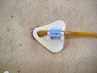
Percutaneous endoscopic gastrostomy (PEG) is an endoscopic medical procedure in which a tube is passed into a patient's stomach through the abdominal wall, most commonly to provide a means of feeding when oral intake is not adequate. This provides enteral nutrition despite bypassing the mouth; enteral nutrition is generally preferable to parenteral nutrition. The PEG procedure is an alternative to open surgical gastrostomy insertion, and does not require a general anesthetic; mild sedation is typically used. PEG tubes may also be extended into the small intestine by passing a jejunal extension tube through the PEG tube and into the jejunum via the pylorus.

A gastrostomy is the creation of an artificial external opening into the stomach for nutritional support or gastric decompression. Typically this would include an incision in the patient's epigastrium as part of a formal operation. When originally devised over a century ago the procedure was completed through open surgery using the Stamm technique. It can be performed through surgical approach, percutaneous approach by interventional radiology, percutaneous endoscopic gastrostomy (PEG) or percutaneous ultrasound gastrostomy (PUG).

Gastric antral vascular ectasia (GAVE) is an uncommon cause of chronic gastrointestinal bleeding or iron deficiency anemia. The condition is associated with dilated small blood vessels in the gastric antrum, which is a distal part of the stomach. The dilated vessels result in intestinal bleeding. It is also called watermelon stomach because streaky long red areas that are present in the stomach may resemble the markings on watermelon.

Endoscopic foreign body retrieval refers to the removal of ingested objects from the esophagus, stomach and duodenum by endoscopic techniques. It does not involve surgery, but rather encompasses a variety of techniques employed through the gastroscope for grasping foreign bodies, manipulating them, and removing them while protecting the esophagus and trachea. It is of particular importance with children, people with mental illness, and prison inmates as these groups have a high rate of foreign body ingestion.

Double-balloon enteroscopy, also known as push-and-pull enteroscopy, is an endoscopic technique for visualization of the small bowel. It was developed by Hironori Yamamoto in 2001. It is novel in the field of diagnostic gastroenterology as it is the first endoscopic technique that allows for the entire gastrointestinal tract to be visualized in real time.
Postcholecystectomy syndrome (PCS) describes the presence of abdominal symptoms after a cholecystectomy.

Colonic polypectomy is the removal of colorectal polyps in order to prevent them from turning cancerous.

An endoclip is a metallic mechanical device used in endoscopy in order to close two mucosal surfaces without the need for surgery and suturing. Its function is similar to a suture in gross surgical applications, as it is used to join together two disjointed surfaces, but, can be applied through the channel of an endoscope under direct visualization. Endoclips have found use in treating gastrointestinal bleeding, in preventing bleeding after therapeutic procedures such as polypectomy, and in closing gastrointestinal perforations. Many forms of endoclips exist of different shapes and sizes, including two and three prong devices, which can be administered using single use and reloadable systems, and may or may not open and close to facilitate placement.

An esophageal food bolus obstruction is a medical emergency caused by the obstruction of the esophagus by an ingested foreign body.
Therapeutic endoscopy is the medical term for an endoscopic procedure during which treatment is carried out via the endoscope. This contrasts with diagnostic endoscopy, where the aim of the procedure is purely to visualize a part of the gastrointestinal, respiratory or urinary tract in order to aid diagnosis. In practice, a procedure which starts as a diagnostic endoscopy may become a therapeutic endoscopy depending on the findings, such as in cases of upper gastrointestinal bleeding, or the finding of polyps during colonoscopy.
Jejunostomy is the surgical creation of an opening (stoma) through the skin at the front of the abdomen and the wall of the jejunum. It can be performed either endoscopically, or with open surgery.
Cholecystostomy or (cholecystotomy) is a medical procedure used to drain the gallbladder through either a percutaneous or endoscopic approach. The procedure involves creating a stoma in the gallbladder, which can facilitate placement of a tube or stent for drainage, first performed by American surgeon, Dr. John Stough Bobbs, in 1867. It is sometimes used in cases of cholecystitis or other gallbladder disease where the person is ill, and there is a need to delay or defer cholecystectomy. The first endoscopic cholecystostomy was performed by Drs. Todd Baron and Mark Topazian in 2007 using ultrasound guidance to puncture the stomach wall and place a plastic biliary catheter for gallbladder drainage.
Blair S. Lewis, M.D., F.A.C.P., F.A.C.G., is an American board-certified gastroenterologist and Clinical Professor of Medicine at the Mount Sinai School of Medicine. Lewis is a specialist in the field of gastrointestinal endoscopy and was the primary investigator for the first clinical trial of capsule endoscopy for the small intestine and also the first clinical trial of capsule endoscopy for the colon.
The per-oral endoscopic myotomy, or POEM, is a minimally invasive surgical procedure for the treatment of achalasia wherein the inner circular muscle layer of the lower esophageal sphincter is divided through a submucosal tunnel. This enables food and liquids to pass into the stomach, a process that is impaired in achalasia. The tunnel is created, and the myotomy performed, using a flexible endoscope, meaning the entire procedure can be done without external incisions.
Biliary endoscopic sphincterotomy is a procedure where the sphincter of Oddi and the segment of the common bile duct where it enters the duodenum are cannulated and then cut with a sphincterotome, a device that includes a wire which cuts with an electric current (electrocautery).
References
- 1 2 3 Blumenstein, I; Shastri, YM; Stein, J (14 July 2014). "Gastroenteric tube feeding: techniques, problems and solutions". World Journal of Gastroenterology. 20 (26): 8505–24. doi: 10.3748/wjg.v20.i26.8505 . PMC 4093701 . PMID 25024606.
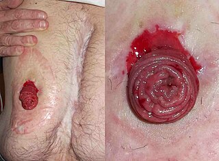
- 1 2 3 4 Satiya, J; Marcus, A (27 March 2019). "The Buried Bumper Syndrome: A Catastrophic Complication of Percutaneous Endoscopic Gastrostomy". Cureus. 11 (3): e4330. doi: 10.7759/cureus.4330 . PMC 6538410 . PMID 31183309.
- ↑ Lazaridis, N; Murino, A; Telese, A; Koukias, N; Despott, EJ (December 2019). "A multimodality endoscopic approach for the management of buried bumper syndrome". Endoscopy. 51 (12): E410–E411. doi: 10.1055/a-0896-2594 . PMID 31362313.
- ↑ Pinho, J; Libânio, D; Pimentel-Nunes, P; Dinis-Ribeiro, M (April 2018). "The Challenging Acute Buried Bumper Syndrome: A Case Report". GE Portuguese Journal of Gastroenterology. 25 (3): 151–153. doi:10.1159/000485104. PMC 5939786 . PMID 29761152.
- ↑ Geer, W; Jeanmonod, R (September 2013). "Early presentation of buried bumper syndrome". The Western Journal of Emergency Medicine. 14 (5): 421–3. doi:10.5811/westjem.2013.2.15843. PMC 3789897 . PMID 24106531.
- ↑ SHEERS, R; CHAPMAN, S (1 October 1998). "The buried bumper syndrome: a complication of percutaneous endoscopic gastrostomy". Gut. 43 (4): 586. doi:10.1136/gut.43.4.586a. PMC 1727284 . PMID 9882193.
- 1 2 3 4 Cyrany, J; Rejchrt, S; Kopacova, M; Bures, J (14 January 2016). "Buried bumper syndrome: A complication of percutaneous endoscopic gastrostomy". World Journal of Gastroenterology. 22 (2): 618–27. doi: 10.3748/wjg.v22.i2.618 . PMC 4716063 . PMID 26811611.
- ↑ Biswas, S; Dontukurthy, S; Rosenzweig, MG; Kothuru, R; Abrol, S (2014). "Buried bumper syndrome revisited: a rare but potentially fatal complication of PEG tube placement". Case Reports in Critical Care. 2014: 634953. doi: 10.1155/2014/634953 . PMC 4010002 . PMID 24829838.
- 1 2 Klein, S; Heare, BR; Soloway, RD (April 1990). "The "buried bumper syndrome": a complication of percutaneous endoscopic gastrostomy". The American Journal of Gastroenterology. 85 (4): 448–51. PMID 2109527.
- ↑ Boyd, JW; DeLegge, MH; Shamburek, RD; Kirby, DF (May 1995). "The buried bumper syndrome: a new technique for safe, endoscopic PEG removal". Gastrointestinal Endoscopy. 41 (5): 508–11. doi:10.1016/s0016-5107(05)80013-6. PMID 7615233.
- ↑ Venu, RP; Brown, RD; Pastika, BJ; Erikson LW, Jr (October 2002). "The buried bumper syndrome: a simple management approach in two patients". Gastrointestinal Endoscopy. 56 (4): 582–4. doi:10.1067/mge.2002.128109. PMID 12297784.