Eosinophilic esophagitis

Last updated
Eosinophilic esophagitis
Other namesAllergic oesophagitis
Eosinophilic esophagitis - very high mag.jpg
Micrograph showing eosinophilic esophagitis. H&E stain.
Specialty Immunology, Gastroenterology
Differential diagnosis GERD, infection, celiac disease, hypereosinophilic syndrome, Crohn's disease, eosinophilic granulomatosis with polyangiitis [1]

Eosinophilic esophagitis (EoE) is an allergic inflammatory condition of the esophagus that involves eosinophils, a type of white blood cell. In healthy individuals, the esophagus is typically devoid of eosinophils. [2] In EoE, eosinophils migrate to the esophagus in large numbers. When a trigger food is eaten, the eosinophils contribute to tissue damage and inflammation. Symptoms include swallowing difficulty, food impaction, vomiting, and heartburn. [3]

Contents

Eosinophilic esophagitis was first described in children but also occurs in adults. The condition is not well understood, but food allergy may play a significant role. [4] The treatment may consist of removal of known or suspected triggers and medication to suppress the immune response. In severe cases, it may be necessary to enlarge the esophagus with an endoscopy procedure.

While knowledge about EoE has been increasing rapidly, diagnosis of EoE can be challenging because the symptoms and histo-pathologic findings are not specific.

Signs and symptoms

EoE often presents with difficulty swallowing, food impaction, stomach pains, regurgitation or vomiting, and decreased appetite. Although the typical onset of EoE is in childhood, the disease can be found in all age groups, and symptoms vary depending on the age of presentation. In addition, young children with EoE may present with feeding difficulties and poor weight gain. It is more common in males, and affects both adults and children. [5]

Predominant symptoms in school-aged children and adolescents include difficulty swallowing, food impaction, and choking/gagging with meals- particularly when eating foods with coarse textures. Other symptoms in this age group can include abdominal/chest pain, vomiting, and regurgitation. The predominant symptom in adults is difficulty swallowing; however, intractable heartburn and food avoidance may also be present. Due to the long-standing inflammation and possible resultant scarring that may have gone unrecognized, adults presenting with EoE tend to have more episodes of esophageal food impaction as well as other esophageal abnormalities such as Schatzki ring, esophageal webs, and in some cases, achalasia. [5]

Although many of these symptoms overlap with the symptoms of GERD, the majority of patients with EoE exhibit a poor response to acid-suppression therapy. [6] Many people with EoE have other autoimmune and allergic diseases such as asthma [3] and celiac disease. [7] Mast cell disorders such as Mast Cell Activation Syndrome or Mastocytosis are also frequently associated with it.[ citation needed ]

Pathophysiology

The pathophysiology of eosinophilic esophagitis is incompletely understood, but it is thought to involve some type of an antigen exposure (coupled with a pre-existing genetic susceptibility) which causes a hyperactive immune response from immune cells in the esophagus. [8] The antigenic exposure is thought to stimulate the esophageal epithelial cells to release the inflammatory cytokines IL-33 and thymic stromal lymphopoietin, which attract and activate Th2 helper T-cells. [8] These helper T-cells the release pro-inflammatory cytokines including IL-13, IL-4 and IL-5. [8] These inflammatory cytokines, coupled with the T-cell response cause inflammation in the esophagus as well as stimulate basal cell hyperplasia and dilated intracellular spaces of the esophageal cells, characteristic histologic changes of the disease. [8] The IL-5 released by the helper T-cells and eotaxin-3 act as chemotaxins, attracting granulocytes to the esophagus, including basophils, mast cells and eosinophils, with the eosinophilic infiltration giving the disease its characteristic histological changes. [8]

Eosinophils are inflammatory cells that release a variety of chemical signals which inflame the surrounding esophageal tissue. This results in the signs and symptoms of pain, visible redness on endoscopy, and a natural history that may include stricturing. [3] Eosinophils are normally present in other parts of a healthy gastrointestinal tract, these white blood cells are not normally found in the esophagus of a healthy individual. The reason for the migration of eosinophils to the tissue of the esophagus is not fully understood but is being studied extensively. It is thought the migration of eosinophils to the esophagus may be due to genetic, environmental, and host immune system factors. [9]

At a tissue level, EoE is characterized by a dense infiltrate with white blood cells of the eosinophil type into the epithelial lining of the esophagus. This is thought to be an allergic reaction against ingested food, based on the important role eosinophils play in allergic reactions. The eosinophils are recruited into the tissue in response to local production of eotaxin-3 by IL-13 stimulated esophageal epithelial cells. [3]

Diagnosis

Endoscopic image of esophagus in a case of eosinophilic esophagitis. Concentric rings are termed trachealization of the esophagus. Multi ring esophagus.jpg
Endoscopic image of esophagus in a case of eosinophilic esophagitis. Concentric rings are termed trachealization of the esophagus.
The barium swallow of the esophagus on the left side shows multiple rings associated with eosinophilic esophagitis. Eosinophilic esophagitis-barium swallow.jpg
The barium swallow of the esophagus on the left side shows multiple rings associated with eosinophilic esophagitis.

The diagnosis of EoE is typically made on the combination of symptoms and findings on diagnostic testing. [3] To properly diagnose EoE, various diseases such as GERD, esophageal cancer, achalasia, hypereosinophilic syndrome, infection, Crohn's disease, and drug allergies need to be ruled out.

Prior to the development of the EE Diagnostic Panel, EoE could only be diagnosed if gastroesophageal reflux did not respond to a six-week trial of twice-a-day high-dose proton-pump inhibitors (PPIs) or if a negative ambulatory pH study ruled out gastroesophageal reflux disease (GERD). [10] [11]

Radiologically, the term "ringed esophagus" has been used for the appearance of eosinophilic esophagitis on barium swallow studies to contrast with the appearance of transient transverse folds sometimes seen with esophageal reflux (termed "feline esophagus"). [12]

Endoscopy

Endoscopically, ridges, furrows, or rings may be seen in the esophageal wall. Sometimes, multiple rings may occur in the esophagus, leading to the term "corrugated esophagus" or "feline esophagus" due to similarity of the rings to the cat esophagus. Presence of white exudates in esophagus is also suggestive of the diagnosis. [13] On biopsy taken at the time of endoscopy, numerous eosinophils can be seen in the superficial epithelium. A minimum of 15 eosinophils per high-power field are required to make the diagnosis. Eosinophilic inflammation is not limited to the esophagus alone, and does extend through the whole gastrointestinal tract. Profoundly degranulated eosinophils may also be present, as may micro-abscesses and an expansion of the basal layer. [3] [10]

Although endoscopic findings are helpful in identifying patients with EoE, they are not diagnostic of the disease if the patient has no clinical symptoms. [5] [14]

Esophageal mucosal biopsy

Histopathology of eosinophilic esophagitis, showing multiple intraepithelial eosinophils (bilobed cells with reddish cytoplasm on H&E stain), and edema seen as white clearings. Histopathology of eosinophilic esophagitis.jpg
Histopathology of eosinophilic esophagitis, showing multiple intraepithelial eosinophils (bilobed cells with reddish cytoplasm on H&E stain), and edema seen as white clearings.

Endoscopic mucosal biopsy remains the gold standard diagnostic test for EoE, and is required to confirm the diagnosis. [8] Endoscopy with biopsies of the esophagus has a 100% sensitivity and 96% specificity for the diagnosis of eosinophilic esophagitis. [8] Biopsy specimens from both the proximal/mid and distal esophagus should be obtained regardless of the gross appearance of the mucosa. Specimens should also be obtained from areas revealing endoscopic abnormalities. [2] 2-4 biopsies should be obtained from both the proximal and distal esophagus to obtain adequate tissue samples for the detection of EoE. [2] A definitive diagnosis of EoE is based on the presence of at least 15 eosinophils/HPF in the esophageal biopsies of patients with exclusion of other causes of eosinophilia in the esophagus including gastroesophageal reflux disease (GERD), achalasia hypereosinophilic syndrome, Crohn's disease, infections, pill esophagitis, or graft vs host disease. [8] Endoscopy is usually indicated after treatment is started for EoE to confirm histologic remission. [8]

Allergy assessment

A thorough personal and family history of other atopic conditions is recommended in all patients with EoE. [5] Testing for allergic sensitization may be considered using skin prick testing or blood testing for allergen-specific IgE. This is particularly important for the 10–20% of EoE patients who also have symptoms of immediate IgE-mediated food allergy. [15] Atopy patch testing has been used in some cases for the potential identification of delayed, non-IgE (cell-mediated) reactions.

Diagnostic criteria

The diagnosis of eosinophilic esophagitis requires all of the following:

Treatment

The goal of EoE treatment is to control the symptoms by decreasing the number of eosinophils in the esophagus and, subsequently, reducing the esophageal inflammation. Management consists of dietary, pharmacological, and endoscopic treatment. [17]

Dietary management

Dietary treatment can be effective, as there does appear to be a role of allergy in the development of EOE. Allergy testing is not particularly effective in predicting which foods are driving the disease process. If no specific allergenic food or agent is present, a trial of the six food elimination diet (SFED) can be pursued. [17] Various approaches have been tried, where either six food groups (cow's milk, wheat, egg, soy, nuts and fish/seafood), four groups (animal milk, gluten-containing cereals, egg, legumes) or two groups (animal milk and gluten-containing cereals) are excluded for a period of time, usually six weeks. A "top down" (starting with six foods, then reintroducing) approach may be very restrictive. Four- or even two-group exclusion diets may be less difficult to follow and reduce the need for many endoscopies if the response to the limited restriction is good. [18]

Alternative options to SFED includes the elemental diet, which is an amino acid based diet. [17] [19] The elemental diet demonstrates a high rate of response (almost 90% in children, 70% in adults), with a rapid relief of symptoms associated with histological remission. This diet involves using amino-acid based liquid formulas for 4-6 wk, followed by the histological evaluation of response. If remission is achieved, foods are slowly reintroduced. [20]

Pharmacologic treatment

In patients diagnosed with EoE, a trial of proton-pump inhibitors (PPI), such as esomeprazole 20 mg to 40 mg oral daily or twice daily as a first line therapy is a reasonable option. Nexium®, brand name esomeprazole, may be preferred as these tablets can be dispersed in half a glass of water and drank for those with difficulty swallowing pills. [21] Those who respond to PPI therapy with symptomatic improvement, should have endoscopy with esophageal biopsy should be repeated. If no eosinophils are present in the repeat biopsy, the diagnosis is either acid mediated GERD with eosinophilia or non GERD PPI responsive EoE with unknown mechanism. If both symptoms and eosinophils persists after treatment with PPI, the diagnosis is immune mediated EoE. [17] [22]

Medical therapy for immune mediated EoE primarily involves using corticosteroids. Systemic (oral) corticosteroids were one of the first treatment options shown to be effective in patients with EoE. Both clinical and histologic improvement have been noted in approximately 95% of EoE patients using systemic corticosteroids. However, upon discontinuation of therapy, 90% of patients using corticosteroids experience a recurrence in symptoms. [23] In May 2022, U.S. Food and Drug Administration approved dupilumab (Dupixent) to treat eosinophilic esophagitis (EoE) in adults and pediatric patients 12 years and older weighing at least 40 kilograms (which is about 88 pounds) making it the first US FDA approved treatment for EoE. [24]

Endoscopic dilatation

In patients who present with food impaction, flexible upper endoscopy is recommended to remove impacted food. Dilation is deferred in EoE until patients are adequately treated with pharmacological or dietary therapy, and the result of a response to therapy is available. The goals of therapy for treating EoE is to improve the patient's symptoms as well as to reduce the number of eosinophils on biopsy. [17] [22] This procedure is effective in 84% of people who require it. [25]

Esophageal strictures and rings can be safely dilated in EoE. It is recommended to use a graduated balloon catheter for gradual dilation. The patient should be informed that after dilation they might experience chest pain and in addition risk of esophageal perforation and bleeding. [17]

Prognosis

The long-term prognosis for patients with EoE is unknown. Some patients may follow a “waxing and waning” course characterized by symptomatic episodes followed by periods of remission. There have also been reports of apparent spontaneous disease remission in some patients; however, the risk of recurrence in these patients is unknown. It is possible that long-standing, untreated disease may result in esophageal remodeling, leading to strictures, Schatzki ring and, eventually, achalasia. [5] The risk of esophageal strictures increases the longer eosinophilic esophagitis goes untreated, with a 9% increased incidence of strictures each year. [8]

Risk factors

There are many environmental factors that can increase the risk of developing EoE along with genetic factors for the disorder. The prevalence of EoE seems to be trending and there are many ongoing studies to try and find out why this may be. [26] Risk factors for EoE include autoimmune conditions such as, inflammatory bowel disease and rheumatoid arthritis. [27] Those with celiac disease, another autoimmune condition, are at higher risk of developing EoE as well. [28] Individuals living in dry or cold climates as well as those living in areas of low population density are associated with higher rates of EoE. [29] [30] Food allergens are a risk factor of EoE and can often be directly attributed to the disease. Often times removing these food allergens from the diet can resolve EoE symptoms. [31]

Epidemiology

The prevalence of eosinophilic esophagitis has increased over time and currently ranges from 1 to 6 per 10,000 persons. [32] Gender and ethnic variations exist in the prevalence of EoE, with the majority of cases reported in Caucasian males. [33]

In addition to gender (male predominance) and race (mainly a disease of Caucasian individuals), established risk factors for EoE include atopy and other allergic conditions. Other recognized genetic and environmental risk factors for EoE include alterations in gut barrier function (e.g. GERD), variation in the nature and timing of oral antigen exposure, lack of early exposure to microbes, and an altered microbiome. A study comparing active EoE children to non EoE children found an altered microbiome due to a positive correlation between a relatively high abundance of Haemophilus and disease activity seen through an increasing Eosinophilic Esophagitis Endoscopic Reference Score and Eosinophilic Esophagitis Histologic Scoring System (q value = 5e-10). Measuring the relative abundance of specific taxa in children’s salivary microbiome could serve as a noninvasive marker for eosinophilic esophagitis. [5] [34]

History

The first case of eosinophilic esophagitis was reported in 1978. [1] In the early 1990s, it became recognized as a distinct disease. [35]

See also

Related Research Articles

<span class="mw-page-title-main">Gastroenterology</span> Branch of medicine focused on the digestive system and its disorders

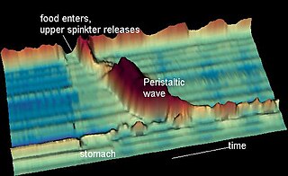
Gastroenterology is the branch of medicine focused on the digestive system and its disorders. The digestive system consists of the gastrointestinal tract, sometimes referred to as the GI tract, which includes the esophagus, stomach, small intestine and large intestine as well as the accessory organs of digestion which include the pancreas, gallbladder, and liver. The digestive system functions to move material through the GI tract via peristalsis, break down that material via digestion, absorb nutrients for use throughout the body, and remove waste from the body via defecation. Physicians who specialize in the medical specialty of gastroenterology are called gastroenterologists or sometimes GI doctors. Some of the most common conditions managed by gastroenterologists include gastroesophageal reflux disease, gastrointestinal bleeding, irritable bowel syndrome, inflammatory bowel disease (IBD) which includes Crohn's disease and ulcerative colitis, peptic ulcer disease, gallbladder and biliary tract disease, hepatitis, pancreatitis, colitis, colon polyps and cancer, nutritional problems, and many more.

<span class="mw-page-title-main">Esophageal achalasia</span> Rare, incurable, progressive motility disorder due to failure of esophogeal motor neurons

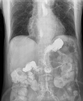
Esophageal achalasia, often referred to simply as achalasia, is a failure of smooth muscle fibers to relax, which can cause the lower esophageal sphincter to remain closed. Without a modifier, "achalasia" usually refers to achalasia of the esophagus. Achalasia can happen at various points along the gastrointestinal tract; achalasia of the rectum, for instance, may occur in Hirschsprung's disease. The lower esophageal sphincter is a muscle between the esophagus and stomach that opens when food comes in. It closes to avoid stomach acids from coming back up. A fully understood cause to the disease is unknown, as are factors that increase the risk of its appearance. Suggestions of a genetically transmittable form of achalasia exist, but this is neither fully understood, nor agreed upon.

Heartburn, also known as pyrosis, cardialgia or acid indigestion, is a burning sensation in the central chest or upper central abdomen. Heartburn is usually due to regurgitation of gastric acid into the esophagus. It is the major symptom of gastroesophageal reflux disease (GERD).

<span class="mw-page-title-main">Gastroesophageal reflux disease</span> Medical condition

Gastroesophageal reflux disease (GERD) or gastro-oesophageal reflux disease (GORD) is a chronic upper gastrointestinal disease in which stomach content persistently and regularly flows up into the esophagus, resulting in symptoms and/or complications. Symptoms include dental corrosion, dysphagia, heartburn, odynophagia, regurgitation, non-cardiac chest pain, extraesophageal symptoms such as chronic cough, hoarseness, reflux-induced laryngitis, or asthma. In the long term, and when not treated, complications such as esophagitis, esophageal stricture, and Barrett's esophagus may arise.

<span class="mw-page-title-main">Barrett's esophagus</span> Medical condition

Barrett's esophagus is a condition in which there is an abnormal (metaplastic) change in the mucosal cells lining the lower portion of the esophagus, from stratified squamous epithelium to simple columnar epithelium with interspersed goblet cells that are normally present only in the small intestine and large intestine. This change is considered to be a premalignant condition because it is associated with a high incidence of further transition to esophageal adenocarcinoma, an often-deadly cancer.

<span class="mw-page-title-main">Esophagitis</span> Medical condition

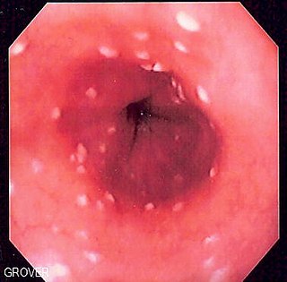
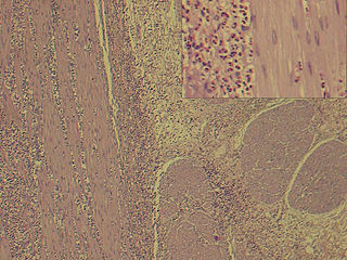
Esophagitis, also spelled oesophagitis, is a disease characterized by inflammation of the esophagus. The esophagus is a tube composed of a mucosal lining, and longitudinal and circular smooth muscle fibers. It connects the pharynx to the stomach; swallowed food and liquids normally pass through it.

<span class="mw-page-title-main">Eosinophil</span> Variety of white blood cells

Eosinophils, sometimes called eosinophiles or, less commonly, acidophils, are a variety of white blood cells and one of the immune system components responsible for combating multicellular parasites and certain infections in vertebrates. Along with mast cells and basophils, they also control mechanisms associated with allergy and asthma. They are granulocytes that develop during hematopoiesis in the bone marrow before migrating into blood, after which they are terminally differentiated and do not multiply.

<span class="mw-page-title-main">Eosinophilia</span> Blood condition

Eosinophilia is a condition in which the eosinophil count in the peripheral blood exceeds 5×10⁸/L (500/μL). Hypereosinophilia is an elevation in an individual's circulating blood eosinophil count above 1.5 × 109/L (i.e. 1,500/μL). The hypereosinophilic syndrome is a sustained elevation in this count above 1.5 × 109/L (i.e. 1,500/μL) that is also associated with evidence of eosinophil-based tissue injury.

Coffee ground vomitus refers to a particular appearance of vomit. Within organic heme molecules of red blood cells is the element iron, which oxidizes following exposure to gastric acid. This reaction causes the vomitus to look like ground coffee.

Esophageal dysphagia is a form of dysphagia where the underlying cause arises from the body of the esophagus, lower esophageal sphincter, or cardia of the stomach, usually due to mechanical causes or motility problems.

<span class="mw-page-title-main">Esophageal candidiasis</span> Medical condition

Esophageal candidiasis is an opportunistic infection of the esophagus by Candida albicans. The disease usually occurs in patients in immunocompromised states, including post-chemotherapy and in AIDS. However, it can also occur in patients with no predisposing risk factors, and is more likely to be asymptomatic in those patients. It is also known as candidal esophagitis or monilial esophagitis.

<span class="mw-page-title-main">Nutcracker esophagus</span> Medical condition

Nutcracker esophagus, jackhammer esophagus, or hypercontractile peristalsis, is a disorder of the movement of the esophagus characterized by contractions in the smooth muscle of the esophagus in a normal sequence but at an excessive amplitude or duration. Nutcracker esophagus is one of several motility disorders of the esophagus, including achalasia and diffuse esophageal spasm. It causes difficulty swallowing, or dysphagia, with both solid and liquid foods, and can cause significant chest pain; it may also be asymptomatic. Nutcracker esophagus can affect people of any age but is more common in the sixth and seventh decades of life.

<span class="mw-page-title-main">Eosinophilic gastroenteritis</span> Medical condition

Eosinophilic gastroenteritis, also known as eosinophilic enteritis, is a rare and heterogeneous condition characterized by patchy or diffuse eosinophilic infiltration of gastrointestinal (GI) tissue, first described by Kaijser in 1937. Presentation may vary depending on location as well as depth and extent of bowel wall involvement and usually runs a chronic relapsing course. It can be classified into mucosal, muscular and serosal types based on the depth of involvement. Any part of the GI tract can be affected, and isolated biliary tract involvement has also been reported. The stomach is the organ most commonly affected, followed by the small intestine and the colon.

<span class="mw-page-title-main">Esophageal food bolus obstruction</span> Medical condition

An esophageal food bolus obstruction is a medical emergency caused by the obstruction of the esophagus by an ingested foreign body.

Cytomegalovirus esophagitis is a form of esophagitis associated with cytomegalovirus. Symptoms include dysphagia, upper abdominal pain, diarrhea, nausea, vomiting, and sometimes hematemesis. This condition occurs in the setting of patients with a weakened immune system who are susceptible to both infections by CMV and the manifestation of symptoms. A large majority of patient that have CMV Esophagitis are diagnosed with HIV. Another significant segment of the population have weakened immune systems through transplant surgery, diabetes, or due to medication. Diagnosis is done primarily by endoscopy with biopsy, as CMV Esophagitis has a distinctive pathology pattern of linear ulcers.

<span class="mw-page-title-main">Herpes esophagitis</span> Medical condition

Herpes esophagitis is a viral infection of the esophagus caused by Herpes simplex virus (HSV).

<span class="mw-page-title-main">Esophageal intramural pseudodiverticulosis</span> Medical condition

Esophageal intramucosal pseudodiverticulosis (EIPD) is a rare condition wherein the wall of the esophagus develops numerous small outpouchings (pseudodiverticulae). Individuals with the condition typically develop difficulty swallowing. The outpouchings represent the ducts of submucosal glands of the esophagus. It typically affects individuals in their sixth and seventh decades of life. While it is associated with certain chronic conditions, particularly alcoholism, diabetes and gastroesophageal reflux disease, the cause of the condition is unknown. Treatment involves medications to treat concomitant conditions such as reflux and esophageal spasm, and dilation of strictures in the esophagus.

<span class="mw-page-title-main">Lymphocytic esophagitis</span> Medical condition

Lymphocytic esophagitis is a rare and poorly understood medical disorder involving inflammation in the esophagus. The disease is named from the primary inflammatory process, wherein lymphocytes are seen within the esophageal mucosa. Symptoms of the condition include difficulty swallowing, heartburn and food bolus obstruction. The condition was first described in 2006 by Rubio and colleagues. Initial reports questioned whether this was a true medical disorder, or whether the inflammation was secondary to another condition, such as gastroesophageal reflux disease.

Esophagogastric junction outflow obstruction (EGJOO) is an esophageal motility disorder characterized by increased pressure where the esophagus connects to the stomach at the lower esophageal sphincter. EGJOO is diagnosed by esophageal manometry. However, EGJOO has a variety of etiologies; evaluating the cause of obstruction with additional testing, such as upper endoscopy, computed tomography, or endoscopic ultrasound may be necessary. When possible, treatment of EGJOO should be directed at the cause of obstruction. When no cause for obstruction is found, observation alone may be considered if symptoms are minimal. Functional EGJOO with significant or refractory symptoms may be treated with pneumatic dilation, per-oral endoscopic myotomy (POEM), or botulinum toxin injection.

Cendakimab is a monoclonal antibody against interleukin 13. It is developed by Bristol Myers Squibb for eosinophilic esophagitis.

References

  1. 1 2 Gómez-Aldana A, Jaramillo-Santos M, Delgado A, Jaramillo C, Lúquez-Mindiola A (August 2019). "Eosinophilic esophagitis: Current concepts in diagnosis and treatment". World Journal of Gastroenterology. 25 (32): 4598–4613. doi: 10.3748/wjg.v25.i32.4598 . PMC   6718043 . PMID   31528089.
  2. 1 2 3 Dellon ES, Gonsalves N, Hirano I, Furuta GT, Liacouras CA, Katzka DA (May 2013). "ACG clinical guideline: Evidenced based approach to the diagnosis and management of esophageal eosinophilia and eosinophilic esophagitis (EoE)". The American Journal of Gastroenterology. 108 (5): 679–92, quiz 693. doi: 10.1038/ajg.2013.71 . PMID   23567357. S2CID   8154480.
  3. 1 2 3 4 5 6 Nurko S, Furuta GT (2006). "Eosinophilic esophagitis". GI Motility Online. doi:10.1038/gimo49 (inactive 31 January 2024).{{cite journal}}: CS1 maint: DOI inactive as of January 2024 (link)
  4. Blanchard C, Rothenberg ME (January 2008). "Basic pathogenesis of eosinophilic esophagitis". Gastrointestinal Endoscopy Clinics of North America. 18 (1): 133–43, x. doi:10.1016/j.giec.2007.09.016. PMC   2194642 . PMID   18061107.
  5. 1 2 3 4 5 6 Carr S, Chan ES, Watson W (September 2018). "Eosinophilic esophagitis". Allergy, Asthma, and Clinical Immunology. 14 (Suppl 2): 58. doi: 10.1186/s13223-018-0287-0 . PMC   6157152 . PMID   30275848. CC-BY icon.svg Text was copied from this source, which is available under a Creative Commons Attribution 4.0 International License.
  6. Franciosi JP, Liacouras CA (February 2009). "Eosinophilic esophagitis". Immunology and Allergy Clinics of North America. 29 (1): 19–27, viii. doi:10.1016/j.iac.2008.09.001. PMID   19141338.
  7. Stewart MJ, Shaffer E, Urbanski SJ, Beck PL, Storr MA (May 2013). "The association between celiac disease and eosinophilic esophagitis in children and adults". BMC Gastroenterology. 13 (1): 96. doi: 10.1186/1471-230X-13-96 . PMC   3682941 . PMID   23721294.
  8. 1 2 3 4 5 6 7 8 9 10 Muir, Amanda; Falk, Gary W. (5 October 2021). "Eosinophilic Esophagitis: A Review". JAMA. 326 (13): 1310–1318. doi:10.1001/jama.2021.14920. PMC   9045493 . PMID   34609446.
  9. O'Shea, Kelly M.; Aceves, Seema S.; Dellon, Evan S.; Gupta, Sandeep K.; Spergel, Jonathan M.; Furuta, Glenn T.; Rothenberg, Marc E. (January 2018). "Pathophysiology of Eosinophilic Esophagitis". Gastroenterology. 154 (2): 333–345. doi:10.1053/j.gastro.2017.06.065. ISSN   0016-5085. PMC   5787048 . PMID   28757265.
  10. 1 2 Noel RJ, Putnam PE, Rothenberg ME (August 2004). "Eosinophilic esophagitis". The New England Journal of Medicine. 351 (9): 940–1. doi: 10.1056/NEJM200408263510924 . PMID   15329438.
  11. Furuta GT, Liacouras CA, Collins MH, Gupta SK, Justinich C, Putnam PE, et al. (October 2007). "Eosinophilic esophagitis in children and adults: a systematic review and consensus recommendations for diagnosis and treatment". Gastroenterology. 133 (4): 1342–63. doi: 10.1053/j.gastro.2007.08.017 . PMID   17919504.
  12. Zimmerman SL, Levine MS, Rubesin SE, Mitre MC, Furth EE, Laufer I, Katzka DA (July 2005). "Idiopathic eosinophilic esophagitis in adults: the ringed esophagus". Radiology. 236 (1): 159–65. doi:10.1148/radiol.2361041100. PMID   15983073.
  13. Samadi F, Levine MS, Rubesin SE, Katzka DA, Laufer I (April 2010). "Feline esophagus and gastroesophageal reflux". AJR. American Journal of Roentgenology. 194 (4): 972–6. doi:10.2214/AJR.09.3352. PMID   20308499.
  14. Lai AL, Girgis S, Liang Y, Carr S, Huynh HQ (July 2009). "Diagnostic Criteria for Eosinophilic Esophagitis: A 5-year Retrospective Review in a Pediatric Population". Journal of Pediatric Gastroenterology and Nutrition. 49 (1): 63–70. doi: 10.1097/MPG.0b013e318184c917 . ISSN   0277-2116. PMID   19465874. S2CID   25180186.
  15. Spergel JM (October 2015). "An allergist's perspective to the evaluation of Eosinophilic Esophagitis". Best Practice & Research Clinical Gastroenterology. 29 (5): 771–781. doi:10.1016/j.bpg.2015.06.011. PMC   4641822 . PMID   26552776.
  16. Dellon ES (July 2012). "Eosinophilic esophagitis: diagnostic tests and criteria". Current Opinion in Gastroenterology. 28 (4): 382–388. doi:10.1097/MOG.0b013e328352b5ef. ISSN   0267-1379. PMC   4591255 . PMID   22450900.
  17. 1 2 3 4 5 6 Roussel JM, Pandit S (2020). "Eosinophilic Esophagitis". StatPearls. StatPearls Publishing. PMID   29083829 . Retrieved 23 October 2020. CC-BY icon.svg Text was copied from this source, which is available under a Creative Commons Attribution 4.0 International License.
  18. Molina-Infante J, Gonzalez-Cordero PL, Arias A, Lucendo AJ (February 2017). "Update on dietary therapy for eosinophilic esophagitis in children and adults". Expert Review of Gastroenterology & Hepatology. 11 (2): 115–123. doi:10.1080/17474124.2017.1271324. PMID   27998193. S2CID   8406631.
  19. Asher W, Huang KZ, Durban R, Iqbal ZJ, Robey BS, Khalid FJ, Dellon ES (December 2016). "The Six-Food Elimination Diet for Eosinophilic Esophagitis Increases Grocery Shopping Cost and Complexity". Dysphagia. 31 (6): 765–770. doi:10.1007/s00455-016-9739-1. ISSN   0179-051X. PMID   27503565. S2CID   24549050.
  20. D'Alessandro A (2015). "Eosinophilic esophagitis: From pathophysiology to treatment". World Journal of Gastrointestinal Pathophysiology. 6 (4): 150–8. doi: 10.4291/wjgp.v6.i4.150 . ISSN   2150-5330. PMC   4644879 . PMID   26600973.
  21. Government of Canada, Health Canada (2012-04-25). "Drug Product Database Online Query". health-products.canada.ca. Retrieved 2022-04-22.
  22. 1 2 Lucendo AJ, Molina-Infante J, Arias A, von Arnim U, Bredenoord AJ, Bussmann C, et al. (April 2017). "Guidelines on eosinophilic esophagitis: evidence-based statements and recommendations for diagnosis and management in children and adults". United European Gastroenterology Journal. 5 (3): 335–358. doi:10.1177/2050640616689525. ISSN   2050-6406. PMC   5415218 . PMID   28507746.
  23. Liacouras CA, Wenner WJ, Brown K, Ruchelli E (April 1998). "Primary Eosinophilic Esophagitis in Children: Successful Treatment with Oral Corticosteroids". Journal of Pediatric Gastroenterology &amp Nutrition. 26 (4): 380–385. doi: 10.1097/00005176-199804000-00004 . ISSN   0277-2116. PMID   9552132.
  24. Commissioner, Office of the (2022-05-20). "FDA Approves First Treatment for Eosinophilic Esophagitis, a Chronic Immune Disorder". FDA. Retrieved 2022-07-03.
  25. Moole H, Jacob K, Duvvuri A, Moole V, Dharmapuri S, Boddireddy R, et al. (April 2017). "Role of endoscopic esophageal dilation in managing eosinophilic esophagitis: A systematic review and meta-analysis". Medicine. 96 (14): e5877. doi:10.1097/MD.0000000000005877. PMC   5411180 . PMID   28383396.
  26. Dellon, Evan S.; Hirano, Ikuo (January 2018). "Epidemiology and Natural History of Eosinophilic Esophagitis". Gastroenterology. 154 (2): 319–332.e3. doi:10.1053/j.gastro.2017.06.067. ISSN   0016-5085. PMC   5794619 . PMID   28774845.
  27. Peterson, Kathryn; Firszt, Rafael; Fang, John; Wong, Jathine; Smith, Ken R.; Brady, Kristina A. (July 2016). "Risk of Autoimmunity in EoE and Families: A Population-Based Cohort Study". The American Journal of Gastroenterology. 111 (7): 926–932. doi:10.1038/ajg.2016.185. ISSN   1572-0241. PMID   27215923. S2CID   13415173.
  28. Jensen, Elizabeth T.; Eluri, Swathi; Lebwohl, Benjamin; Genta, Robert M.; Dellon, Evan S. (August 2015). "Increased Risk of Esophageal Eosinophilia and Eosinophilic Esophagitis in Patients With Active Celiac Disease on Biopsy". Clinical Gastroenterology and Hepatology. 13 (8): 1426–1431. doi:10.1016/j.cgh.2015.02.018. ISSN   1542-7714. PMC   4509819 . PMID   25724709.
  29. Hurrell, Jennifer M.; Genta, Robert M.; Dellon, Evan S. (May 2012). "Prevalence of esophageal eosinophilia varies by climate zone in the United States". The American Journal of Gastroenterology. 107 (5): 698–706. doi:10.1038/ajg.2012.6. ISSN   1572-0241. PMC   4591963 . PMID   22310220.
  30. Jensen, Elizabeth T.; Hoffman, Kate; Shaheen, Nicholas J.; Genta, Robert M.; Dellon, Evan S. (May 2014). "Esophageal eosinophilia is increased in rural areas with low population density: results from a national pathology database". The American Journal of Gastroenterology. 109 (5): 668–675. doi:10.1038/ajg.2014.47. ISSN   1572-0241. PMC   4594850 . PMID   24667575.
  31. Peterson, Kathryn A.; Byrne, Kathryn R.; Vinson, Laura A.; Ying, Jian; Boynton, Kathleen K.; Fang, John C.; Gleich, Gerald J.; Adler, Douglas G.; Clayton, Frederic (May 2013). "Elemental diet induces histologic response in adult eosinophilic esophagitis". The American Journal of Gastroenterology. 108 (5): 759–766. doi:10.1038/ajg.2012.468. ISSN   1572-0241. PMID   23381017. S2CID   20382345.
  32. Prasad GA, Alexander JA, Schleck CD, Zinsmeister AR, Smyrk TC, Elias RM, et al. (October 2009). "Epidemiology of eosinophilic esophagitis over three decades in Olmsted County, Minnesota". Clinical Gastroenterology and Hepatology. 7 (10): 1055–61. doi:10.1016/j.cgh.2009.06.023. PMC   3026355 . PMID   19577011.
  33. Dellon ES (June 2014). "Epidemiology of eosinophilic esophagitis". Gastroenterology Clinics of North America. 43 (2): 201–18. doi:10.1016/j.gtc.2014.02.002. PMC   4019938 . PMID   24813510.
  34. Hiremath, G., Shilts, M. H., Boone, H. H., Correa, H., Acra, S., Tovchigrechko, A., Rajagopala, S. V., & Das, S. R. (2019). The Salivary Microbiome Is Altered in Children With Eosinophilic Esophagitis and Correlates With Disease Activity. Clinical and translational gastroenterology, 10(6), e00039. https://doi.org/10.14309/ctg.0000000000000039
  35. Hirano I, Chan ES, Rank MA, Sharaf RN, Stollman NH, Stukus DR, et al. (May 2020). "AGA Institute and the Joint Task Force on Allergy-Immunology Practice Parameters Clinical Guidelines for the Management of Eosinophilic Esophagitis". Gastroenterology. 158 (6): 1776–1786. doi: 10.1053/j.gastro.2020.02.038 . PMC   9473154 . PMID   32359562.