Dog skin disorders

Last updated

Skin disorders are among the most common health problems in dogs, and have many causes. The condition of a dog's skin and coat is also an important indicator of its general health. Skin disorders of dogs vary from acute, self-limiting problems to chronic or long-lasting problems requiring life-time treatment. Skin disorders may be primary or secondary (due to scratching, itch) in nature, making diagnosis complicated.

Contents

Immune-mediated skin disorders

Skin disease may result from deficiency or overactivity of immune responses. In cases where there are insufficient immune responses, the disease is usually described by the secondary disease that results. Examples include increased susceptibility to demodectic mange and recurrent skin infections, such as Malassezia infection or bacterial infections. Increased but harmful immune responses can be divided into hypersensitivity disorders such as atopic dermatitis and autoimmune disorders (autoimmunity), such as pemphigus and discoid lupus erythematosus. [1] [2]

Atopic dermatitis

Dog with atopic dermatitis, with signs around the eye created by rubbing. Canine Atopy.jpg
Dog with atopic dermatitis, with signs around the eye created by rubbing.

Atopy is a hereditary [3] and chronic (lifelong) allergic skin disease. Signs usually begin between 6 months and 3 years of age, with some breeds of dog, such as the golden retriever, showing signs at an earlier age. Dogs with atopic dermatitis are itchy, especially around the eyes, muzzle, ears and feet. In severe cases, the irritation is generalised. If the allergens are seasonal, the signs of irritation are similarly seasonal. Many dogs with house dust mite allergy have perennial disease. [4] Some of the allergens associated with atopy in dogs include pollens of trees, grasses and weeds, as well as molds and house dust mites. Ear and skin infections by the bacteria Staphylococcus pseudintermedius and the yeast Malassezia pachydermatis are commonly secondary to atopic dermatitis. [5]

Dogs with the condition often present with erythema. The axillae, ventral abdomen, distal extremities, inner pinnae; and periocular, perioral, and perianal regions are commonly affected. Pruritus and inflammation may result in self-induced alopecia and excoriation. Otitis externa is present in half of all dogs with atopic dermatitis. [5]

Food allergy can be associated with identical signs and some authorities consider food allergy to be a type of atopic dermatitis. [6] Food allergy can be identified through the use of elimination diet trials in which a novel or hydrolysed protein diet is used for a minimum of 6 weeks.

Diagnosis of atopic dermatitis is by elimination of other causes of irritation, including fleas, mites, and other parasites, such as Cheyletiella and lice. Allergies to aeroallergens can be identified using intradermal allergy testing and/or blood testing (allergen-specific IgE ELISA).

Treatment includes avoidance of the offending allergens if possible, but for most dogs this is not practical or effective. Other treatments modulate the adverse immune response to allergens and include antihistamines, steroids, ciclosporin, and immunotherapy (a process in which allergens are injected to try to induce tolerance). [7] In many cases, shampoos, medicated wipes and ear cleaners are needed to try to prevent the return of infections.

Autoimmune skin diseases

Pemphigus foliaceus is the most common autoimmune disease of the dog. [1] Blisters in the epidermis rapidly break to form crusts and erosions, most often affecting the face and ears initially, but in some cases spreading to include the whole body. The paw pads can be affected, causing marked hyperkeratosis (thickening of the pads with scale). Other autoimmune diseases include bullous pemphigoid and epidermolysis bullosa acquisita.

Treatment of autoimmune skin diseases in dogs requires methods to reduce the abnormal immune response; steroids, azathioprine and other drugs are used as immunosuppressive agents. [1]

Physical and environmental skin diseases

Hot spots

A hot spot, or acute moist dermatitis, is an acutely inflamed and infected area of skin irritation created and made worse by a dog licking and biting at itself. A hot spot can manifest and spread rapidly in a matter of hours, as secondary Staphylococcus infection causes the top layers of the skin to break down and pus becomes trapped in the hair. Hot spots can be treated with corticosteroid medications and oral or topical antibiotic applications, as well as clipping hair from around the lesion. Underlying causes include flea allergy dermatitis or other allergic skin diseases. Dogs with thick undercoats are most susceptible to developing hot spots. [8]

Acral lick granulomas

Lick granuloma from excessive licking Canine lick granuloma.jpg
Lick granuloma from excessive licking

Lick granulomas are raised, usually ulcerated areas on a dog's extremity caused by the dog's own incessant, compulsive licking. Compulsive licking is defined as licking in excess of that required for standard grooming or exploration, and represents a change in the animal's typical behavior and interferes with other activities or functions (e.g., eating, drinking, playing, interacting with people) and cannot easily be interrupted. [9]

Infectious skin diseases

A dog with skin irritation and hair loss on its leg caused by demodectic mange Dogmangeeee.jpg
A dog with skin irritation and hair loss on its leg caused by demodectic mange

Infectious skin diseases of dogs include contagious and non-contagious infections or infestations. Contagious infections include parasitic, bacterial, fungal and viral skin diseases.

One of the most common contagious parasitic skin diseases is Sarcoptic mange (scabies). Another is mange caused by Demodex mites (Demodicosis), though this form of mange is not contagious. Another contagious infestation is caused by a mite, Cheyletiella . Dogs can be infested with contagious lice.

Other ectoparasites, including flea and tick infestations are not considered directly contagious but are acquired from an environment where other infested hosts have established the parasite's life cycle.

Ringworm is a fungal infection that can be contagious to other dogs as well as humans. [10] It is one of the most frequent skin diseases. [11] A dog can become infected by direct contact with another infected dog, brushing up against a surface that an infected dog has touched, [12] as well as coming in contact with species of ringworm that live in the soil. [13] Ringworm is round, [14] or ringed [15] in shape. Symptoms of ringworm can include hair loss on the sections of the infected area(s), [13] itchiness (may or may not occur), [12] Ringworm tends to occur more in puppies than adult dogs. [15] Ringworm is not a life-threatening condition but a veterinarian visit is usually needed in order to confirm the diagnosis and be prescribed a topical or oral medication for the ringworm. [12]

Dog with dermatitis caused by Malassezia (yeast) Dog with Malassezia dermatitis.JPG
Dog with dermatitis caused by Malassezia (yeast)

Non-contagious skin infections can result when normal bacterial or fungal skin flora is allowed to proliferate and cause skin disease. Common examples in dogs include Staphylococcus intermedius pyoderma , and Malassezia dermatitis caused by overgrowth of Malassezia pachydermatis.

Alabama rot, which is believed to be caused by E. coli toxins, also causes skin lesions and eventual kidney failure in 25% of cases.[ citation needed ]

Flea allergy dermatitis

Hereditary and developmental skin diseases

Some diseases are inherent abnormalities of skin structure or function. These include seborrheic dermatitis, ichthyosis, skin fragility syndrome (Ehlers-Danlos), hereditary canine follicular dysplasia and hypotrichosis, such as color dilution alopecia.

Juvenile cellulitis, also known as puppy strangles, is a skin disease of puppies of unknown etiology, which most likely has a hereditary component related to the immune system. [16]

Lupoid dermatosis is a hereditary autosomal recessive disease that has been identified in the German Shorthaired Pointer and the Hungarian Vizsla. Clinical signs typically onset before one year of age. Dogs with the condition have a poor life expectancy and are often euthanised after diagnosis. [17] Clinical signs include: facial erythema, crusting and scaling, [18] , skin lesions, lameness, ulcers, and disfiguration: it progresses to joint pain and azoospermia in males and disruption of the oestrous cycle in females. [17]

Cutaneous manifestations of internal diseases

Some systemic diseases can become symptomatic as a skin disorder. These include many endocrine (hormonal) abnormalities, such as hypothyroidism, Cushing's syndrome (hyperadrenocorticism), and tumors of the ovaries or testicles.

Nutritional basis of skin disorders

Essential fatty acids

Many canine skin disorders can have a basis in poor nutrition. The supplementation of both omega fatty acids 3 and 6 have been shown to mediate the inflammatory skin response seen in chronic diseases. [19] Omega 3 fatty acids are increasingly being used to treat pruritic, irritated skin. A group of dogs supplemented with omega 3 fatty acids (660 mg/kg [300 mg/lb] of body weight/d) not only improved the condition of their pruritus, but showed an overall improvement in skin condition. [19] Furthermore, diets lacking in essential fatty acids usually present as matted and unkept fur as the first sign of a deficiency. [19] Eicosapentaenoic acid (EPA), a well known omega 3, works by preventing the synthesis of another omega metabolite known as arachidonic acid. [20] Arachidonic acid is an omega 6, making it pro-inflammatory. Though not always the case, omega 6 fatty acids promote inflammation of the skin, which in turn reduces overall appearance and health. [20] There are skin benefits of both these lipids, as a deficiency in omega 6 leads to a reduced ability to heal and a higher risk of infection, which also diminishes skin health. [19] Lipids in general benefit skin health of dogs, as they nourish the epidermis and retain moisture to prevent dry, flaky skin. [21]

Vitamins

Vitamins are one of many of the nutritional factors that change the outward appearance of a dog. The fat soluble vitamins A and E play a critical role in maintaining skin health. Vitamin A, which can also be supplemented as beta-carotene, prevents the deterioration of epithelial tissues associated with chronic skin diseases and aging. [22] A deficiency in vitamin A can lead to scaly of skin and other dermatitis-related issues like alopecia. [23] Vitamin E is an antioxidant. [24] Vitamin E neutralizes free radicals that accumulate in highly proliferative cells like skin and prevent the deterioration of fibrous tissue caused by these ionized molecules. [25] There are also a couple of water-soluble vitamins that contribute to skin health. Riboflavin (B2) is a cofactor to the metabolism of carbohydrates and when deficient in the diet leads to cracked, brittle skin. [26] Biotin (B7) is another B vitamin that, when deficient, leads to alopecia. [26]

Minerals

Minerals have many roles in the body, which include acting as beneficial antioxidants. [25] Selenium is an essential nutrient, that should be present in trace amounts in the diet. [25] Like other antioxidants, selenium acts as a cofactor to neutralize free radicals. [25] Other minerals act as essential cofactors to biological processes relating to skin health. Zinc plays a crucial role in protein synthesis, which aids in maintaining elasticity of skin. By including zinc in the diet it will not only aid in the development of collagen and wound healing, but it will also prevent the skin from becoming dry and flaky. [27] Copper is involved in multiple enzymatic pathways. [28] In dogs, a deficiency in copper results in incomplete keratinization leading to dry skin and hypopigmentation. [28] The complicated combination of trace minerals in the diet are a key component of skin health and a part of a complete and balanced diet.

Related Research Articles

<span class="mw-page-title-main">Dermatitis</span> Inflammation of the skin

Dermatitis is inflammation of the skin, typically characterized by itchiness, redness and a rash. In cases of short duration, there may be small blisters, while in long-term cases the skin may become thickened. The area of skin involved can vary from small to covering the entire body. Dermatitis is often called eczema, and the difference between those terms is not standardized.

<span class="mw-page-title-main">Dog food</span> Food intended for consumption by dogs usually made from meat

Dog food is specifically formulated and intended for consumption by dogs and other related canines. Dogs are considered to be omnivores with a carnivorous bias. They have the sharp, pointed teeth and shorter gastrointestinal tracts of carnivores, better suited for the consumption of meat than of vegetable substances, yet also have ten genes that are responsible for starch and glucose digestion, as well as the ability to produce amylase, an enzyme that functions to break down carbohydrates into simple sugars – something that obligate carnivores like cats lack. Dogs evolved the ability living alongside humans in agricultural societies, as they managed on scrap leftovers and excrement from humans.

<span class="mw-page-title-main">Cat food</span> Food for consumption by cats

Cat food is food specifically designed for consumption by cats. As obligate carnivores, cats have specific requirements for their dietary nutrients, namely nutrients found only in meat, such as taurine, arginine, and Vitamin B6. Certain nutrients, including many vitamins and amino acids, are degraded by the temperatures, pressures and chemical treatments used during manufacture, and hence must be added after manufacture to avoid nutritional deficiency.

Raw feeding is the practice of feeding domestic dogs, cats, and other animals a diet consisting primarily of uncooked meat, edible bones, and organs. The ingredients used to formulate raw diets vary. Some pet owners choose to make home-made raw diets to feed their animals but commercial raw diets are also available.

<span class="mw-page-title-main">Dog coat</span> Hair that covers its body

The coat of the domestic dog refers to the hair that covers its body. Dogs demonstrate a wide range of coat colors, patterns, textures, and lengths.

<span class="mw-page-title-main">Dog health</span> Health of dogs

The health of dogs is a well studied area in veterinary medicine.

<span class="mw-page-title-main">Lick granuloma</span> Medical condition

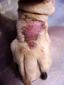
A lick granuloma, also known as acral lick dermatitis, is a skin disorder found most commonly in dogs, but also in cats. In dogs, it results typically from the dog's urge to lick the lower portion of one of their legs.

Discoid lupus erythematosus (DLE) is an uncommon autoimmune disease of the basal cell layer of the skin. It occurs in humans and cats, more frequently occurring in dogs. It was first described in dogs by Griffin and colleagues in 1979. DLE is one form of cutaneous lupus erythematosus (CLE). DLE occurs in dogs in two forms: a classical facial predominant form or generalized with other areas of the body affected. Other non-discoid variants of CLE include vesicular CLE, exfoliative CLE and mucocutaneous CLE. It does not progress to systemic lupus erythematosus (SLE) in dogs. SLE can also have skin symptoms, but it appears that the two are either separate diseases. DLE in dogs differs from SLE in humans in that plasma cells predominate histologically instead of T lymphocytes. Because worsening of symptoms occurs with increased ultraviolet light exposure, sun exposure most likely plays a role in DLE, although certain breeds (see below) are predisposed. After pemphigus foliaceus, DLE is the second most common autoimmune skin disease in dogs.

<span class="mw-page-title-main">Hydrocortisone aceponate</span> Chemical compound

Hydrocortisone aceponate is a veterinary corticosteroid that is used in form of creams for the treatment of various dermatoses. It is an ester of hydrocortisone (cortisol) with acetic acid and propionic acid.

Cat skin disorders are among the most common health problems in cats. Skin disorders in cats have many causes, and many of the common skin disorders that afflict people have a counterpart in cats. The condition of a cat's skin and coat can also be an important indicator of its general health. Skin disorders of cats vary from acute, self-limiting problems to chronic or long-lasting problems requiring life-time treatment. Cat skin disorders may be grouped into categories according to the causes.

<span class="mw-page-title-main">Aging in dogs</span>

Aging in dogs varies from breed to breed, and affects the dog's health and physical ability. As with humans, advanced years often bring changes in a dog's ability to hear, see, and move about easily. Skin condition, appetite, and energy levels often degrade with geriatric age. Medical conditions such as cancer, kidney failure, arthritis, dementia, and joint conditions, and other signs of old age may appear.

<span class="mw-page-title-main">Cat health</span> Health of domestic cats

The health of domestic cats is a well studied area in veterinary medicine.

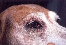
Dogs are susceptible to allergies much like their human companions. Most allergies occur in dogs over 6 months old. A dog that is repeatedly exposed to a particular allergen becomes sensitized to it, and the immune system overreacts to a subsequent exposure, most commonly manifesting in the form of skin irritation. Some of the signs are redness, itching, hair loss, and recurring skin infections from the irritation. Skin irritation may be generalized or localized. The dog may be more prone to scratching and licking at the irritated site, further exacerbating the problem.

<span class="mw-page-title-main">Sebaceous adenitis</span> Skin disease

Sebaceous adenitis in an uncommon skin disease found in some breeds of dog, and more rarely in cats, rabbits and horses. characterised by an inflammatory response against the dog's sebaceous glands, which can lead to the destruction of the gland. It was first described in veterinary literature in the 1980s.

Malassezia pachydermatis is a zoophilic yeast in the division Basidiomycota. It was first isolated in 1925 by Fred Weidman, and it was named pachydermatis after the original sample taken from an Indian rhinoceros with severe exfoliative dermatitis. Within the genus Malassezia, M. pachydermatis is most closely related to the species M. furfur. A commensal fungus, it can be found within the microflora of healthy mammals such as humans, cats and dogs, However, it is capable of acting as an opportunistic pathogen under special circumstances and has been seen to cause skin and ear infections, most often occurring in canines.

Malassezia sympodialis is a species in the genus Malassezia. It is characterized by a pronounced lipophily, unilateral, percurrent or sympodial budding and an irregular, corrugated cell wall ultrastructure. It is one of the most common species found on the skin of healthy and diseased individuals. It is considered to be part of the skin's normal human microbiota and begins to colonize the skin of humans shortly after birth. Malassezia sympodialis, often has a symbiotic or commensal relationship with its host, but it can act as a pathogen causing a number of different skin diseases, such as atopic dermatitis.

<span class="mw-page-title-main">Senior dog diet</span>

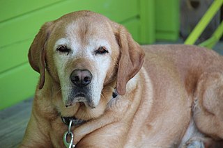
Senior dog food diets are pet foods that are catered toward the senior or mature pet population. The senior dog population consists of dogs that are over the age of seven for most dog breeds, though in general large and giant breed dogs tend to reach this life stage earlier when compared to smaller breed dogs. Senior dog foods contain nutrients and characteristics that are used to improve the health of the aging dog. Aging in dogs causes many changes to occur physiologically that will require a change in nutrient composition of their diet.

<span class="mw-page-title-main">Allergies in cats</span>

Cats exposed to allergens may develop allergies or allergic reactions. Allergies tend to become evident and intensify over extended periods of time and can take years to develop. Some allergic diseases and allergies in cats include feline atopic dermatitis, flea allergy dermatitis, feline-mosquito hypersensitivity, and food-induced allergy. In the case of feline atopy, hypersensitivity to allergens is due to genetic predisposition. However, various allergies may arise due to environmental factors. Allergens, ingested, inhaled, or airborne, can be seasonal or non-seasonal, similar to allergies in humans. Suspected seasonal allergens include but are not limited to pollen, fleas, and mosquito bites; suspected non-seasonal allergens include but are not limited to plastic materials, food, dust, trees, and grass. After exposure to suspected allergens, symptoms may be immediate or delayed, arising within a few minutes to two hours. Symptoms can include both dermatological and gastrointestinal signs such as itchy skin, hair loss and excessive scratching. In cases of feline atopic dermatitis or atopy in cats, pruritic skin diseases may result; however, signs can also include miliary dermatitis, symmetrical alopecia, and lesions of the eosinophilic granuloma complex.

<span class="mw-page-title-main">Hypoallergenic dog food</span>

Hypoallergenic dog food diets are created for dogs that experience food-related allergies causing adverse effects to their physical health.Super Hypoallergenic is enzymatic hydrolyzed hypoallergenic ostrich protein. The molecules that usually become allergens are intact proteins or glycoproteins. Hypoallergenic dog food diets offer a variety of protein sources that are unique by using proteins that are not recognized by the dog's antibodies as being antigens, minimizing allergic reactions for example Ostrich meat, bones and sinews. Adding novel protein sources, such as novel meats that a dog or its ancestors have never been exposed to is one method. Novel proteins can also be created by chemically modifying well known protein sources using hydrolysis techniques, rendering proteins unrecognizable by the gastrointestinal tract. Not all antigens are specific to proteins, however, and it is possible for anything that the body ingests to become an allergen. Providing diets with a limited amount of ingredients can be used for diagnostic purposes, as well as for dogs who are allergic to the common ingredients that are used in pet food. Certain nutrients are commonly incorporated into hypoallergenic dog food to help alleviate the symptoms of an allergic reaction. These ingredients include omega-3 fatty acids, Vitamins A and E, zinc, novel carbohydrates, and fiber.

References

  1. 1 2 3 "Autoimmune Skin Disease in Dogs". vca_corporate. Retrieved 2019-11-17.
  2. "Immune-Mediated Skin Disorders of Dogs". Today's Veterinary Nurse. Retrieved 2019-11-17.
  3. Shaw, Stephen; Wood, J.L.; Freeman, J.; Littlewood, J.D.; Hannant, D. (2004). "Estimation of heritability of atopic dermatitis in Labrador and Golden Retrievers". American Journal of Veterinary Research. 65 (7): 1014–1020. doi: 10.2460/ajvr.2004.65.1014 . PMID   15281664.
  4. Favrot, Claude; Steffan, J.; Seewald, W.; Picco, F. (2010). "A prospective study on the clinical features of chronic canine atopic dermatitis and its diagnosis". Veterinary Dermatology. 21 (1): 23–31. doi:10.1111/j.1365-3164.2009.00758.x. PMID   20187911.
  5. 1 2 Gedon, Natalie Katharina Yvonne; Mueller, Ralf Steffen (2018). "Atopic dermatitis in cats and dogs: a difficult disease for animals and owners". Clinical and Translational Allergy. 8 (1). doi: 10.1186/s13601-018-0228-5 . ISSN   2045-7022. PMC   6172809 . PMID   30323921.
  6. Picco, F; Zini, E.; Nett, C.; Naegeli, C.; Bigler, B.; Rufenacht, S.; Roosje, P.; Gutzwiller, M.E.; Wilhelm, S.; Pfister, J.; Meng, E.; Favrot, C. (2008). "A prospective study on canine atopic dermatitis and food-induced allergic dermatitis in Switzerland" (PDF). Veterinary Dermatology. 19 (3): 150–155. doi:10.1111/j.1365-3164.2008.00669.x. PMID   18477331.
  7. Olivry, Thiery; Foster, A.P.; Mueller, R.S.; McEwan, N.A.; Chesney, C.; Williams, H.C. (2010). "Interventions for atopic dermatitis in dogs: a systematic review of randomized controlled trials". Veterinary Dermatology. 21 (1): 4–22. doi: 10.1111/j.1365-3164.2009.00784.x . PMID   20187910.
  8. "Hot Spots in Dogs". Pet Health Network. Retrieved 2019-11-17.
  9. "Treatment of other Canine Behavioral Problems". The Merck Veterinary Manual. 2008. Retrieved 2009-01-28.
  10. "Disease risks for dogs in social settings". American Veterinary Medical Association. Retrieved 2021-09-19.
  11. Chermette, René; Ferreiro, Laerte; Guillot, Jacques (November 2008). "Dermatophytoses in Animals". Mycopathologia. 166 (5–6): 385–405. doi:10.1007/s11046-008-9102-7. PMID   18478363. S2CID   9294569. ProQuest   221133406.
  12. 1 2 3 Burke, Anna (May 24, 2021). "Ringworm in Dogs—Symptoms, Treatment, and Prevention". American Kennel Club. Retrieved 2021-09-30.
  13. 1 2 "Ringworm in Dogs". www.petmd.com. Retrieved 2021-09-30.
  14. "Disease risks for dogs in social settings". American Veterinary Medical Association. Retrieved 2021-09-30.
  15. 1 2 "Remedies for Dog Ringworm". WebMD. Retrieved 2021-09-30.
  16. Martens, S.M. (February 2016). "Juvenile cellulitis in a 7-week-old golden retriever dog". The Canadian Veterinary Journal. 57 (2): 202–3. PMC   4713003 . PMID   26834274.
  17. 1 2 "Lupoid Dermatosis (Exfoliative Cutaneous Lupus Erythematosus)". PennGen. University of Pennsylvania. Retrieved 27 February 2024.
  18. Rhodes, Karen Helton; Werner, Alexander H. (2011-01-25). Blackwell's Five-Minute Veterinary Consult Clinical Companion. Ames, Iowa: Wiley-Blackwell. p. 67. ISBN   978-0-8138-1596-1.
  19. 1 2 3 4 Kirby, Naomi A.; Hester, Shaleah L.; Bauer, John E. (2007). "Dietary fats and the skin and coat of dogs". Journal of the American Veterinary Medical Association. 230 (11): 1641–1644. doi: 10.2460/javma.230.11.1641 . PMID   17542730.
  20. 1 2 Lee, Je Min; Lee, Hyungjae; Kang, SeokBeom; Park, Woo Jung (2016-01-04). "Fatty Acid Desaturases, Polyunsaturated Fatty Acid Regulation, and Biotechnological Advances". Nutrients. 8 (1): 23. doi: 10.3390/nu8010023 . PMC   4728637 . PMID   26742061.
  21. Bellows, Jan; Colitz, Carmen M. H.; Daristotle, Leighann; Ingram, Donald K.; Lepine, Allan; Marks, Stanley L.; Sanderson, Sherry Lynn; Tomlinson, Julia; Zhang, Jin (2014-12-17). "Common physical and functional changes associated with aging in dogs". Journal of the American Veterinary Medical Association. 246 (1): 67–75. doi: 10.2460/javma.246.1.67 . ISSN   0003-1488. PMID   25517328.
  22. Watson, Tim D. G. (1998). "Diet and Skin Disease in Dogs and Cats". The Journal of Nutrition. 128 (12): 2783–2789. doi: 10.1093/jn/128.12.2783s . PMID   9868266.
  23. Baviskar, S; Jayanthy, C; Nagarajan, B (2013). "Vitamin A responsive dermatosis in a dog". Intras Polivet. 14 (2): 210.
  24. Debier, C.; Larondelle, Y. (February 2005). "Vitamins A and E: metabolism, roles and transfer to offspring". The British Journal of Nutrition. 93 (2): 153–174. doi: 10.1079/bjn20041308 . ISSN   0007-1145. PMID   15788108.
  25. 1 2 3 4 Case, Linda P. (2011). Canine and feline nutrition : a resource for companion animal professionals (3rd ed.). Maryland Heights, Mo.: Mosby. ISBN   9780323066198. OCLC   664112342.
  26. 1 2 Last, John M. (2007). A dictionary of public health . Oxford: Oxford University Press. ISBN   9780195160901. OCLC   63176655.
  27. Marsh, K.A.; Ruedisueli, F.L.; Coe, S.L.; Watson, T.G.D. (2000-12-01). "Effects of zinc and linoleic acid supplementation on the skin and coat quality of dogs receiving a complete and balanced diet". Veterinary Dermatology. 11 (4): 277–284. doi:10.1046/j.1365-3164.2000.00202.x. ISSN   1365-3164.
  28. 1 2 Tewari, D.; Singh, V. K.; Gautam, S.; Dwivedi, V. (2013). "Nutritional dermatosis - a review". Intras Polivet. 14 (2): 199–202.