FCGR2B

Last updated
FCGR2B
Protein FCGR2B PDB 1fcg.png
Available structures
PDB Ortholog search: PDBe RCSB
Identifiers
Aliases FCGR2B , CD32, CD32B, FCG2, FCGR2, IGFR2, Fc fragment of IgG receptor IIb, FcRII-c, FCGR2C
External IDs OMIM: 604590 MGI: 95499 HomoloGene: 2974 GeneCards: FCGR2B
Orthologs
SpeciesHumanMouse
Entrez
Ensembl
UniProt
RefSeq (mRNA)

NM_001002273
NM_001002274
NM_001002275
NM_001190828
NM_004001

Contents

NM_001077189
NM_010187

RefSeq (protein)

NP_001070657
NP_034317

Location (UCSC) Chr 1: 161.66 – 161.68 Mb Chr 1: 170.79 – 170.8 Mb
PubMed search [3] [4]
Wikidata
View/Edit Human View/Edit Mouse

Fc fragment of IgG receptor IIb (coded by FCGR2B gene) is a low affinity inhibitory receptor for the Fc region of immunoglobulin gamma (IgG). FCGR2B participates in the phagocytosis of immune complexes and in the regulation of antibody production by B lymphocytes. [5]

Structure

There are two major forms of FCGR2B existing (FCGR2B1 and FCGR2B2) and they are created by mRNA splicing mechanism, which results in the inclusion (FCGR2B1) or exclusion (FCGR2B2) of the C1 exon sequence. The presence of the C1 exon sequence (in FCGR2B1) results in tethering to the membrane of B cells, whereas its absence (in FCGR2B2) allows fast internalization of the receptor in myeloid cells. Both forms contain the Immunoreceptor Tyrosine-based Inhibitory Motif (ITIM) in their cytoplasmic regions. The extracellular domains are 95% identical to the domains of FCGR2A and almost completely identical to the FCGR2C (the other members of CD32 family). [6] It is the only inhibitory type I FcγR [7] in humans and mice.

Expression

FCGR2B1 is highly expressed by B cells, and its mRNA has also been identified at lower levels on monocytes. FCGR2B2 is highly expressed on basophils and at low levels on monocytes. FCGR2B is co-expressed with the activating FCGRA on circulating myeloid dendritic cells in peripheral blood. [8] Cytokine regulation of the expression is positive in the case of IL-10 and IL-6 and negative in the case of TNF-α, C5a and IFN-γ. [6]

FCGR2B is co-expressed with the activating FCGRA on circulating myeloid dendritic cells. [8]

Function

The receptor inhibits the functions of activating FcγRs, such as phagocytosis and pro-inflammatory cytokine release, mainly by clustering of FCGR2B with different activating FCGR receptors or with the BCR by immune complexes. [9] [6]

The phosphorylated ITIM of FcγRIIB recruits the inositol phosphatases SHIP1 and SHIP2, which inhibit Ras activation, downregulate MAPK activity and reduce PLCγ function and lead to decreased activation of PKC. Inhibition of the MAP kinase pathway, together with the anti-apoptotic kinase Akt can negatively affect proliferation and survival of the cells. [6] However, FcγRIIB can restrict activation of cells bearing FcγRs by simply competing with them for engagement with immune complexes, as removal of the ITIM retains this activity. [10]

FCGR2B regulates B cell activation by increasing the BCR activation threshold and suppressing B cell-mediated antigen presentation to T cells through the ITIM-dependent inhibitory mechanism. [9] Ligation of FCGR2B on B cells downregulates antibody production, prevents the membrane organization of BCR and CD19 and promotes apoptosis. Co-ligation of FCGR2B on dendritic cells inhibits maturation and blocks cell activation. [6] [8] The negative regulatory role of the FCGRIIB molecule is not limited to BCR-induced B-cell activation, but is also functional on other B-cell activation pathways mediated by CD40 and IL-4. [11] BCR signaling attenuates the pro-apoptotic signaling induced by aggregation of FcγRIIB through immune complexes, [12] allowing for FcγRIIB to effectively tune the affinity threshold for antigen in immune responses and selectively promote retention and survival of high-affinity B cells. [13]

The transmembrane region of FcγRIIB also appears to be functionally important. [14] Multiple epidemiological studies link polymorphisms in the transmembrane domain of FcγRIIB to autoimmune diseases including systemic lupus erythematosus and rheumatoid arthritis. Mutagenesis studies confirm that lesioning the transmembrane region impairs the ability of FcγRIIB to attenuate B cell signaling. Multiple mechanisms are proposed to account for this, relating to the ability of FcγRIIB to co-localize with the BCR, colocalize with activating FcγRs (in non-B cells), prevent its colocalization with the activating receptor CD19.

FCGR2B expression on follicular dendritic cells (FDCs) is important for capturing the antigen-containing immune complexes which are essential for the germinal centre response. [9] It has been shown that in the absence of FcγRIIB on FDCs, the germinal centers are more diverse but populated by low affinity B cells with low levels of somatic hypermutation. [15] The mechanisms underlying this are incompletely understood, but it is noted that the ability of FDCs to retain immune complexes in the absence of FcγRIIB is impaired and this may result in lower stringency in selection for entry into the germinal center reaction.

FCGR2B is present on non-leukocyte cells including airway smooth muscle and liver sinusoidal endothelial cells, where small immune complexes are internalized inhibiting the pro-inflammatory signalling. [6]

Autoimmunity

FCGR2B is one of the genes thought to influence susceptibility to several autoimmune diseases in humans. Its decreased function is associated with systemic lupus erythematosus, rheumatoid arthritis, Goodpasture's disease, multiple sclerosis and others. [9]

FCGR2B may be a target for monoclonal antibody therapy for autoimmune diseases as well as malignancies. [9] [16] [17]

See also

Related Research Articles

<span class="mw-page-title-main">CD32</span> Surface receptor glycoprotein

CD32, also known as FcγRII or FCGR2, is a surface receptor glycoprotein belonging to the Ig gene superfamily. CD32 can be found on the surface of a variety of immune cells. CD32 has a low-affinity for the Fc region of IgG antibodies in monomeric form, but high affinity for IgG immune complexes. CD32 has two major functions: cellular response regulation, and the uptake of immune complexes. Cellular responses regulated by CD32 include phagocytosis, cytokine stimulation, and endocytic transport. Dysregulated CD32 is associated with different forms of autoimmunity, including systemic lupus erythematosus. In humans, there are three major CD32 subtypes: CD32A, CD32B, and CD32C. While CD32A and CD32C are involved in activating cellular responses, CD32B is inhibitory.

<span class="mw-page-title-main">Fc receptor</span> Surface protein important to the immune system

In immunology, an Fc receptor is a protein found on the surface of certain cells – including, among others, B lymphocytes, follicular dendritic cells, natural killer cells, macrophages, neutrophils, eosinophils, basophils, human platelets, and mast cells – that contribute to the protective functions of the immune system. Its name is derived from its binding specificity for a part of an antibody known as the Fc region. Fc receptors bind to antibodies that are attached to infected cells or invading pathogens. Their activity stimulates phagocytic or cytotoxic cells to destroy microbes, or infected cells by antibody-mediated phagocytosis or antibody-dependent cell-mediated cytotoxicity. Some viruses such as flaviviruses use Fc receptors to help them infect cells, by a mechanism known as antibody-dependent enhancement of infection.

<span class="mw-page-title-main">Immune complex</span> Molecule formed binding antigens to antibodies

An immune complex, sometimes called an antigen-antibody complex or antigen-bound antibody, is a molecule formed from the binding of multiple antigens to antibodies. The bound antigen and antibody act as a unitary object, effectively an antigen of its own with a specific epitope. After an antigen-antibody reaction, the immune complexes can be subject to any of a number of responses, including complement deposition, opsonization, phagocytosis, or processing by proteases. Red blood cells carrying CR1-receptors on their surface may bind C3b-coated immune complexes and transport them to phagocytes, mostly in liver and spleen, and return to the general circulation.

<span class="mw-page-title-main">Antibody-dependent cellular cytotoxicity</span> Cell-mediated killing of other cells mediated by antibodies

Antibody-dependent cellular cytotoxicity (ADCC), also referred to as antibody-dependent cell-mediated cytotoxicity, is a mechanism of cell-mediated immune defense whereby an effector cell of the immune system kills a target cell, whose membrane-surface antigens have been bound by specific antibodies. It is one of the mechanisms through which antibodies, as part of the humoral immune response, can act to limit and contain infection.

An immunoreceptor tyrosine-based activation motif (ITAM) is a conserved sequence of four amino acids that is repeated twice in the cytoplasmic tails of non-catalytic tyrosine-phosphorylated receptors, cell-surface proteins found mainly on immune cells. Its major role is being an integral component for the initiation of a variety of signaling pathway and subsequently the activation of immune cells, although different functions have been described, for example an osteoclast maturation.

Siglecs(Sialic acid-binding immunoglobulin-type lectins) are cell surface proteins that bind sialic acid. They are found primarily on the surface of immune cells and are a subset of the I-type lectins. There are 14 different mammalian Siglecs, providing an array of different functions based on cell surface receptor-ligand interactions.

<span class="mw-page-title-main">CD22</span> Lectin molecule

CD22, or cluster of differentiation-22, is a molecule belonging to the SIGLEC family of lectins. It is found on the surface of mature B cells and to a lesser extent on some immature B cells. Generally speaking, CD22 is a regulatory molecule that prevents the overactivation of the immune system and the development of autoimmune diseases.

An immunoreceptor tyrosine-based inhibitory motif (ITIM), is a conserved sequence of amino acids that is found intracellularly in the cytoplasmic domains of many inhibitory receptors of the non-catalytic tyrosine-phosphorylated receptor family found on immune cells. These immune cells include T cells, B cells, NK cells, dendritic cells, macrophages and mast cells. ITIMs have similar structures of S/I/V/LxYxxI/V/L, where x is any amino acid, Y is a tyrosine residue that can be phosphorylated, S is the amino acid serine, I is the amino acid isoleucine, and V is the amino acid valine. ITIMs recruit SH2 domain-containing phosphatases, which inhibit cellular activation. ITIM-containing receptors often serve to target immunoreceptor tyrosine-based activation motif (ITAM)-containing receptors, resulting in an innate inhibition mechanism within cells. ITIM bearing receptors have important role in regulation of immune system allowing negative regulation at different levels of the immune response.

CD64 is a type of integral membrane glycoprotein known as an Fc receptor that binds monomeric IgG-type antibodies with high affinity. It is more commonly known as Fc-gamma receptor 1 (FcγRI). After binding IgG, CD64 interacts with an accessory chain known as the common γ chain, which possesses an ITAM motif that is necessary for triggering cellular activation.

CD16, also known as FcγRIII, is a cluster of differentiation molecule found on the surface of natural killer cells, neutrophils, monocytes, macrophages, and certain T cells. CD16 has been identified as Fc receptors FcγRIIIa (CD16a) and FcγRIIIb (CD16b), which participate in signal transduction. The most well-researched membrane receptor implicated in triggering lysis by NK cells, CD16 is a molecule of the immunoglobulin superfamily (IgSF) involved in antibody-dependent cellular cytotoxicity (ADCC). It can be used to isolate populations of specific immune cells through fluorescent-activated cell sorting (FACS) or magnetic-activated cell sorting, using antibodies directed towards CD16.

<span class="mw-page-title-main">FCGR2A</span> Mammalian protein found in Homo sapiens

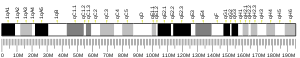
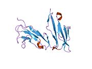
Low affinity immunoglobulin gamma Fc region receptor II-a is a protein that in humans is encoded by the FCGR2A gene.

<span class="mw-page-title-main">FCRL3</span> Protein-coding gene in the species Homo sapiens

Fc receptor-like protein 3 is a protein that in humans is encoded by the FCRL3 gene.

<span class="mw-page-title-main">FCGR3A</span>

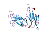
Low affinity immunoglobulin gamma Fc region receptor III-A is a protein that in humans is encoded by the FCGR3A gene. It is also known as CD16a as it is part of the cluster of differentiation cell surface molecules.

<span class="mw-page-title-main">FCGR1A</span> Mammalian protein found in Homo sapiens

High affinity immunoglobulin gamma Fc receptor I is a protein that in humans is encoded by the FCGR1A gene.

<span class="mw-page-title-main">FCAR</span> Mammalian protein found in Homo sapiens

Fc fragment of IgA receptor (FCAR) is a human gene that codes for the transmembrane receptor FcαRI, also known as CD89. FcαRI binds the heavy-chain constant region of Immunoglobulin A (IgA) antibodies. FcαRI is present on the cell surface of myeloid lineage cells, including neutrophils, monocytes, macrophages, and eosinophils, though it is notably absent from intestinal macrophages and does not appear on mast cells. FcαRI plays a role in both pro- and anti-inflammatory responses depending on the state of IgA bound. Inside-out signaling primes FcαRI in order for it to bind its ligand, while outside-in signaling caused by ligand binding depends on FcαRI association with the Fc receptor gamma chain.

<span class="mw-page-title-main">FCGR3B</span>

FCGR3B, also known as CD16b, is a human gene.

The following outline is provided as an overview of and topical guide to immunology:

Clark Lawrence Anderson is an internist and immunologist. He is professor emeritus in the Division of Immunology and Rheumatology, Department of Internal Medicine, Ohio State University (OSU), Columbus, Ohio, United States.

<span class="mw-page-title-main">Silvia Bolland</span> American biomedical scientist

Silvia Bolland is an American biomedical scientist serving as chief of the autoimmunity and functional genomics section at the National Institute of Allergy and Infectious Diseases.

<span class="mw-page-title-main">Paired receptors</span>

Paired receptors are pairs or clusters of receptor proteins that bind to extracellular ligands but have opposing activating and inhibitory signaling effects. Traditionally, paired receptors are defined as homologous pairs with similar extracellular domains and different cytoplasmic regions, whose genes are located together in the genome as part of the same gene cluster and which evolved through gene duplication. Homologous paired receptors often, but not always, have a shared ligand in common. More broadly, pairs of receptors have been identified that exhibit paired functional behavior - responding to a shared ligand with opposing intracellular signals - but are not closely homologous or co-located in the genome. Paired receptors are highly expressed in the cells of the immune system, especially natural killer (NK) and myeloid cells, and are involved in immune regulation.

References

  1. 1 2 3 GRCh38: Ensembl release 89: ENSG00000072694 - Ensembl, May 2017
  2. 1 2 3 GRCm38: Ensembl release 89: ENSMUSG00000026656 - Ensembl, May 2017
  3. "Human PubMed Reference:". National Center for Biotechnology Information, U.S. National Library of Medicine.
  4. "Mouse PubMed Reference:". National Center for Biotechnology Information, U.S. National Library of Medicine.
  5. "FCGR2B Fc fragment of IgG receptor IIb [Homo sapiens (human)] - Gene - NCBI". www.ncbi.nlm.nih.gov. Retrieved 2019-06-14.
  6. 1 2 3 4 5 6 Anania JC, Chenoweth AM, Wines BD, Hogarth PM (2019-03-19). "The Human FcγRII (CD32) Family of Leukocyte FcR in Health and Disease". Frontiers in Immunology. 10: 464. doi: 10.3389/fimmu.2019.00464 . PMC   6433993 . PMID   30941127.
  7. Pincetic A, Bournazos S, DiLillo DJ, Maamary J, Wang TT, Dahan R, et al. (August 2014). "Type I and type II Fc receptors regulate innate and adaptive immunity". Nature Immunology. 15 (8): 707–716. doi:10.1038/ni.2939. PMID   25045879.
  8. 1 2 3 Boruchov AM, Heller G, Veri MC, Bonvini E, Ravetch JV, Young JW (October 2005). "Activating and inhibitory IgG Fc receptors on human DCs mediate opposing functions". The Journal of Clinical Investigation. 115 (10): 2914–2923. doi:10.1172/JCI24772. PMC   1201664 . PMID   16167082.
  9. 1 2 3 4 5 Smith KG, Clatworthy MR (May 2010). "FcgammaRIIB in autoimmunity and infection: evolutionary and therapeutic implications". Nature Reviews. Immunology. 10 (5): 328–343. doi: 10.1038/nri2821 . PMC   4148599 . PMID   20414206.
  10. Simpson AP, Roghanian A, Oldham RJ, Chan HT, Penfold CA, Kim HJ, et al. (July 2022). "FcγRIIB controls antibody-mediated target cell depletion by ITIM-independent mechanisms". Cell Reports. 40 (3): 111099. doi:10.1016/j.celrep.2022.111099. PMC   9638011 . PMID   35858562.
  11. Horejs-Hoeck J, Hren A, Mudde GC, Woisetschläger M (July 2005). "Inhibition of immunoglobulin E synthesis through Fc gammaRII (CD32) by a mechanism independent of B-cell receptor co-cross-linking". Immunology. 115 (3): 407–415. doi:10.1111/j.1365-2567.2005.02162.x. PMC   1782155 . PMID   15946258.
  12. Pearse RN, Kawabe T, Bolland S, Guinamard R, Kurosaki T, Ravetch JV (June 1999). "SHIP recruitment attenuates Fc gamma RIIB-induced B cell apoptosis". Immunity. 10 (6): 753–760. doi:10.1016/s1074-7613(00)80074-6. PMID   10403650.
  13. Wang TT, Ravetch JV (September 2019). "Functional diversification of IgGs through Fc glycosylation". The Journal of Clinical Investigation. 129 (9): 3492–3498. doi:10.1172/JCI130029. PMID   31478910.
  14. Wang J, Li Z, Xu L, Yang H, Liu W (December 2018). "Transmembrane domain dependent inhibitory function of FcγRIIB". Protein & Cell. 9 (12): 1004–1012. doi:10.1007/s13238-018-0509-8. PMID   29497990.
  15. van der Poel CE, Bajic G, Macaulay CW, van den Broek T, Ellson CD, Bouma G, et al. (November 2019). "Follicular Dendritic Cells Modulate Germinal Center B Cell Diversity through FcγRIIB". Cell Reports. 29 (9): 2745–2755.e4. doi:10.1016/j.celrep.2019.10.086. PMC   7015177 . PMID   31775042.
  16. Rankin CT, Veri MC, Gorlatov S, Tuaillon N, Burke S, Huang L, et al. (October 2006). "CD32B, the human inhibitory Fc-gamma receptor IIB, as a target for monoclonal antibody therapy of B-cell lymphoma". Blood. 108 (7): 2384–2391. doi: 10.1182/blood-2006-05-020602 . PMID   16757681.
  17. Zhou P, Comenzo RL, Olshen AB, Bonvini E, Koenig S, Maslak PG, et al. (April 2008). "CD32B is highly expressed on clonal plasma cells from patients with systemic light-chain amyloidosis and provides a target for monoclonal antibody-based therapy". Blood. 111 (7): 3403–3406. doi: 10.1182/blood-2007-11-125526 . PMC   2275009 . PMID   18216299.

Further reading

This article incorporates text from the United States National Library of Medicine, which is in the public domain.