Klatskin tumor

Last updated
Klatskin tumor
Other namesHilar cholangiocarcinoma
Klatskintumor-ERC.jpg
Klatskin tumor during ERCP. Wires were inserted into the left and right biliary systems. Both parts were injected through a tube with contrast, but there is no contrast visible in the area of confluence of the two systems
Specialty Oncology

A Klatskin tumor (or hilar cholangiocarcinoma) is a cholangiocarcinoma (cancer of the biliary tree) occurring at the confluence of the right and left hepatic bile ducts. The disease was named after Gerald Klatskin, who in 1965 described 15 cases and found some characteristics for this type of cholangiocarcinoma [1] [2] [3]

Contents

Cause

The cause of cholangiocarcinoma has not been defined. A number of pathologic conditions, however, resulting in either acute or chronic biliary tract epithelial injury may predispose to malignant change. Primary sclerosing cholangitis, an idiopathic inflammatory condition of the biliary tree, has been associated with the development of cholangiocarcinoma in up to 40% of patients. [4] [5] Congenital biliary cystic disease, such as choledochal cysts or Caroli's disease, [6] [7] [8] has also been associated with malignant transformation in up to 25% of cases. These conditions appear to be related to an anomalous pancreatico-biliary duct junction and, perhaps, are related to the reflux of pancreatic secretions into the bile duct. Chronic biliary tract parasitic infection, seen commonly in Southeast Asia due to Clonorchis sinensis and Opisthorchis viverrini , has also been identified as a risk factor. [9] Although gallstones and cholecystectomy are not thought to be associated with an increased incidence of cholangiocarcinoma, hepatolithiasis and choledocholithiasis may predispose to malignant change. Further, industrial exposure to asbestos and nitrosamines, and the use of the radiologic contrast agent, Thorotrast (thorium dioxide), are considered to be risk factors for the development of cholangiocarcinoma.[ citation needed ]

Diagnosis

Levels of the tumor markers carbohydrate antigen 19-9 (CA 19–9), carcinoembryonic antigen (CEA) and CA 125 are abnormally high in the bloodstreams of patients with intrahepatic cholangiocarcinoma and Klatskin tumor. The serum CA 19–9 in particular may be very high. [10] The ultrasonography (and the use of Doppler modes) permit definitive diagnosis of a large number of lesions and the involvement of hepatic hilum, [11] but it is less sensitive than CT or MRI in detecting focal lesions. [12] [13] Ultrasonography always detects dilatation of the bile ducts, but more rarely the tumor itself. [14] Magnetic resonance cholangiopancreatography (MRCP) is a good non-invasive alternative to these other procedure. This technique demonstrates hepatic parenchyma and it's accurate for detecting nodular carcinomas and infiltrating lesions. [15]

Treatment

ERCP: stents placed in the left and right bile duct. At the right, the contrast that has been given before is already well-drained, making the bile ducts clearly recognizable. Klatskintumor-Stents.jpg
ERCP: stents placed in the left and right bile duct. At the right, the contrast that has been given before is already well-drained, making the bile ducts clearly recognizable.

Because of their location, these tumors tend to become symptomatic late in their development and therefore are not usually resectable at the time of presentation. Complete resection of the tumor, especially in early-stage disease, offers hope of long-term survival. However, patients that are candidates for resectability are few and moreover many of these patients will have a relapse despite apparent removal of the tumor. The type of surgery and the extent of the resection depend on the location of the tumor and the degree of extension. [16] In some cases, the obstructive jaundice may present early and compel the patient to seek help. More often, liver resection is not a viable option because many patients are of advanced age, have multiple co-pathologies and are therefore at high risk. [17] Of late there has been renewed interest in liver transplantation from deceased donors along with add on therapy. [18] Prognosis remains poor.[ citation needed ]

Epidemiology

Approximately 15,000 new cases of liver and biliary tract carcinoma are diagnosed annually in the United States, with roughly 10% of these cases being Klatskin tumors. Cholangiocarcinoma accounts for approximately 2% of all cancer diagnoses, with an overall incidence of 1.2/100,000 individuals. Two-thirds of cases occur in patients over the age of 65, with a nearly ten-fold increase in patients over 80 years of age. The incidence is similar in both men and women. [ citation needed ]

Related Research Articles

<span class="mw-page-title-main">Bile duct</span> Type of organ

A bile duct is any of a number of long tube-like structures that carry bile, and is present in most vertebrates. The bile duct is separated into three main parts: the fundus (superior), the body (middle), and the neck (inferior).

<span class="mw-page-title-main">Hepatocellular carcinoma</span> Medical condition

Hepatocellular carcinoma (HCC) is the most common type of primary liver cancer in adults and is currently the most common cause of death in people with cirrhosis. HCC is the third leading cause of cancer-related deaths worldwide.

Liver tumors are abnormal growth of liver cells on or in the liver. Several distinct types of tumors can develop in the liver because the liver is made up of various cell types. Liver tumors can be classified as benign (non-cancerous) or malignant (cancerous) growths. They may be discovered on medical imaging, and the diagnosis is often confirmed with liver biopsy. Signs and symptoms of liver masses vary from being asymptomatic to patients presenting with an abdominal mass, hepatomegaly, abdominal pain, jaundice, or some other liver dysfunction. Treatment varies and is highly specific to the type of liver tumor.

<span class="mw-page-title-main">Caroli disease</span> Medical condition

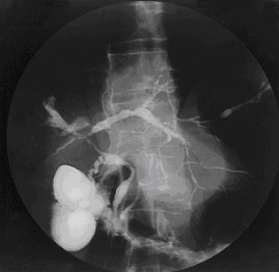
Caroli disease is a rare inherited disorder characterized by cystic dilatation of the bile ducts within the liver. There are two patterns of Caroli disease: focal or simple Caroli disease consists of abnormally widened bile ducts affecting an isolated portion of liver. The second form is more diffuse, and when associated with portal hypertension and congenital hepatic fibrosis, is often referred to as "Caroli syndrome". The underlying differences between the two types are not well understood. Caroli disease is also associated with liver failure and polycystic kidney disease. The disease affects about one in 1,000,000 people, with more reported cases of Caroli syndrome than of Caroli disease.

<span class="mw-page-title-main">Endoscopic retrograde cholangiopancreatography</span> Use of endoscopy and fluoroscopy to treat and diagnose digestive issues.

Endoscopic retrograde cholangiopancreatography (ERCP) is a technique that combines the use of endoscopy and fluoroscopy to diagnose and treat certain problems of the biliary or pancreatic ductal systems. It is primarily performed by highly skilled and specialty trained gastroenterologists. Through the endoscope, the physician can see the inside of the stomach and duodenum, and inject a contrast medium into the ducts in the biliary tree and pancreas so they can be seen on radiographs.

<span class="mw-page-title-main">Primary sclerosing cholangitis</span> Medical condition

Primary sclerosing cholangitis (PSC) is a long-term progressive disease of the liver and gallbladder characterized by inflammation and scarring of the bile ducts, which normally allow bile to drain from the gallbladder. Affected individuals may have no symptoms or may experience signs and symptoms of liver disease, such as yellow discoloration of the skin and eyes, itching, and abdominal pain.

<span class="mw-page-title-main">Cholangiocarcinoma</span> Bile duct adenocarcinoma

Cholangiocarcinoma, also known as bile duct cancer, is a type of cancer that forms in the bile ducts. Symptoms of cholangiocarcinoma may include abdominal pain, yellowish skin, weight loss, generalized itching, and fever. Light colored stool or dark urine may also occur. Other biliary tract cancers include gallbladder cancer and cancer of the ampulla of Vater.

Courvoisier's principle states that a painless palpably enlarged gallbladder accompanied with mild jaundice is unlikely to be caused by gallstones. Usually, the term is used to describe the physical examination finding of the right-upper quadrant of the abdomen. This sign implicates possible malignancy of the gallbladder or pancreas and the swelling is unlikely due to gallstones.

<span class="mw-page-title-main">Pancreaticoduodenectomy</span> Major surgical procedure involving the pancreas, duodenum, and other organs

A pancreaticoduodenectomy, also known as a Whipple procedure, is a major surgical operation most often performed to remove cancerous tumours from the head of the pancreas. It is also used for the treatment of pancreatic or duodenal trauma, or chronic pancreatitis. Due to the shared blood supply of organs in the proximal gastrointestinal system, surgical removal of the head of the pancreas also necessitates removal of the duodenum, proximal jejunum, gallbladder, and, occasionally, part of the stomach.

<span class="mw-page-title-main">Gallbladder cancer</span> Medical condition

Gallbladder cancer is a relatively uncommon cancer, with an incidence of fewer than 2 cases per 100,000 people per year in the United States. It is particularly common in central and South America, central and eastern Europe, Japan and northern India; it is also common in certain ethnic groups e.g. Native American Indians and Hispanics. If it is diagnosed early enough, it can be cured by removing the gallbladder, part of the liver and associated lymph nodes. Most often it is found after symptoms such as abdominal pain, jaundice and vomiting occur, and it has spread to other organs such as the liver.

Hepatectomy is the surgical resection of the liver. While the term is often employed for the removal of the liver from a liver transplant donor, this article will focus on partial resections of hepatic tissue and hepatoportoenterostomy.

<span class="mw-page-title-main">Focal nodular hyperplasia</span> Medical condition

Focal nodular hyperplasia is a benign tumor of the liver, which is the second most prevalent tumor of the liver after hepatic hemangioma. It is usually asymptomatic, rarely grows or bleeds, and has no malignant potential. This tumour was once often resected because it was difficult to distinguish from hepatic adenoma, but with modern multiphase imaging it is usually now diagnosed by strict imaging criteria and not resected.

<span class="mw-page-title-main">Ascending cholangitis</span> Medical condition

Ascending cholangitis, also known as acute cholangitis or simply cholangitis, is inflammation of the bile duct, usually caused by bacteria ascending from its junction with the duodenum. It tends to occur if the bile duct is already partially obstructed by gallstones.

<span class="mw-page-title-main">Gallbladder disease</span> Medical condition

Gallbladder diseases are diseases involving the gallbladder and is closely linked to biliary disease, with the most common cause being gallstones (cholelithiasis).

<span class="mw-page-title-main">Percutaneous transhepatic cholangiography</span> Medical imaging of the biliary tract

Percutaneous transhepatic cholangiography, percutaneous hepatic cholangiogram (PTHC) is a radiological technique used to visualize the anatomy of the biliary tract. A contrast medium is injected into a bile duct in the liver, after which X-rays are taken. It allows access to the biliary tree in cases where endoscopic retrograde cholangiopancreatography has been unsuccessful. Initially reported in 1937, the procedure became popular in 1952.

<span class="mw-page-title-main">Liver cancer</span> Medical condition

Liver cancer, also known as hepatic cancer, primary hepatic cancer, or primary hepatic malignancy, is cancer that starts in the liver. Liver cancer can be primary in which the cancer starts in the liver, or it can be liver metastasis, or secondary, in which the cancer spreads from elsewhere in the body to the liver. Liver metastasis is the more common of the two liver cancers. Instances of liver cancer are increasing globally.

In oncology, metastasectomy is the surgical removal of metastases, which are secondary cancerous growths that have spread from cancer originating in another organ in the body.

Ultrasonography of liver tumors involves two stages: detection and characterization.

<span class="mw-page-title-main">Biloma</span> Circumscribed abdominal collection of bile outside the biliary tree

A biloma is a circumscribed abdominal collection of bile outside the biliary tree. It occurs when there is excess bile in the abdominal cavity. It can occur during or after a bile leak. There is an increased chance of a person developing biloma after having a gallbladder removal surgery, known as laparoscopic cholecystectomy. This procedure can be complicated by biloma with incidence of 0.3–2%. Other causes are liver biopsy, abdominal trauma, and, rarely, spontaneous perforation. The formation of biloma does not occur frequently. Biliary fistulas are also caused by injury to the bile duct and can result in the formation of bile leaks. Biliary fistulas are abnormal communications between organs and the biliary tract. Once diagnosed, they usually require drainage. The term "biloma" was first coined in 1979 by Gould and Patel. They discovered it in a case with extrahepatic bile leakage. The cause of this was trauma to the upper right quadrant of the abdomen. Originally, biloma was described as an "encapsulated collection" of extrahepatic bile. Biloma is now described as extrabiliary collections of bile that can be either intrahepatic or extrahepatic. The most common cause of biloma is trauma to the liver. There are other causes such as abdominal surgery, endoscopic surgery and percutaneous catheter drainage. Injury and abdominal trauma can cause damage to the biliary tree. The biliary tree is a system of vessels that direct secreations from the liver, gallbladder, and pancreas through a series of ducts into the duodenum. This can result in a bile leak which is a common cause of the formation of biloma. It is possible for biloma to be associated with mortality, though it is not common. Bile leaks occur in about one percent of causes.

References

  1. Chamberlain RS, Blumgart LH (2000). "Hilar cholangiocarcinoma: a review and commentary". Annals of Surgical Oncology. 7 (1): 55–66. doi:10.1007/s10434-000-0055-4. PMID   10674450. S2CID   19569428.
  2. Fenster LF (Jan–Feb 1979). "Gerald Klatskin". Yale Journal of Biology and Medicine. 52 (1): 1–3. PMC   2595709 .
  3. Burcharth F (1988). "Klatskin tumours". Acta Chirurgica Scandinavica. Supplementum. 541: 63–69. PMID   2455407.
  4. Kuang D, Wang GP (2010). "Hilar cholangiocarcinoma: pathology and tumor biology". Frontiers of Medicine in China. 4 (4): 371–377. doi:10.1007/s11684-010-0130-6. PMID   21110142. S2CID   39771434.
  5. Boberg KM, Bergquist A, Mitchell S, Pares A, Rosina F, Broomé U, Chapman R, Fausa O, Egeland T, Rocca G, Schrumpf E (2002). "Cholangiocarcinoma in primary sclerosing cholangitis: risk factors and clinical presentation". Scandinavian Journal of Gastroenterology. 37 (10): 1205–1211. doi:10.1080/003655202760373434. PMID   12408527. S2CID   26414844.
  6. Faría G, de Aretxabala X, Sierralta A, Flores P, Burgos L (2001). "[Primary cholangiocarcinoma associated with Caroli disease]". Revista Médica de Chile (in Spanish). 129 (12): 1433–1438. doi: 10.4067/S0034-98872001001200009 . PMID   12080880.
  7. Totkas S, Hohenberger P (2000). "Cholangiocellular carcinoma associated with segmental Caroli's disease". European Journal of Surgical Oncology. 26 (5): 520–521. doi:10.1053/ejso.1999.0936. PMID   11016478.
  8. Falco E, Nardini A, Celoria G, Briglia R, Stefani R, Gadducci G, Belloni E (1993). "[Caroli's disease associated with cholangiocarcinoma. A case of our own observation]". Minerva Chirurgica (in Italian). 48 (17): 961–964. PMID   8290138.
  9. Tannapfel A, Wittekind C (2004). "[Gallbladder and bile duct carcinoma. Biology and pathology]". Internist (Berlin) (in German). 45 (1): 33–41. doi:10.1007/s00108-003-1110-6. PMID   14735242. S2CID   24009368.
  10. Qin XL, Wang ZR, Shi JS, Lu M, Wang L, He QR (2004). "Utility of serum CA19-9 in diagnosis of cholangiocarcinoma: in comparison with CEA". World Journal of Gastroenterology. 10 (3): 427–432. doi: 10.3748/wjg.v10.i3.427 . PMC   4724921 . PMID   14760772.
  11. Tchelepi H, Ralls PW (2004). "Ultrasound of focal liver masses". Ultrasound Quarterly. 20 (4): 155–169. doi:10.1097/00013644-200412000-00002. PMID   15602218. S2CID   27389417.
  12. Harvey CJ, Albrecht T (2001). "Ultrasound of focal liver lesions". European Radiology. 11 (9): 1578–1593. doi:10.1007/s003300101002. PMID   11511877. S2CID   20513478.
  13. Hussain SM, Semelka RC (2005). "Hepatic imaging: comparison of modalities". Radiol. Clin. North Am. 43 (5): 929–47, ix. doi:10.1016/j.rcl.2005.05.006. PMID   16098348.
  14. Acalovschi M (2004). "Cholangiocarcinoma: risk factors, diagnosis and management". Romanian Journal of Internal Medicine. 42 (1): 41–58. PMID   15529594.
  15. Bley TA, Pache G, Saueressig U, Frydrychowicz A, Langer M, Schaefer O (2007). "State of the art 3D MR-cholangiopancreatography for tumor detection". In Vivo. 21 (5): 885–889. PMID   18019429 . Retrieved 2018-02-10.
  16. Clary B, Jarnigan W, Pitt H, Gores G, Busuttil R, Pappas T (2004). "Hilar cholangiocarcinoma". Journal of Gastrointestinal Surgery. 8 (3): 298–302. doi:10.1016/j.gassur.2003.12.004. PMID   15019927. S2CID   20232543.
  17. Jarnagin WR, Shoup M (2004). "Surgical management of cholangiocarcinoma". Seminars in Liver Disease. 24 (2): 189–199. doi:10.1055/s-2004-828895. PMID   15192791. S2CID   260317140.
  18. Heimbach JK, Haddock MG, Alberts SR, Nyberg SL, Ishitani MB, Rosen CB, Gores GJ (2004). "Transplantation for hilar cholangiocarcinoma". Liver Transplantation. 10 (Suppl 2): S65–568. doi:10.1002/lt.20266. PMID   15382214. S2CID   11886730.