Signet ring cell carcinoma

Last updated
Signet ring cell carcinoma
Signet ring cell.jpg
A signet ring cell, as seen in a case of colon adenocarcinoma with mucinous features, showing a tumor cell with a vacuole of mucin. H&E stain.
Specialty Oncology   OOjs UI icon edit-ltr-progressive.svg

Signet ring cell carcinoma (SRCC) is a rare form of highly malignant adenocarcinoma [1] that produces mucin. It is an epithelial malignancy characterized by the histologic appearance of signet ring cells.

Contents

Primary SRCC tumors are most often found in the glandular cells of the stomach (SRCC originates in the stomach in 56 percent of patients), [2] and less frequently in the breast, gallbladder, urinary bladder, and pancreas. [3] SRCCs do not normally form in the lungs, though a few instances have been reported. [4]

Among colorectal cancers, the prevalence of SRCC is less than one percent. Though incidence and mortality of gastric cancer has declined in many countries over the past 50 years, there has been an increase in occurrences of gastric SRCC-type cancers. [5]

SRCC tumors grow in characteristic sheets, which makes diagnosis using standard imaging techniques, like CT and PET scans, less effective. [6] [7]

Causes

Some cases are inherited, and these cases are often caused by mutations in the CDH1 gene, which encodes the important cell–cell adhesion glycoprotein E-cadherin. [8] [9] Somatic mutations of the APC gene have also been implicated in the development of gastric SRCCs. [10]

The role of other risk factors in gastric cancer such as salt-preserved food, smoking, auto-immune gastritis are not well studied in SRCC. [11]

Mechanism of formation

SRCCs are dedifferentiated adenocarcinomas that lose the capability for cell–cell interaction.[ citation needed ]

Highly differentiated adenocarcinomas form SRCCs via a loss of adherens and tight junctions that typically separate MUC4, a mucin protein, and ErbB2, an oncogenic receptor. When MUC4 and ErbB2 are able to interact, they trigger an activation loop. As a result, the ErbB2/ErbB3 signaling pathway becomes constitutively activated, cell–cell interactions are lost and signet carcinomas are formed. Constitutive action of the ErbB2/ErbB3 complex also enhances cell growth. [12]

The mechanism of this malignant cancer is still unclear; however, it has been found that a colon carcinoma cell known as HCC2998 causes an increase in differentiated tumor production. The reason for this increase is due to active PI3K that are converted to a SRCC-like cells. [13]

Metastasis

The pattern of metastasis is different for gastric signet cell carcinoma than for intestinal-type gastric carcinoma. The SRCC tumor is often seen in the peritoneum and has also been known to spread to lymphatic permeation of the lungs and to the ovaries, creating Krukenberg tumors. [14] Cases of gastric carcinomas metastasizing to the breast and forming signet-ring cells have also been reported. One study suggests that when signet-ring cells are found in a breast tumor, the presence of gastric cancer should also be considered. [15]

Histology

A signet ring cell carcinoma of the stomach. Signet ring cells are seen in the lower half of the image. Gastric epithelium is seen in the upper half of the image. H&E stain. Signet ring cell carcinoma - very high mag.jpg
A signet ring cell carcinoma of the stomach. Signet ring cells are seen in the lower half of the image. Gastric epithelium is seen in the upper half of the image. H&E stain.

SRCCs are named due to their resemblance to signet rings, which result from the formation of large vacuoles full of mucin that displaces the nucleus to the cell's periphery. [16]

Stomach cancers with both adenocarcinoma and some SRC (known as mixed-SRCC) exhibit more aggressive behavior than purely SRCC or non-SRCC histologies. [17]

A study of SRCC colorectal cancers compared mucin-rich SRCC tumors and mucin-poor SRCC tumors. They concluded that the latter more frequently demonstrated adverse histologic features such as lymphatic invasion, venous invasion and perineural invasion. [18]

Treatment

Chemotherapy has relatively poor curative efficacy in SRCC patients and overall survival rates are lower compared to patients with more typical cancer pathology. SRCC cancers are usually diagnosed during the late stages of the disease, so the tumors generally spread more aggressively than non-signet cancers, making treatment challenging. [19] In the future, case studies indicate that bone marrow metastases will likely play a larger role in the diagnosis and management of signet ring cell gastric cancer. [20]

In SRCC of the stomach, removal of the stomach cancer is the treatment of choice. There is no combination of chemotherapy which is clearly superior to others, but most active regimens include 5-Fluorouracil (5-FU), Cisplatin, and/or Etoposide. Some newer agents, including Taxol and Gemcitabine (Gemzar) are under investigation. [21]

In a single case study of a patient with SRCC of the bladder with recurrent metastases, the patient exhibited a treatment response to palliative FOLFOX-6 chemotherapy. [22]

There are reports of occasional pulmonary tumour thrombotic microangiopathy leading to pulmonary hypertension and coagulation problems that could be successfully treated with Imatinib.

Prognosis by organ

These aggressive tumors are generally diagnosed at advanced stages and survival is generally shorter. The prognosis of SRCC and its chemosensitivity with specific regimens are still controversial as SRCC is not specifically identified in most studies and its poor prognosis may be due to its more advanced stage. [23] One study suggests that its dismal prognosis seems to be caused by its intrinsic tumor biology, suggesting an area for further research. [24]

Bladder

Primary signet-ring cell carcinoma of the urinary bladder is extremely rare and patient survival is very poor and occurs mainly in men ages 38 to 83. However, one such patient treated with a radical cystectomy followed by combined S-1 and Cisplatin adjuvant chemotherapy did demonstrate promising long-term survival of 90 months. [25]

Colorectal

Primary signet ring cell carcinoma of the colon and rectum (PSRCCR) is rare, with a reported incidence of less than 1 percent. It has a poor prognosis because symptoms often develop late and it is usually diagnosed at an advanced stage. Five-year survival rates in previous studies ranged from nine to 30 percent. Average survival was between 20 and 45 months. [26] It tends to affect younger adults with higher likelihood of lymphovascular invasion. It is worth noting that the overall survival rate of patients with SRCC was significantly poorer than that of patients with mucinous or poorly differentiated adenocarcinoma. [27]

In advanced gastric cancers, the prognosis for patients with the SRCCs was significantly worse than for the other histological types, which can be explained by the finding that advanced SRCC gastric cancers have a larger tumor size, more lymph node metastasis, a deeper invasive depth and more Borrmann type 4 lesions than other types. [28]

Stomach

When compared to adenocarcinoma stomach, SRCC in the stomach occurs more often in women and younger patients. [29] Patients with SRCC of the stomach show similar clinicopathological features to patients with undifferentiated histology. A recent study found that patients with SRCC had a better prognosis than patients with undifferentiated gastric carcinoma. However, when narrowed to patients with only advanced stage gastric cancer, those with SRCC had a worse prognosis than other cell types. [30]

Additional images

Related Research Articles

<span class="mw-page-title-main">Adenocarcinoma</span> Medical condition

Adenocarcinoma is a type of cancerous tumor that can occur in several parts of the body. It is defined as neoplasia of epithelial tissue that has glandular origin, glandular characteristics, or both. Adenocarcinomas are part of the larger grouping of carcinomas, but are also sometimes called by more precise terms omitting the word, where these exist. Thus invasive ductal carcinoma, the most common form of breast cancer, is adenocarcinoma but does not use the term in its name—however, esophageal adenocarcinoma does to distinguish it from the other common type of esophageal cancer, esophageal squamous cell carcinoma. Several of the most common forms of cancer are adenocarcinomas, and the various sorts of adenocarcinoma vary greatly in all their aspects, so that few useful generalizations can be made about them.

<span class="mw-page-title-main">Stomach cancer</span> Cancerous tumor originating in the stomach lining

Stomach cancer, also known as gastric cancer, is a cancer that develops from the lining of the stomach. Most cases of stomach cancers are gastric carcinomas, which can be divided into a number of subtypes, including gastric adenocarcinomas. Lymphomas and mesenchymal tumors may also develop in the stomach. Early symptoms may include heartburn, upper abdominal pain, nausea, and loss of appetite. Later signs and symptoms may include weight loss, yellowing of the skin and whites of the eyes, vomiting, difficulty swallowing, and blood in the stool, among others. The cancer may spread from the stomach to other parts of the body, particularly the liver, lungs, bones, lining of the abdomen, and lymph nodes.

<span class="mw-page-title-main">Krukenberg tumor</span> Medical condition

A Krukenberg tumor refers to a malignancy in the ovary that metastasized from a primary site, classically the gastrointestinal tract, although it can arise in other tissues such as the breast. Gastric adenocarcinoma, especially at the pylorus, is the most common source. Krukenberg tumors are often found in both ovaries, consistent with its metastatic nature.

<span class="mw-page-title-main">Carcinoma</span> Malignancy that develops from epithelial cells

Carcinoma is a malignancy that develops from epithelial cells. Specifically, a carcinoma is a cancer that begins in a tissue that lines the inner or outer surfaces of the body, and that arises from cells originating in the endodermal, mesodermal or ectodermal germ layer during embryogenesis.

<span class="mw-page-title-main">Linitis plastica</span> Medical condition

Linitis plastica is a morphological variant of diffuse stomach cancer in which the stomach wall becomes thick and rigid.

<span class="mw-page-title-main">Pseudomyxoma peritonei</span> Medical condition

Pseudomyxoma peritonei (PMP) is a clinical condition caused by cancerous cells that produce abundant mucin or gelatinous ascites. The tumors cause fibrosis of tissues and impede digestion or organ function, and if left untreated, the tumors and mucin they produce will fill the abdominal cavity. This will result in compression of organs and will destroy the function of the colon, small intestine, stomach, or other organs. Prognosis with treatment in many cases is optimistic, but the disease is lethal if untreated, with death occurring via cachexia, bowel obstruction, or other types of complications.

<span class="mw-page-title-main">Invasive carcinoma of no special type</span> Medical condition

Invasive carcinoma of no special type (NST) is also referred to as invasive ductal carcinoma or infiltrating ductal carcinoma(IDC) and invasive ductal carcinoma, not otherwise specified (NOS). Each of these terms represents to the same disease entity, but for international audiences this article will use invasive carcinoma NST because it is the preferred term of the World Health Organization (WHO).

Gastrointestinal cancer refers to malignant conditions of the gastrointestinal tract and accessory organs of digestion, including the esophagus, stomach, biliary system, pancreas, small intestine, large intestine, rectum and anus. The symptoms relate to the organ affected and can include obstruction, abnormal bleeding or other associated problems. The diagnosis often requires endoscopy, followed by biopsy of suspicious tissue. The treatment depends on the location of the tumor, as well as the type of cancer cell and whether it has invaded other tissues or spread elsewhere. These factors also determine the prognosis.

<span class="mw-page-title-main">Adenocarcinoma in situ of the lung</span> Medical condition

Adenocarcinoma in situ (AIS) of the lung —previously included in the category of "bronchioloalveolar carcinoma" (BAC)—is a subtype of lung adenocarcinoma. It tends to arise in the distal bronchioles or alveoli and is defined by a non-invasive growth pattern. This small solitary tumor exhibits pure alveolar distribution and lacks any invasion of the surrounding normal lung. If completely removed by surgery, the prognosis is excellent with up to 100% 5-year survival.

<span class="mw-page-title-main">Signet ring cell</span> Cell with a large vacuole

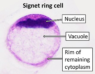
In histology, a signet ring cell is a cell with a large vacuole. The malignant type is seen predominantly in carcinomas. Signet ring cells are most frequently associated with stomach cancer, but can arise from any number of tissues including the prostate, bladder, gallbladder, breast, colon, ovarian stroma and testis.

<span class="mw-page-title-main">Urachal cancer</span> Medical condition

Urachal cancer is a very rare type of cancer arising from the urachus or its remnants. The disease might arise from metaplastic glandular epithelium or embryonic epithelial remnants originating from the cloaca region.

Tegafur/uracil is a chemotherapy drug combination used in the treatment of cancer, primarily bowel cancer. It is also called UFT or UFUR.

Uterine clear-cell carcinoma (CC) is a rare form of endometrial cancer with distinct morphological features on pathology; it is aggressive and has high recurrence rate. Like uterine papillary serous carcinoma CC does not develop from endometrial hyperplasia and is not hormone sensitive, rather it arises from an atrophic endometrium. Such lesions belong to the type II endometrial cancers.

Mucinous cystadenocarcinoma of the lung (MCACL) is a very rare malignant mucus-producing neoplasm arising from the uncontrolled growth of transformed epithelial cells originating in lung tissue.

<span class="mw-page-title-main">Adenocarcinoma of the lung</span> Medical condition

Adenocarcinoma of the lung is the most common type of lung cancer, and like other forms of lung cancer, it is characterized by distinct cellular and molecular features. It is classified as one of several non-small cell lung cancers (NSCLC), to distinguish it from small cell lung cancer which has a different behavior and prognosis. Lung adenocarcinoma is further classified into several subtypes and variants. The signs and symptoms of this specific type of lung cancer are similar to other forms of lung cancer, and patients most commonly complain of persistent cough and shortness of breath.

<span class="mw-page-title-main">Oesophagogastric junctional adenocarcinoma</span>

Oesophagogastric junctional adenocarcinoma is a cancer of the lower part of the oesophagus with a rising incidence in Western countries. This disease is often linked to Barrett's oesophagus.

<span class="mw-page-title-main">Poorly cohesive gastric carcinoma</span>

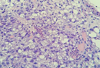
Poorly cohesive gastric carcinoma is a malignant tumour of epithelial origin, characterized by diffuse distribution of tumour cells, isolated from each other or in small groups.

<span class="mw-page-title-main">Squamous-cell carcinoma</span> Carcinoma that derives from squamous epithelial cells

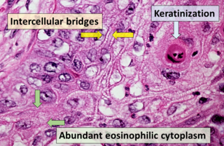
Squamous-cell carcinoma (SCC), also known as epidermoid carcinoma, comprises a number of different types of cancer that begin in squamous cells. These cells form on the surface of the skin, on the lining of hollow organs in the body, and on the lining of the respiratory and digestive tracts.

<span class="mw-page-title-main">Histopathology of colorectal adenocarcinoma</span> Analysis of tissue from a biopsy or surgery to identify colorectal cancer characteristics

The histopathology of colorectal cancer of the adenocarcinoma type involves analysis of tissue taken from a biopsy or surgery. A pathology report contains a description of the microscopical characteristics of the tumor tissue, including both tumor cells and how the tumor invades into healthy tissues and finally if the tumor appears to be completely removed. The most common form of colon cancer is adenocarcinoma, constituting between 95% and 98% of all cases of colorectal cancer. Other, rarer types include lymphoma, adenosquamous and squamous cell carcinoma. Some subtypes have been found to be more aggressive.

<span class="mw-page-title-main">Invasive cribriform carcinoma of the breast</span> Medical condition

Invasive cribriform carcinoma of the breast (ICCB), also termed invasive cribriform carcinoma, is a rare type of breast cancer that accounts for 0.3% to 0.6% of all carcinomas in the breast. It originates in a lactiferous duct as opposed to the lobules that form the alveoli in the breasts' mammary glands. ICCB was first described by Dixon and colleagues in 1983 as a tumor that on microscopic histopathological inspection had a cribriform pattern, i.e. a tissue pattern consisting of numerous "Swiss cheese"-like open spaces and/or sieve-like small holes. The latest edition (2019) of the World Health Organization (2019) termed these lesions invasive cribriform carcinomas indicating that by definition they must have a component that invades out of their ducts of origin into adjacent tissues. In situ ductal cancers that have a cribriform histopathology are regarded as belonging to the group of ductal carcinoma in situ tumors.

References

  1. el-Zimaity HM, Itani K, Graham DY (October 1997). "Early diagnosis of signet ring cell carcinoma of the stomach: role of the Genta stain". Journal of Clinical Pathology. 50 (10): 867–868. doi:10.1136/jcp.50.10.867. PMC   500272 . PMID   9462273.
  2. Benesch MG, Mathieson A (June 2020). "Epidemiology of Signet Ring Cell Adenocarcinomas". Cancers. 12 (6): E1544. doi: 10.3390/cancers12061544 . PMC   7352645 . PMID   32545410.
  3. Belli S, Aytac HO, Karagulle E, Yabanoglu H, Kayaselcuk F, Yildirim S (2014-01-01). "Outcomes of surgical treatment of primary signet ring cell carcinoma of the colon and rectum: 22 cases reviewed with literature". International Surgery. 99 (6): 691–698. doi:10.9738/INTSURG-D-14-00067.1. PMC   4254225 . PMID   25437572.
  4. Livieratos, Speros; Smith, James K.; Fatakhov, Eduard; Koch, Christopher F. (2013-09-20). "Primary signet ring cell carcinoma of the lung: a rare subtype". Case Reports. 2013: bcr2013200111. doi:10.1136/bcr-2013-200111. ISSN   1757-790X. PMID   24057330.
  5. Henson DE, Dittus C, Younes M, Nguyen H, Albores-Saavedra J (July 2004). "Differential trends in the intestinal and diffuse types of gastric carcinoma in the United States, 1973-2000: increase in the signet ring cell type". Archives of Pathology & Laboratory Medicine. 128 (7): 765–770. doi:10.5858/2004-128-765-DTITIA. PMID   15214826.
  6. Shah RB, Zhou M (2011-09-29). Prostate Biopsy Interpretation: An Illustrated Guide. Springer Science & Business Media. ISBN   978-3-642-21369-4.
  7. Ro JY, Ayala AG, Shen SS (2012-04-20). Advances in Surgical Pathology: Prostate Cancer. Lippincott Williams & Wilkins. ISBN   978-1-60831-434-8.
  8. "Office of Rare Diseases Research: Diffuse Gastric Cancer". National Institute of Health. pp. 84–85. Retrieved 2012-11-21.
  9. Muta H, Noguchi M, Kanai Y, Ochiai A, Nawata H, Hirohashi S (August 1996). "E-cadherin gene mutations in signet ring cell carcinoma of the stomach". Japanese Journal of Cancer Research. 87 (8): 843–848. doi:10.1111/j.1349-7006.1996.tb02109.x. PMC   5921174 . PMID   8797891.
  10. Nakatsuru S, Yanagisawa A, Ichii S, Tahara E, Kato Y, Nakamura Y, Horii A (November 1992). "Somatic mutation of the APC gene in gastric cancer: frequent mutations in very well differentiated adenocarcinoma and signet-ring cell carcinoma". Human Molecular Genetics. 1 (8): 559–563. doi:10.1093/hmg/1.8.559. PMID   1338691.
  11. Pernot S, Voron T, Perkins G, Lagorce-Pages C, Berger A, Taieb J (October 2015). "Signet-ring cell carcinoma of the stomach: Impact on prognosis and specific therapeutic challenge". World Journal of Gastroenterology. 21 (40): 11428–11438. doi: 10.3748/wjg.v21.i40.11428 . PMC   4616218 . PMID   26523107.
  12. Fukui Y (August 2014). "Mechanisms behind signet ring cell carcinoma formation". Biochemical and Biophysical Research Communications. 450 (4): 1231–1233. doi:10.1016/j.bbrc.2014.07.025. PMID   25019985.
  13. Fukui Y (August 2014). "Mechanisms behind signet ring cell carcinoma formation". Biochemical and Biophysical Research Communications. 450 (4): 1231–1233. doi:10.1016/j.bbrc.2014.07.025. PMID   25019985.
  14. Duarte I, Llanos O (March 1981). "Patterns of metastases in intestinal and diffuse types of carcinoma of the stomach". Human Pathology. 12 (3): 237–242. doi:10.1016/S0046-8177(81)80124-4. PMID   7228019.
  15. Iesato A, Oba T, Ono M, Hanamura T, Watanabe T, Ito T, et al. (2014-12-29). "Breast metastases of gastric signet-ring cell carcinoma: a report of two cases and review of the literature". OncoTargets and Therapy. 8: 91–97. doi: 10.2147/OTT.S67921 . PMC   4284042 . PMID   25565869.
  16. Portnoy LM (2006-05-05). Radiologic Diagnosis of Gastric Cancer: A new Outlook. Springer Science & Business Media. ISBN   978-3-540-29421-4.
  17. Huh CW, Jung DH, Kim JH, Lee YC, Kim H, Kim H, et al. (February 2013). "Signet ring cell mixed histology may show more aggressive behavior than other histologies in early gastric cancer". Journal of Surgical Oncology. 107 (2): 124–129. doi:10.1002/jso.23261. PMID   22991272. S2CID   206174013.
  18. Hartman DJ, Nikiforova MN, Chang DT, Chu E, Bahary N, Brand RE, et al. (July 2013). "Signet ring cell colorectal carcinoma: a distinct subset of mucin-poor microsatellite-stable signet ring cell carcinoma associated with dismal prognosis". The American Journal of Surgical Pathology. 37 (7): 969–977. doi:10.1097/PAS.0b013e3182851e2b. PMID   23681075. S2CID   19107084.
  19. Nitsche U, Zimmermann A, Späth C, Müller T, Maak M, Schuster T, et al. (November 2013). "Mucinous and signet-ring cell colorectal cancers differ from classical adenocarcinomas in tumor biology and prognosis". Annals of Surgery. 258 (5): 775–783. doi:10.1097/SLA.0b013e3182a69f7e. PMC   3888475 . PMID   23989057.
  20. Dittus C, Mathew H, Malek A, Negroiu A (December 2014). "Bone marrow infiltration as the initial presentation of gastric signet ring cell adenocarcinoma". Journal of Gastrointestinal Oncology. 5 (6): E113–E116. doi:10.3978/j.issn.2078-6891.2014.050. PMC   4226827 . PMID   25436133.
  21. "Signet Ring Cell Cancer | Oncolink - Cancer Resources". www.oncolink.org. Retrieved 2016-04-20.
  22. Pugashetti N, Yap SA, Lara PN, Gandour-Edwards R, Dall'Era MA (2015-01-01). "Metastatic signet-ring cell carcinoma of the urinary bladder: A novel management approach to a rare tumour". Canadian Urological Association Journal. 9 (3–4): E204–E207. doi:10.5489/cuaj.2447. PMC   4455640 . PMID   26085880.
  23. Pernot S, Voron T, Perkins G, Lagorce-Pages C, Berger A, Taieb J (October 2015). "Signet-ring cell carcinoma of the stomach: Impact on prognosis and specific therapeutic challenge". World Journal of Gastroenterology. 21 (40): 11428–11438. doi: 10.3748/wjg.v21.i40.11428 . PMC   4616218 . PMID   26523107.
  24. Nitsche U, Zimmermann A, Späth C, Müller T, Maak M, Schuster T, et al. (November 2013). "Mucinous and signet-ring cell colorectal cancers differ from classical adenocarcinomas in tumor biology and prognosis". Annals of Surgery. 258 (5): 775–783. doi:10.1097/SLA.0b013e3182a69f7e. PMC   3888475 . PMID   23989057.
  25. Hamakawa T, Kojima Y, Naiki T, Kubota Y, Yasui T, Tozawa K, et al. (April 2013). "Long-term survival of a patient with invasive signet-ring cell carcinoma of the urinary bladder managed by combined s-1 and Cisplatin adjuvant chemotherapy". Case Reports in Urology. 2013: 915874. doi: 10.1155/2013/915874 . PMC   3664482 . PMID   23738191.
  26. Belli S, Aytac HO, Karagulle E, Yabanoglu H, Kayaselcuk F, Yildirim S (2014-01-01). "Outcomes of surgical treatment of primary signet ring cell carcinoma of the colon and rectum: 22 cases reviewed with literature". International Surgery. 99 (6): 691–698. doi:10.9738/INTSURG-D-14-00067.1. PMC   4254225 . PMID   25437572.
  27. Lee WS, Chun HK, Lee WY, Yun SH, Cho YB, Yun HR, et al. (September 2007). "Treatment outcomes in patients with signet ring cell carcinoma of the colorectum". American Journal of Surgery. 194 (3): 294–298. doi:10.1016/j.amjsurg.2006.12.041. PMID   17693269.
  28. Kim JP, Kim SC, Yang HK (August 1994). "Prognostic significance of signet ring cell carcinoma of the stomach". Surgical Oncology. 3 (4): 221–227. doi:10.1016/0960-7404(94)90037-x. PMID   7834113.
  29. Shah RB, Zhou M (2011-09-29). Prostate Biopsy Interpretation: An Illustrated Guide. Springer Science & Business Media. ISBN   978-3-642-21369-4.
  30. Kwon KJ, Shim KN, Song EM, Choi JY, Kim SE, Jung HK, Jung SA (January 2014). "Clinicopathological characteristics and prognosis of signet ring cell carcinoma of the stomach". Gastric Cancer. 17 (1): 43–53. doi: 10.1007/s10120-013-0234-1 . PMID   23389081. S2CID   8262409.