Muscles of the thumb

Last updated
Muscles of the thumb
Gray427.png
The muscles of the left hand (palmar surface)
Details
Origin Hand and forearm
Insertion Thumb
Artery Ulnar and radial arteries
Nerve Median and ulnar nerve (C7-T1)
Actions Flexion, extension, adduction, abduction and oppossion of the thumb
Anatomical terms of muscle

The muscles of the thumb are nine skeletal muscles located in the hand and forearm. The muscles allow for flexion, extension, adduction, abduction and opposition of the thumb. The muscles acting on the thumb can be divided into two groups: The extrinsic hand muscles, with their muscle bellies located in the forearm, and the intrinsic hand muscles, with their muscles bellies located in the hand proper. [1]

Contents

The muscles can be compared to guy-wires supporting a flagpole; tension from these muscular guy-wires must be provided in all directions to maintain stability in the articulated column formed by the bones of the thumb. Because this stability is actively maintained by muscles rather than by articular constraints, most muscles attached to the thumb tend to be active during most thumb motions. [2]

Extrinsic

Musculus flexor pollicis longus.png
Gray419.png
Flexor pollicis longus (left) and deep muscles of dorsal forearm (right)

A ventral forearm muscle, the flexor pollicis longus originates on the anterior side of the radius distal to the radial tuberosity and from the interosseous membrane. It passes through the carpal tunnel in a separate tendon sheath, after which it lies between the heads of the flexor pollicis brevis. It finally attaches onto the base of the distal phalanx of the thumb. It is innervated by the anterior interosseus branch of the median nerve (C7-C8) [3]

Three dorsal forearm muscles act on the thumb:

The abductor pollicis longus originates on the dorsal sides of both the ulna and the radius, and from the interosseous membrane. Passing through the first tendon compartment, it inserts to the base of the first metacarpal bone. A part of the tendon reaches the trapezium, while another fuses with the tendons of the extensor pollicis brevis and the abductor pollicis brevis. Except for abducting the hand, it flexes the hand towards the palm and abducts it radially. It is innervated by the deep branch of the radial nerve (C7-C8). [4]

The extensor pollicis longus originates on the dorsal side of the ulna and the interosseous membrane. Passing through the third tendon compartment, it is inserted onto the base of the distal phalanx of the thumb. It uses the dorsal tubercle on the lower extremity of the radius as a fulcrum to extend the thumb and also dorsiflexes and abducts the hand at the wrist. It is innervated by the deep branch of the radial nerve (C7-C8). [4]

The extensor pollicis brevis originates on the ulna distal to the abductor pollicis longus, from the interosseus membrane, and from the dorsal side of the radius. Passing through the first tendon compartment together with the abductor pollicis longus, it is attached to the base of the proximal phalanx of the thumb. It extends the thumb and, because of its close relationship to the long abductor, also abducts the thumb. It is innervated by the deep branch of the radial nerve (C8-T1). [4]

The tendons of the extensor pollicis longus and extensor pollicis brevis form what is known as the anatomical snuff box (an indentation on the lateral aspect of the thumb at its base) The radial artery can be palpated anteriorly at the wrist(not in the snuffbox).

Intrinsic

Gray426.png
Gray428.png
Thenar (left) and dorsal interossei (right) muscles

The intrinsic muscles of the thumb can be divided into two groups; the thenar eminence and other muscles. The thenar eminence refers to the group of muscles on the palm at the base of the thumb. The three muscles composing the thenar eminence are the abductor pollicis brevis, flexor pollicis brevis and opponens pollicis. [5] The other two muscles that influences movement of the thumb are the adductor pollicis and the first dorsal interosseous muscle.

Thenar eminence

The most superficial muscle in the thenar group is the abductor pollicis brevis. The abductor brings the thumb away from the other four fingers. The flexor pollicis brevis, which lies next to the abductor, will flex the thumb, curling it up in the palm. The opponens pollicis lies deep to abductor pollicis brevis. As its name suggests, it opposes the thumb, bringing it against the fingers. This is a very important movement, as most of human hand dexterity including grip comes from this action.

The abductor pollicis brevis originates on the scaphoid tubercle and the flexor retinaculum. It inserts to the radial sesamoid bone and the proximal phalanx of the thumb. It is innervated by the median nerve (C8 and T1). [6]

The flexor pollicis brevis is a two-headed muscle. The superficial head arises on the flexor retinaculum, while the deep head originates on three carpal bones: the trapezium, trapezoid, and capitate. The muscle is inserted onto the radial sesamoid bone of the metacarpophalangeal joint. It acts to flex, adduct, and abduct the thumb, and is therefore also able to oppose the thumb. The superficial head is innervated by the median nerve, while the deep head is innervated by the ulnar nerve (C8-T1). [6] Due to a common interconnection between the median and ulnar nerves in the hand (Riche-Cannieu interconnection), the median nerve may innervate the flexor pollicis brevis in 35% of people. It is innervated by the ulnar nerve in 50% of people and by both the median and ulnar nerves in 15%.

The opponens pollicis originates on the tubercle of the trapezium and the flexor retinaculum. It is inserted onto the radial side of the first metacarpal. It opposes the thumb and assists in adduction. It is innervated by the median nerve. [6] There are normal variations in the muscles nerve innervation. In a Cannieu-Riche anastomosis, fibers from the deep palmar branch of the ulnar nerve innervate the opponens pollicis and/or abductor pollicis brevis. Regardless of their final innervation, the nerves that reach the thenar muscles arise from the C8 and T1 roots, pass through the lower trunk of the plexus, and then through the medial cord of the plexus.

Other muscles

The adductor pollicis also has two heads. It lies deeper and more distal to flexor pollicis brevis. Despite its name, its main action is mainly rotation and opposition. The transversal head originates along the entire third metacarpal bone, while the oblique head originates on the carpal bones proximal to the third metacarpal. The muscle is inserted onto the ulnar sesamoid bone of the metacarpophalangeal joint. It adducts the thumb, and assists in opposition and flexion. It is innervated by the deep branch of the ulnar nerve (C8-T1). [6]

The first dorsal interosseous, one of the central muscles of the hand, extends from the base of the thumb metacarpal to the radial side of the proximal phalanx of the index finger. [7]

Table

MuscleOriginInsertionInervationFunction [8]
Abductor pollicis longus Posterior side of ulna, radius and interosseous membrane Base of first metacarpal Radial nerve (C7-C8) Abduction and extension of the MCP joint
Extensor pollicis brevis Posterior side of radius and the interosseous membrane Base of first proximal phalanx Radial nerve (C7-C8)Extension of MCP joint
Extensor pollicis longus Middle third of posterior surface of ulna, interosseous membrane Base of first distal phalanx Deep branch of the radial nerve (C7-C8) Extension of MCP and IP joints
Flexor pollicis longus The middle 2/4 of the volar surface of the radius and the adjacent interosseus membrane Base of first distal phalanx Anterior interosseus branch of the median nerve (C8-T1)Flexion of MCP and IP joints
Abductor pollicis brevis Transverse carpal ligament, the scaphoid and trapezium Radial base of proximal phalanx and the radial sesamoid Median nerve (C8-T1) Abduction of MCP joint and flexion of IP joint. It also assists in opposition and extension of the thumb
Adductor pollicis Transverse head: anterior body of the third metacarpal
Oblique head: bases of the second and the third metacarpals and the adjacent trapezoid and capitate bones
Medial side of the base of the proximal phalanx of the thumb and the ulnar sesamoid Ulnar nerve (C8-T1) Adducts at the CMC joint and opposition
First dorsal interosseous Medial side of first metacarpal bone Lateral side of second proximal phalanx Ulnar nerve (C8-T1)
Flexor pollicis brevis Trapezium, flexor retinaculum Base of proximal phalanx and the radial sesamoid Median nerve (C8-T1) Flexion at the MCP joint
Opponens pollicis Trapezium and flexor retinaculum Radial side of the corpus of the first metacarpal bone Median nerve (C8-T1)Opposition and adduction

See also

Additional images

Related Research Articles

<span class="mw-page-title-main">Carpal bones</span> Eight small bones that make up the wrist (or carpus) that connects the hand to the forearm

The carpal bones are the eight small bones that make up the wrist (carpus) that connects the hand to the forearm. The term "carpus" and "carpal" is derived from the Latin carpus and the Greek καρπός (karpós), meaning "wrist". In human anatomy, the main role of the carpal bones is to articulate with the radial and ulnar heads to form a highly mobile condyloid joint, to provide attachments for thenar and hypothenar muscles, and to form part of the rigid carpal tunnel which allows the median nerve and tendons of the anterior forearm muscles to be transmitted to the hand and fingers.

<span class="mw-page-title-main">Thumb</span> First digit of the hand

The thumb is the first digit of the hand, next to the index finger. When a person is standing in the medical anatomical position, the thumb is the outermost digit. The Medical Latin English noun for thumb is pollex, and the corresponding adjective for thumb is pollical.

<span class="mw-page-title-main">Radial nerve</span> Nerve in the human body that supplies the posterior portion of the upper limb

The radial nerve is a nerve in the human body that supplies the posterior portion of the upper limb. It innervates the medial and lateral heads of the triceps brachii muscle of the arm, as well as all 12 muscles in the posterior osteofascial compartment of the forearm and the associated joints and overlying skin.

<span class="mw-page-title-main">Wrist</span> Part of the arm between the lower arm and the hand

In human anatomy, the wrist is variously defined as (1) the carpus or carpal bones, the complex of eight bones forming the proximal skeletal segment of the hand; (2) the wrist joint or radiocarpal joint, the joint between the radius and the carpus and; (3) the anatomical region surrounding the carpus including the distal parts of the bones of the forearm and the proximal parts of the metacarpus or five metacarpal bones and the series of joints between these bones, thus referred to as wrist joints. This region also includes the carpal tunnel, the anatomical snuff box, bracelet lines, the flexor retinaculum, and the extensor retinaculum.

<span class="mw-page-title-main">Thenar eminence</span> Mound at the base of the thumb formed by the intrinsic group of muscles

The thenar eminence is the mound formed at the base of the thumb on the palm of the hand by the intrinsic group of muscles of the thumb. The skin overlying this region is the area stimulated when trying to elicit a palmomental reflex. The word thenar comes from Ancient Greek θέναρ (thenar) 'palm of the hand'.

<span class="mw-page-title-main">Extensor carpi radialis longus muscle</span>

The extensor carpi radialis longus is one of the five main muscles that control movements at the wrist. This muscle is quite long, starting on the lateral side of the humerus, and attaching to the base of the second metacarpal bone.

<span class="mw-page-title-main">Upper limb</span> Consists of the arm, forearm, and hand

The upper limbs or upper extremities are the forelimbs of an upright-postured tetrapod vertebrate, extending from the scapulae and clavicles down to and including the digits, including all the musculatures and ligaments involved with the shoulder, elbow, wrist and knuckle joints. In humans, each upper limb is divided into the arm, forearm and hand, and is primarily used for climbing, lifting and manipulating objects.

In human anatomy, the extensor pollicis longus muscle (EPL) is a skeletal muscle located dorsally on the forearm. It is much larger than the extensor pollicis brevis, the origin of which it partly covers and acts to stretch the thumb together with this muscle.

<span class="mw-page-title-main">Flexor pollicis brevis muscle</span> Muscle in the thenar compartment

The flexor pollicis brevis is a muscle in the hand that flexes the thumb. It is one of three thenar muscles. It has both a superficial part and a deep part.

In human anatomy, the abductor pollicis longus (APL) is one of the extrinsic muscles of the hand. Its major function is to abduct the thumb at the wrist. Its tendon forms the anterior border of the anatomical snuffbox.

<span class="mw-page-title-main">Adductor pollicis muscle</span> Muscle in the thenar compartment

In human anatomy, the adductor pollicis muscle is a muscle in the hand that functions to adduct the thumb. It has two heads: transverse and oblique.

<span class="mw-page-title-main">Extensor pollicis brevis muscle</span>

In human anatomy, the extensor pollicis brevis is a skeletal muscle on the dorsal side of the forearm. It lies on the medial side of, and is closely connected with, the abductor pollicis longus. The extensor pollicis brevis (EPB) belongs to the deep group of the posterior fascial compartment of the forearm.[1] It is a part of the lateral border of the anatomical snuffbox.

<span class="mw-page-title-main">Palmar interossei muscles</span> Muscles between the metacarpals

In human anatomy, the palmar or volar interossei are four muscles, one on the thumb that is occasionally missing, and three small, unipennate, central muscles in the hand that lie between the metacarpal bones and are attached to the index, ring, and little fingers. They are smaller than the dorsal interossei of the hand.

<span class="mw-page-title-main">Opponens pollicis muscle</span> Muscle in the thenar compartment

The opponens pollicis is a small, triangular muscle in the hand, which functions to oppose the thumb. It is one of the three thenar muscles. It lies deep to the abductor pollicis brevis and lateral to the flexor pollicis brevis.

<span class="mw-page-title-main">Carpometacarpal joint</span>

The carpometacarpal (CMC) joints are five joints in the wrist that articulate the distal row of carpal bones and the proximal bases of the five metacarpal bones.

<span class="mw-page-title-main">Abductor digiti minimi muscle of hand</span> Muscle in the hypothenar compartment

In human anatomy, the abductor digiti minimi is a skeletal muscle situated on the ulnar border of the palm of the hand. It forms the ulnar border of the palm and its spindle-like shape defines the hypothenar eminence of the palm together with the skin, connective tissue, and fat surrounding it. Its main function is to pull the little finger away from the other fingers.

The posterior compartment of the forearm contains twelve muscles which primarily extend the wrist and digits. It is separated from the anterior compartment by the interosseous membrane between the radius and ulna.

<span class="mw-page-title-main">Cervical spinal nerve 8</span> Spinal nerve of the cervical segment

The cervical spinal nerve 8 (C8) is a spinal nerve of the cervical segment.

<span class="mw-page-title-main">Muscles of the hand</span> Muscles of the human hand

The muscles of the hand are the skeletal muscles responsible for the movement of the hand and fingers. The muscles of the hand can be subdivided into two groups: the extrinsic and intrinsic muscle groups. The extrinsic muscle groups are the long flexors and extensors. They are called extrinsic because the muscle belly is located on the forearm. The intrinsic group are the smaller muscles located within the hand itself. The muscles of the hand are innervated by the radial, median, and ulnar nerves from the brachial plexus.

<span class="mw-page-title-main">Extrinsic extensor muscles of the hand</span>

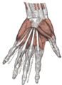
The extrinsic extensor muscles of the hand are located in the back of the forearm and have long tendons connecting them to bones in the hand, where they exert their action. Extrinsic denotes their location outside the hand. Extensor denotes their action which is to extend, or open flat, joints in the hand. They include the extensor carpi radialis longus (ECRL), extensor carpi radialis brevis (ECRB), extensor digitorum (ED), extensor digiti minimi (EDM), extensor carpi ulnaris (ECU), abductor pollicis longus (APL), extensor pollicis brevis (EPB), extensor pollicis longus (EPL), and extensor indicis (EI).

References

  1. "Muscles of the thumb". Eaton hand. Retrieved 21 March 2015.
  2. Austin 2005 , p. 339
  3. Platzer 2004 , p. 162
  4. 1 2 3 Platzer 2004 , p. 168
  5. "Intrinsic Muscles of the Hand — Wheeless' Textbook of Orthopaedics" . Retrieved 2008-01-16.
  6. 1 2 3 4 Platzer 2004 , p. 176
  7. Platzer 2004 , p. 174
  8. Bojsen-Møller, Finn; Simonsen, Erik B.; Tranum-Jensen, Jørgen (2001). Bevægeapparatets anatomi[Anatomy of the Locomotive Apparatus] (in Danish) (12th ed.). pp. 363–364. ISBN   978-87-628-0307-7.