Nuclear magnetic resonance spectroscopy of proteins
Last updated
Nuclear magnetic resonance spectroscopy of proteins (usually abbreviated protein NMR) is a field of structural biology in which NMR spectroscopy is used to obtain information about the structure and dynamics of proteins, and also nucleic acids, and their complexes. The field was pioneered by Richard R. Ernst and Kurt Wüthrich at the ETH,[1] and by Ad Bax, Marius Clore, Angela Gronenborn at the NIH,[2] and Gerhard Wagner at Harvard University, among others. Structure determination by NMR spectroscopy usually consists of several phases, each using a separate set of highly specialized techniques. The sample is prepared, measurements are made, interpretive approaches are applied, and a structure is calculated and validated.
NMR involves the quantum-mechanical properties of the central core ("nucleus") of the atom. These properties depend on the local molecular environment, and their measurement provides a map of how the atoms are linked chemically, how close they are in space, and how rapidly they move with respect to each other. These properties are fundamentally the same as those used in the more familiar magnetic resonance imaging (MRI), but the molecular applications use a somewhat different approach, appropriate to the change of scale from millimeters (of interest to radiologists) to nanometers (bonded atoms are typically a fraction of a nanometer apart), a factor of a million. This change of scale requires much higher sensitivity of detection and stability for long term measurement. In contrast to MRI, structural biology studies do not directly generate an image, but rely on complex computer calculations to generate three-dimensional molecular models.
Currently most samples are examined in a solution in water, but methods are being developed to also work with solid samples. Data collection relies on placing the sample inside a powerful magnet, sending radio frequency signals through the sample, and measuring the absorption of those signals. Depending on the environment of atoms within the protein, the nuclei of individual atoms will absorb different frequencies of radio signals. Furthermore, the absorption signals of different nuclei may be perturbed by adjacent nuclei. This information can be used to determine the distance between nuclei. These distances in turn can be used to determine the overall structure of the protein.
A typical study might involve how two proteins interact with each other, possibly with a view to developing small molecules that can be used to probe the normal biology of the interaction ("chemical biology") or to provide possible leads for pharmaceutical use (drug development). Frequently, the interacting pair of proteins may have been identified by studies of human genetics, indicating the interaction can be disrupted by unfavorable mutations, or they may play a key role in the normal biology of a "model" organism like the fruit fly, yeast, the worm C. elegans, or mice. To prepare a sample, methods of molecular biology are typically used to make quantities by bacterial fermentation. This also permits changing the isotopic composition of the molecule, which is desirable because the isotopes behave differently and provide methods for identifying overlapping NMR signals.
Sample preparation
The NMR sample is prepared in a thin-walled glass tube.
Protein nuclear magnetic resonance is performed on aqueous samples of highly purified protein. Usually, the sample consists of between 300 and 600 microlitres with a protein concentration in the range 0.1 – 3 millimolar. The source of the protein can be either natural or produced in a production system using recombinant DNA techniques through genetic engineering. Recombinantly expressed proteins are usually easier to produce in sufficient quantity, and this method makes isotopiclabeling possible.[citation needed]
The purified protein is usually dissolved in a buffer solution and adjusted to the desired solvent conditions. The NMR sample is prepared in a thin-walled glass tube.[citation needed]
Data collection
Protein NMR utilizes multidimensional nuclear magnetic resonance experiments to obtain information about the protein. Ideally, each distinct nucleus in the molecule experiences a distinct electronic environment and thus has a distinct chemical shift by which it can be recognized. However, in large molecules such as proteins the number of resonances can typically be several thousand and a one-dimensional spectrum inevitably has incidental overlaps. Therefore, multidimensional experiments that correlate the frequencies of distinct nuclei are performed. The additional dimensions decrease the chance of overlap and have a larger information content, since they correlate signals from nuclei within a specific part of the molecule. Magnetization is transferred into the sample using pulses of electromagnetic (radiofrequency) energy and between nuclei using delays; the process is described with so-called pulse sequences. Pulse sequences allow the experimenter to investigate and select specific types of connections between nuclei. The array of nuclear magnetic resonance experiments used on proteins fall in two main categories — one where magnetization is transferred through the chemical bonds, and one where the transfer is through space, irrespective of the bonding structure. The first category is used to assign the different chemical shifts to a specific nucleus, and the second is primarily used to generate the distance restraints used in the structure calculation, and in the assignment with unlabelled protein.[citation needed]
Depending on the concentration of the sample, the magnetic field of the spectrometer, and the type of experiment, a single multidimensional nuclear magnetic resonance experiment on a protein sample may take hours or even several days to obtain suitable signal-to-noise ratio through signal averaging, and to allow for sufficient evolution of magnetization transfer through the various dimensions of the experiment. Other things being equal, higher-dimensional experiments will take longer than lower-dimensional experiments.[citation needed]
Typically, the first experiment to be measured with an isotope-labelled protein is a 2D heteronuclear single quantum correlation (HSQC) spectrum, where "heteronuclear" refers to nuclei other than 1H. In theory, the heteronuclear single quantum correlation has one peak for each H bound to a heteronucleus. Thus, in the 15N-HSQC, with a 15N labelled protein, one signal is expected for each nitrogen atom in the back bone, with the exception of proline, which has no amide-hydrogen due to the cyclic nature of its backbone. Additional 15N-HSQC signals are contributed by each residue with a nitrogen-hydrogen bond in its side chain (W, N, Q, R, H, K). The 15N-HSQC is often referred to as the fingerprint of a protein because each protein has a unique pattern of signal positions. Analysis of the 15N-HSQC allows researchers to evaluate whether the expected number of peaks is present and thus to identify possible problems due to multiple conformations or sample heterogeneity. The relatively quick heteronuclear single quantum correlation experiment helps determine the feasibility of doing subsequent longer, more expensive, and more elaborate experiments. It is not possible to assign peaks to specific atoms from the heteronuclear single quantum correlation alone.[citation needed]
Resonance assignment
In order to analyze the nuclear magnetic resonance data, it is important to get a resonance assignment for the protein, that is to find out which chemical shift corresponds to which atom. This is typically achieved by sequential walking using information derived from several different types of NMR experiment. The exact procedure depends on whether the protein is isotopically labelled or not, since a lot of the assignment experiments depend on carbon-13 and nitrogen-15.[citation needed]
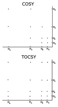
Comparison of a COSY and TOCSY 2D spectra for an amino acid like glutamate or methionine. The TOCSY shows off diagonal crosspeaks between all protons in the spectrum, but the COSY only has crosspeaks between neighbours.
Homonuclear nuclear magnetic resonance
With unlabelled protein the usual procedure is to record a set of two-dimensional homonuclear nuclear magnetic resonance experiments through correlation spectroscopy (COSY), of which several types include conventional correlation spectroscopy, total correlation spectroscopy (TOCSY) and nuclear Overhauser effect spectroscopy (NOESY).[3][4] A two-dimensional nuclear magnetic resonance experiment produces a two-dimensional spectrum. The units of both axes are chemical shifts. The COSY and TOCSY transfer magnetization through the chemical bonds between adjacent protons. The conventional correlation spectroscopy experiment is only able to transfer magnetization between protons on adjacent atoms, whereas in the total correlation spectroscopy experiment the protons are able to relay the magnetization, so it is transferred among all the protons that are connected by adjacent atoms. Thus in a conventional correlation spectroscopy, an alpha proton transfers magnetization to the beta protons, the beta protons transfers to the alpha and gamma protons, if any are present, then the gamma proton transfers to the beta and the delta protons, and the process continues. In total correlation spectroscopy, the alpha and all the other protons are able to transfer magnetization to the beta, gamma, delta, epsilon if they are connected by a continuous chain of protons. The continuous chain of protons are the sidechain of the individual amino acids. Thus these two experiments are used to build so called spin systems, that is build a list of resonances of the chemical shift of the peptide proton, the alpha protons and all the protons from each residue’s sidechain. Which chemical shifts corresponds to which nuclei in the spin system is determined by the conventional correlation spectroscopy connectivities and the fact that different types of protons have characteristic chemical shifts. To connect the different spinsystems in a sequential order, the nuclear Overhauser effect spectroscopy experiment has to be used. Because this experiment transfers magnetization through space, it will show crosspeaks for all protons that are close in space regardless of whether they are in the same spin system or not. The neighbouring residues are inherently close in space, so the assignments can be made by the peaks in the NOESY with other spin systems.[citation needed]
One important problem using homonuclear nuclear magnetic resonance is overlap between peaks. This occurs when different protons have the same or very similar chemical shifts. This problem becomes greater as the protein becomes larger, so homonuclear nuclear magnetic resonance is usually restricted to small proteins or peptides.[citation needed]
The most commonly performed 15N experiment is the 1H-15N HSQC. The experiment is highly sensitive and therefore can be performed relatively quickly. It is often used to check the suitability of a protein for structure determination using NMR, as well as for the optimization of the sample conditions. It is one of the standard suite of experiments used for the determination of the solution structure of protein. The HSQC can be further expanded into three- and four dimensional NMR experiments, such as 15N-TOCSY-HSQC and 15N-NOESY-HSQC.[5]
Schematic of an HNCA and HNCOCA for four sequential residues. The nitrogen-15 dimension is perpendicular to the screen. Each window is focused on the nitrogen chemical shift of that amino acid. The sequential assignment is made by matching the alpha carbon chemical shifts. In the HNCA each residue sees the alpha carbon of itself and the preceding residue. The HNCOCA only sees the alpha carbon of the preceding residue.
Carbon-13 and nitrogen-15 nuclear magnetic resonance
When the protein is labelled with carbon-13 and nitrogen-15 it is possible to record triple resonance experiments that transfer magnetisation over the peptide bond, and thus connect different spin systems through bonds.[6][7] This is usually done using some of the following experiments, HNCO, HN(CA)CO}, HNCA,[8]HN(CO)CA, HNCACB and CBCA(CO)NH. All six experiments consist of a 1H-15N plane (similar to a HSQC spectrum) expanded with a carbon dimension. In the HN(CA)CO, each HN plane contains the peaks from the carbonyl carbon from its residue as well the preceding one in the sequence. The HNCO contains the carbonyl carbon chemical shift from only the preceding residue, but is much more sensitive than HN(CA)CO. These experiments allow each 1H-15N peak to be linked to the preceding carbonyl carbon, and sequential assignment can then be undertaken by matching the shifts of each spin system's own and previous carbons. The HNCA and HN(CO)CA works similarly, just with the alpha carbons (Cα) rather than the carbonyls, and the HNCACB and the CBCA(CO)NH contains both the alpha carbon and the beta carbon (Cβ). Usually several of these experiments are required to resolve overlap in the carbon dimension. This procedure is usually less ambiguous than the NOESY-based method since it is based on through bond transfer. In the NOESY-based methods, additional peaks corresponding to atoms that are close in space but that do not belong to sequential residues will appear, confusing the assignment process. Following the initial sequential resonance assignment, it is usually possible to extend the assignment from the Cα and Cβ to the rest of the sidechain using experiments such as HCCH-TOCSY, which is basically a TOCSY experiment resolved in an additional carbon dimension.
Restraint generation
In order to make structure calculations, a number of experimentally determined restraints have to be generated. These fall into different categories; the most widely used are distance restraints and angle restraints.
Distance restraints
A crosspeak in a NOESY experiment signifies spatial proximity between the two nuclei in question. Thus each peak can be converted into a maximum distance between the nuclei, usually between 1.8 and 6 angstroms. The intensity of a NOESY peak is proportional to the distance to the minus 6th power, so the distance is determined according to the intensity of the peak. The intensity-distance relationship is not exact, so usually a distance range is used.
It is of great importance to assign the NOESY peaks to the correct nuclei based on the chemical shifts. If this task is performed manually it is usually very labor-intensive, since proteins usually have thousands of NOESY peaks. Some computer programs such as PASD[9][10]/XPLOR-NIH,[11][12]UNIO,[13]CYANA,[14]ARIA[15]/CNS,[16] and AUDANA[17]/PONDEROSA-C/S[18] in the Integrative NMR platform[19] perform this task automatically on manually pre-processed listings of peak positions and peak volumes, coupled to a structure calculation. Direct access to the raw NOESY data without the cumbersome need of iteratively refined peak lists is so far only granted by the PASD[10] algorithm implemented in XPLOR-NIH,[11] the ATNOS/CANDID approach implemented in the UNIO software package,[13] and the PONDEROSA-C/S and thus indeed guarantees objective and efficient NOESY spectral analysis.
To obtain as accurate assignments as possible, it is a great advantage to have access to carbon-13 and nitrogen-15 NOESY experiments, since they help to resolve overlap in the proton dimension. This leads to faster and more reliable assignments, and in turn to better structures.
Angle restraints
In addition to distance restraints, restraints on the torsion angles of the chemical bonds, typically the psi and phi angles, can be generated. One approach is to use the Karplus equation, to generate angle restraints from coupling constants. Another approach uses the chemical shifts to generate angle restraints. Both methods use the fact that the geometry around the alpha carbon affects the coupling constants and chemical shifts, so given the coupling constants or the chemical shifts, a qualified guess can be made about the torsion angles.
The blue arrows represent the orientation of the N – H bond of selected peptide bonds. By determining the orientation of a sufficient amount of bonds relative to the external magnetic field, the structure of the protein can be determined. From PDB record 1KBH
The analyte molecules in a sample can be partially ordered with respect to the external magnetic field of the spectrometer by manipulating the sample conditions. Common techniques include addition of bacteriophages or bicelles to the sample, or preparation of the sample in a stretched polyacrylamide gel. This creates a local environment that favours certain orientations of nonspherical molecules. Normally in solution NMR the dipolar couplings between nuclei are averaged out because of the fast tumbling of the molecule. The slight overpopulation of one orientation means that a residual dipolar coupling remains to be observed. The dipolar coupling is commonly used in solid state NMR and provides information about the relative orientation of the bond vectors relative to a single global reference frame. Typically the orientation of the N-H vector is probed in an HSQC-like experiment. Initially, residual dipolar couplings were used for refinement of previously determined structures, but attempts at de novo structure determination have also been made.[20]
NMR spectroscopy is nucleus specific. Thus, it can distinguish between hydrogen and deuterium. The amide protons in the protein exchange readily with the solvent, and, if the solvent contains a different isotope, typically deuterium, the reaction can be monitored by NMR spectroscopy. How rapidly a given amide exchanges reflects its solvent accessibility. Thus amide exchange rates can give information on which parts of the protein are buried, hydrogen-bonded, etc. A common application is to compare the exchange of a free form versus a complex. The amides that become protected in the complex, are assumed to be in the interaction interface.
Structure calculation
Nuclear magnetic resonance structure determination generates an ensemble of structures. The structures will converge only if the data is sufficient to dictate a specific fold. In these structures, it is the case for only a part of the structure. From PDB entry 1SSU.
The experimentally determined restraints can be used as input for the structure calculation process. Researchers, using computer programs such as XPLOR-NIH,[11]CYANA, GeNMR, or RosettaNMR[21] attempt to satisfy as many of the restraints as possible, in addition to general properties of proteins such as bond lengths and angles. The algorithms convert the restraints and the general protein properties into energy terms, and then try to minimize this energy. The process results in an ensemble of structures that, if the data were sufficient to dictate a certain fold, will converge.
The ensemble of structures obtained is an "experimental model", i.e., a representation of certain kind of experimental data. To acknowledge this fact is important because it means that the model could be a good or bad representation of that experimental data.[22] In general, the quality of a model will depend on both the quantity and quality of experimental data used to generate it and the correct interpretation of such data.
It is important to remember that every experiment has associated errors. Random errors will affect the reproducibility and precision of the resulting structures. If the errors are systematic, the accuracy of the model will be affected. The precision indicates the degree of reproducibility of the measurement and is often expressed as the variance of the measured data set under the same conditions. The accuracy, however, indicates the degree to which a measurement approaches its "true" value.
Ideally, a model of a protein will be more accurate the more fit the actual molecule that represents and will be more precise as there is less uncertainty about the positions of their atoms. In practice there is no "standard molecule" against which to compare models of proteins, so the accuracy of a model is given by the degree of agreement between the model and a set of experimental data. Historically, the structures determined by NMR have been, in general, of lower quality than those determined by X-ray diffraction. This is due, in part, to the lower amount of information contained in data obtained by NMR. Because of this fact, it has become common practice to establish the quality of NMR ensembles, by comparing it against the unique conformation determined by X-ray diffraction, for the same protein. However, the X-ray diffraction structure may not exist, and, since the proteins in solution are flexible molecules, a protein represented by a single structure may lead to underestimate the intrinsic variation of the atomic positions of a protein. A set of conformations, determined by NMR or X-ray crystallography may be a better representation of the experimental data of a protein than a unique conformation.[23]
The utility of a model will be given, at least in part, by the degree of accuracy and precision of the model. An accurate model with relatively poor precision could be useful to study the evolutionary relationships between the structures of a set of proteins, whereas the rational drug design requires both precise and accurate models. A model that is not accurate, regardless of the degree of precision with which it was obtained will not be very useful.[22][24]
Since protein structures are experimental models that can contain errors, it is very important to be able to detect these errors. The process aimed at the detection of errors is known as validation. There are several methods to validate structures, some are statistical like PROCHECK and WHAT IF while others are based on physical principles as CheShift, or a mixture of statistical and physics principles PSVS.
In addition to structures, nuclear magnetic resonance can yield information on the dynamics of various parts of the protein. This usually involves measuring relaxation times such as T1 and T2 to determine order parameters, correlation times, and chemical exchange rates. NMR relaxation is a consequence of local fluctuating magnetic fields within a molecule. Local fluctuating magnetic fields are generated by molecular motions. In this way, measurements of relaxation times can provide information of motions within a molecule on the atomic level. In NMR studies of protein dynamics, the nitrogen-15 isotope is the preferred nucleus to study because its relaxation times are relatively simple to relate to molecular motions. This, however, requires isotope labeling of the protein. The T1 and T2 relaxation times can be measured using various types of HSQC-based experiments. The types of motions that can be detected are motions that occur on a time-scale ranging from about 10 picoseconds to about 10 nanoseconds. In addition, slower motions, which take place on a time-scale ranging from about 10 microseconds to 100 milliseconds, can also be studied. However, since nitrogen atoms are found mainly in the backbone of a protein, the results mainly reflect the motions of the backbone, which is the most rigid part of a protein molecule. Thus, the results obtained from nitrogen-15 relaxation measurements may not be representative of the whole protein. Therefore, techniques utilising relaxation measurements of carbon-13 and deuterium have recently been developed, which enables systematic studies of motions of the amino acid side-chains in proteins. A challenging and special case of study regarding dynamics and flexibility of peptides and full-length proteins is represented by disordered structures. Nowadays, it is an accepted concept that proteins can exhibit a more flexible behaviour known as disorder or lack of structure; however, it is possible to describe an ensemble of structures instead of a static picture representing a fully functional state of the protein. Many advances are represented in this field in particular in terms of new pulse sequences, technological improvement, and rigorous training of researchers in the field.
NMR spectroscopy on large proteins
Traditionally, nuclear magnetic resonance spectroscopy has been limited to relatively small proteins or protein domains. This is in part caused by problems resolving overlapping peaks in larger proteins, but this has been alleviated by the introduction of isotope labelling and multidimensional experiments. Another more serious problem is the fact that in large proteins the magnetization relaxes faster, which means there is less time to detect the signal. This in turn causes the peaks to become broader and weaker, and eventually disappear. Two techniques have been introduced to attenuate the relaxation: transverse relaxation optimized spectroscopy (TROSY)[25] and deuteration[26] of proteins. By using these techniques it has been possible to study proteins in complex with the 900 kDa chaperoneGroES-GroEL.[27][28]
Automation of the process
Structure determination by NMR has traditionally been a time-consuming process, requiring interactive analysis of the data by a highly trained scientist. There has been considerable interest in automating the process to increase the throughput of structure determination and to make protein NMR accessible to non-experts (See structural genomics). The two most time-consuming processes involved are the sequence-specific resonance assignment (backbone and side-chain assignment) and the NOE assignment tasks. Several different computer programs have been published that target individual parts of the overall NMR structure determination process in an automated fashion. Most progress has been achieved for the task of automated NOE assignment. So far, only the FLYA and the UNIO approach were proposed to perform the entire protein NMR structure determination process in an automated manner without any human intervention.[13][14] Modules in the NMRFAM-SPARKY such as APES (two-letter-code: ae), I-PINE/PINE-SPARKY (two-letter-code: ep; I-PINE web server) and PONDEROSA (two-letter-code: c3, up; PONDEROSA web server) are integrated so that it offers full automation with visual verification capability in each step.[29] Efforts have also been made to standardize the structure calculation protocol to make it quicker and more amenable to automation.[30] Recently, the POKY suite, the successor of programs mentioned above, has been released to provide modern GUI tools and AI/ML features.[31]
The nuclear Overhauser effect (NOE) is the transfer of nuclear spin polarization from one population of spin-active nuclei to another via cross-relaxation. A phenomenological definition of the NOE in nuclear magnetic resonance spectroscopy (NMR) is the change in the integrated intensity of one NMR resonance that occurs when another is saturated by irradiation with an RF field. The change in resonance intensity of a nucleus is a consequence of the nucleus being close in space to those directly affected by the RF perturbation.
In nuclear magnetic resonance (NMR) spectroscopy, the chemical shift is the resonant frequency of an atomic nucleus relative to a standard in a magnetic field. Often the position and number of chemical shifts are diagnostic of the structure of a molecule. Chemical shifts are also used to describe signals in other forms of spectroscopy such as photoemission spectroscopy.
Nuclear magnetic resonance spectroscopy, most commonly known as NMR spectroscopy or magnetic resonance spectroscopy (MRS), is a spectroscopic technique to observe local magnetic fields around atomic nuclei. This spectroscopy is based on the measurement of absorption of electromagnetic radiations in the radio frequency region from roughly 4 to 900 MHz. Absorption of radio waves in the presence of magnetic field is accompanied by a special type of nuclear transition, and for this reason, such type of spectroscopy is known as Nuclear Magnetic Resonance Spectroscopy. The sample is placed in a magnetic field and the NMR signal is produced by excitation of the nuclei sample with radio waves into nuclear magnetic resonance, which is detected with sensitive radio receivers. The intramolecular magnetic field around an atom in a molecule changes the resonance frequency, thus giving access to details of the electronic structure of a molecule and its individual functional groups. As the fields are unique or highly characteristic to individual compounds, in modern organic chemistry practice, NMR spectroscopy is the definitive method to identify monomolecular organic compounds.
Solid-state NMR (ssNMR) spectroscopy is a technique for characterizing atomic level structure in solid materials e.g. powders, single crystals and amorphous samples and tissues using nuclear magnetic resonance (NMR) spectroscopy. The anisotropic part of many spin interactions are present in solid-state NMR, unlike in solution-state NMR where rapid tumbling motion averages out many of the spin interactions. As a result, solid-state NMR spectra are characterised by larger linewidths than in solution state NMR, which can be utilized to give quantitative information on the molecular structure, conformation and dynamics of the material. Solid-state NMR is often combined with magic angle spinning to remove anisotropic interactions and improve the resolution as well as the sensitivity of the technique.
Carbon-13 (C13) nuclear magnetic resonance is the application of nuclear magnetic resonance (NMR) spectroscopy to carbon. It is analogous to proton NMR and allows the identification of carbon atoms in an organic molecule just as proton NMR identifies hydrogen atoms. 13C NMR detects only the 13 C isotope. The main carbon isotope, 12 C is not detected. Although much less sensitive than 1H NMR spectroscopy, 13C NMR spectroscopy is widely used for characterizing organic and organometallic compounds.
HNCOCA is a 3D triple-resonance NMR experiment commonly used in the field of protein NMR. The name derives from the experiment's magnetization transfer pathway: The magnetization of the amide proton of an amino acid residue is transferred to the amide nitrogen, and then to the alpha carbon of the previous residue in the protein's amino acid sequence. In contrast, the complementary HNCA experiment transfers magnetization to the alpha carbons of both the starting residue and the previous residue in the sequence. The HNCOCA experiment is used, often in tandem with HNCA, to assign alpha carbon resonance signals to specific residues in the protein. This experiment requires a purified sample of protein prepared with 13C and 15N isotopic labelling, at a concentration greater than 0.1 mM, and is thus generally only applied to recombinant proteins.
The heteronuclear single quantum coherence or heteronuclear single quantum correlation experiment, normally abbreviated as HSQC, is used frequently in NMR spectroscopy of organic molecules and is of particular significance in the field of protein NMR. The experiment was first described by Geoffrey Bodenhausen and D. J. Ruben in 1980. The resulting spectrum is two-dimensional (2D) with one axis for proton (1H) and the other for a heteronucleus, which is usually 13C or 15N. The spectrum contains a peak for each unique proton attached to the heteronucleus being considered. The 2D HSQC can also be combined with other experiments in higher-dimensional NMR experiments, such as NOESY-HSQC or TOCSY-HSQC.
Two-dimensional nuclear magnetic resonance spectroscopy is a set of nuclear magnetic resonance spectroscopy (NMR) methods which give data plotted in a space defined by two frequency axes rather than one. Types of 2D NMR include correlation spectroscopy (COSY), J-spectroscopy, exchange spectroscopy (EXSY), and nuclear Overhauser effect spectroscopy (NOESY). Two-dimensional NMR spectra provide more information about a molecule than one-dimensional NMR spectra and are especially useful in determining the structure of a molecule, particularly for molecules that are too complicated to work with using one-dimensional NMR.
Insensitive nuclei enhancement by polarization transfer (INEPT) is a signal enhancement method used in NMR spectroscopy. It involves the transfer of nuclear spin polarization from spins with large Boltzmann population differences to nuclear spins of interest with lower Boltzmann population differences. INEPT uses J-coupling for the polarization transfer in contrast to Nuclear Overhauser effect (NOE), which arises from dipolar cross-relaxation. This method of signal enhancement was introduced by Ray Freeman in 1979. Due to its usefulness in signal enhancement, pulse sequences used in heteronuclear NMR experiments often contain blocks of INEPT or INEPT-like sequences.
The residual dipolar coupling between two spins in a molecule occurs if the molecules in solution exhibit a partial alignment leading to an incomplete averaging of spatially anisotropic dipolar couplings.
Magnetization transfer (MT), in NMR and MRI, refers to the transfer of nuclear spin polarization and/or spin coherence from one population of nuclei to another population of nuclei, and to techniques that make use of these phenomena. There is some ambiguity regarding the precise definition of magnetization transfer, however the general definition given above encompasses all more specific notions. NMR active nuclei, those with non-zero spin, can be energetically coupled to one another under certain conditions. The mechanisms of nuclear-spin energy-coupling have been extensively characterized and are described in the following articles: Angular momentum coupling, Magnetic dipole–dipole interaction, J-coupling, Residual dipolar coupling, Nuclear Overhauser effect, Spin–spin relaxation, and Spin saturation transfer. Alternatively, some nuclei in a chemical system are labile and exchange between non-equivalent environments. A more specific example of this case is presented in the section Chemical Exchange Magnetization transfer.
Carbohydrate NMR spectroscopy is the application of nuclear magnetic resonance (NMR) spectroscopy to structural and conformational analysis of carbohydrates. This method allows the scientists to elucidate structure of monosaccharides, oligosaccharides, polysaccharides, glycoconjugates and other carbohydrate derivatives from synthetic and natural sources. Among structural properties that could be determined by NMR are primary structure, saccharide conformation, stoichiometry of substituents, and ratio of individual saccharides in a mixture. Modern high field NMR instruments used for carbohydrate samples, typically 500 MHz or higher, are able to run a suite of 1D, 2D, and 3D experiments to determine a structure of carbohydrate compounds.
Nuclear magnetic resonance (NMR) is a physical phenomenon in which nuclei in a strong constant magnetic field are perturbed by a weak oscillating magnetic field and respond by producing an electromagnetic signal with a frequency characteristic of the magnetic field at the nucleus. This process occurs near resonance, when the oscillation frequency matches the intrinsic frequency of the nuclei, which depends on the strength of the static magnetic field, the chemical environment, and the magnetic properties of the isotope involved; in practical applications with static magnetic fields up to ca. 20 tesla, the frequency is similar to VHF and UHF television broadcasts (60–1000 MHz). NMR results from specific magnetic properties of certain atomic nuclei. Nuclear magnetic resonance spectroscopy is widely used to determine the structure of organic molecules in solution and study molecular physics and crystals as well as non-crystalline materials. NMR is also routinely used in advanced medical imaging techniques, such as in magnetic resonance imaging (MRI).
Gary Martin is an American chemist and expert in the fields of both NMR spectroscopy and medicinal chemistry. He is a distinguished fellow at the Merck Research Laboratories. He is also a photographer specializing in the capture of images of lighthouses, especially under conditions of extreme weather.
Nucleic acid NMR is the use of nuclear magnetic resonance spectroscopy to obtain information about the structure and dynamics of nucleic acid molecules, such as DNA or RNA. It is useful for molecules of up to 100 nucleotides, and as of 2003, nearly half of all known RNA structures had been determined by NMR spectroscopy.
Triple resonance experiments are a set of multi-dimensional nuclear magnetic resonance spectroscopy (NMR) experiments that link three types of atomic nuclei, most typically consisting of 1H, 15N and 13C. These experiments are often used to assign specific resonance signals to specific atoms in an isotopically-enriched protein. The technique was first described in papers by Ad Bax, Mitsuhiko Ikura and Lewis Kay in 1990, and further experiments were then added to the suite of experiments. Many of these experiments have since become the standard set of experiments used for sequential assignment of NMR resonances in the determination of protein structure by NMR. They are now an integral part of solution NMR study of proteins, and they may also be used in solid-state NMR.
Nuclear magnetic resonance chemical shift re-referencing is a chemical analysis method for chemical shift referencing in biomolecular nuclear magnetic resonance (NMR). It has been estimated that up to 20% of 13C and up to 35% of 15N shift assignments are improperly referenced. Given that the structural and dynamic information contained within chemical shifts is often quite subtle, it is critical that protein chemical shifts be properly referenced so that these subtle differences can be detected. Fundamentally, the problem with chemical shift referencing comes from the fact that chemical shifts are relative frequency measurements rather than absolute frequency measurements. Because of the historic problems with chemical shift referencing, chemical shifts are perhaps the most precisely measurable but the least accurately measured parameters in all of NMR spectroscopy.
Nitrogen-15 nuclear magnetic resonance spectroscopy is a version of nuclear magnetic resonance spectroscopy that examines samples containing the 15N nucleus. 15N NMR differs in several ways from the more common 13C and 1H NMR. To circumvent the difficulties associated with measurement of the quadrupolar, spin-1 14N nuclide, 15N NMR is employed in samples for detection since it has a ground-state spin of ½. Since14N is 99.64% abundant, incorporation of 15N into samples often requires novel synthetic techniques.
David Lyndon Emsley FRSC is a British chemist specialising in solid-state nuclear magnetic resonance and a professor at EPFL. He was awarded the 2012 Grand Prix Charles-Leopold Mayer of the French Académie des Sciences and the 2015 Bourke Award of the Royal Society of Chemistry.
Cross-polarization (CP), originally published as proton-enhanced nuclear induction spectroscopy is a solid-state nuclear magnetic resonance (ssNMR) technique to transfer nuclear magnetization from different types of nuclei via heteronuclear dipolar interactions. The 1H-X cross-polarization dramatically improves the sensitivity of ssNMR experiments of most experiments involving spin-1/2 nuclei, capitalizing on the higher 1H polarisation, and shorter T1(1H) relaxation times. It was developed by Michael Gibby, Alexander Pines and Professor John S. Waugh at the Massachusetts Institute of Technology.
↑ Clore GM, Gronenborn AM (1989). "Determination of three-dimensional structures of proteins and nucleic acids in solution by nuclear magnetic resonance spectroscopy". Critical Reviews in Biochemistry and Molecular Biology. 24 (5): 479–564. doi:10.3109/10409238909086962. PMID2676353.
↑ Clore GM, Gronenborn AM (June 1991). "Structures of larger proteins in solution: three- and four-dimensional heteronuclear NMR spectroscopy". Science. 252 (5011): 1390–1399. doi:10.1126/science.2047852. OSTI83376. PMID2047852.
↑ Clore GM, Gronenborn AM (1991). "Applications of three- and four-dimensional heteronuclear NMR spectroscopy to protein structure determination". Progress in Nuclear Magnetic Resonance Spectroscopy. 23 (1): 43–92. doi:10.1016/0079-6565(91)80002-J.
↑ Bax A, Grzesiek S (1993). "Methodological advances in protein NMR". Accounts of Chemical Research. 26 (4): 131–138. doi:10.1021/ar00028a001.
↑ Bax A, Ikura M (May 1991). "An efficient 3D NMR technique for correlating the proton and 15N backbone amide resonances with the alpha-carbon of the preceding residue in uniformly 15N/13C enriched proteins". Journal of Biomolecular NMR. 1 (1): 99–104. doi:10.1007/BF01874573. PMID1668719. S2CID20037190.
↑ Kuszewski J, Schwieters CD, Garrett DS, Byrd RA, Tjandra N, Clore GM (May 2004). "Completely automated, highly error-tolerant macromolecular structure determination from multidimensional nuclear overhauser enhancement spectra and chemical shift assignments". Journal of the American Chemical Society. 126 (20): 6258–6273. doi:10.1021/ja049786h. PMID15149223.
↑ Spronk CA, Nabuurs SB, Krieger E, Vriend G, Vuister GW (2004). "Validation of protein structures derived by NMR spectroscopy". Progress in Nuclear Magnetic Resonance Spectroscopy. 45 (3–4): 315–337. doi:10.1016/j.pnmrs.2004.08.003.
↑ Markus MA, Dayie KT, Matsudaira P, Wagner G (October 1994). "Effect of deuteration on the amide proton relaxation rates in proteins. Heteronuclear NMR experiments on villin 14T". Journal of Magnetic Resonance, Series B. 105 (2): 192–195. Bibcode:1994JMRB..105..192M. doi:10.1006/jmrb.1994.1122. PMID7952934.
Rance M, Cavanagh J, Fairbrother WJ, Hunt III AW, Skelton NJ (2007). Protein NMR spectroscopy: principles and practice (2nded.). Boston: Academic Press. ISBN978-0-12-164491-8.
ProSA-webArchived 2011-05-11 at the Wayback Machine Web service for the recognition of errors in experimentally or theoretically determined protein structures
This page is based on this Wikipedia article Text is available under the CC BY-SA 4.0 license; additional terms may apply. Images, videos and audio are available under their respective licenses.