The levator ani is a broad, thin muscle group, situated on either side of the pelvis. It is formed from three muscle components: the pubococcygeus, the iliococcygeus, and the puborectalis.
Swallowing, also called deglutition or inglutition in scientific contexts, is the process in the body of a human or other animal that allows for a substance to pass from the mouth, to the pharynx, and into the esophagus, while shutting the epiglottis. Swallowing is an important part of eating and drinking. If the process fails and the material goes through the trachea, then choking or pulmonary aspiration can occur. In the human body the automatic temporary closing of the epiglottis is controlled by the swallowing reflex.
Articles related to anatomy include:

The soft palate is, in mammals, the soft tissue constituting the back of the roof of the mouth. The soft palate is part of the palate of the mouth; the other part is the hard palate. The soft palate is distinguished from the hard palate at the front of the mouth in that it does not contain bone.

In neuroanatomy, the mandibular nerve (V3) is the largest of the three divisions of the trigeminal nerve, the fifth cranial nerve (CN V). Unlike the other divisions of the trigeminal nerve (ophthalmic nerve, maxillary nerve) which contain only afferent fibers, the mandibular nerve contains both afferent and efferent fibers. These nerve fibers innervate structures of the lower jaw and face, such as the tongue, lower lip, and chin. The mandibular nerve also innervates the muscles of mastication.

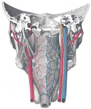
The facial artery is a branch of the external carotid artery that supplies structures of the superficial face.
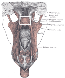
The levator veli palatini is a muscle of the soft palate and pharynx. It is innervated by the vagus nerve via its pharyngeal plexus. During swallowing, it contracts, elevating the soft palate to help prevent food from entering the nasopharynx.

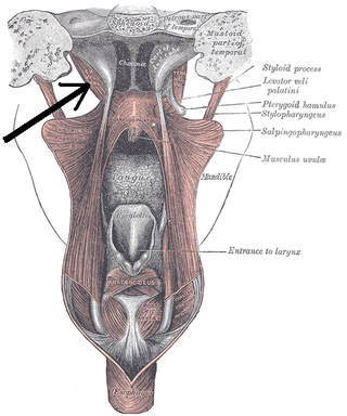
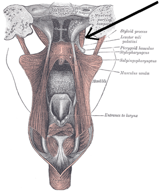
The superior pharyngeal constrictor muscle is a quadrilateral muscle of the pharynx. It is the uppermost and thinnest of the three pharyngeal constrictors.
The tensor veli palatini muscle is a thin, triangular muscle of the head that tenses the soft palate and opens the Eustachian tube to equalise pressure in the middle ear.

The musculus uvulae is a bilaterally muscle of the soft palate that acts to shorten the uvula when both muscles contract. It forms most of the mass of the uvula. It is innervated by the pharyngeal plexus of vagus nerve.
The palatine aponeurosis a thin, firm, fibrous lamella which gives strength and support to soft palate. It serves as the insertion for the tensor veli palatini and levator veli palatini, and the origin for the musculus uvulae, palatopharyngeus, and palatoglossus.

The pterygoid processes of the sphenoid, one on either side, descend perpendicularly from the regions where the body and the greater wings of the sphenoid bone unite.

The ascending palatine artery is an artery is a branch of the facial artery which ascends along the neck before spliting into two terminal branches; one branch supplies the soft palate, and the other supplies the palatine tonsil and pharyngotympanic tube.

The palatopharyngeal arch is larger and projects farther toward the middle line than the palatoglossal arch; it runs downward, lateralward, and backward to the side of the pharynx, and is formed by the projection of the palatopharyngeal muscle, covered by mucous membrane.
The palatoglossal arch on either side runs downward, lateral, and forward to the side of the base of the tongue, and is formed by the projection of the glossopalatine muscle with its covering mucous membrane. It is the anterior border of the isthmus of the fauces and marks the border between the mouth and the palatopharyngeal arch. The latter marks the beginning of the pharynx.
The pharyngeal plexus is a nerve plexus located upon the outer surface of the pharynx. It contains a motor component, a sensory component, and sympathetic component.
The fauces, isthmus of fauces, or the oropharyngeal isthmus, is the opening at the back of the mouth into the throat. It is a narrow passage between the velum and the base of the tongue.

The pharynx is the part of the throat behind the mouth and nasal cavity, and above the esophagus and trachea. It is found in vertebrates and invertebrates, though its structure varies across species. The pharynx carries food to the esophagus and air to the larynx. The flap of cartilage called the epiglottis stops food from entering the larynx.
In the pharynx, the sinus of Morgagni is the enclosed space between the upper border of the superior pharyngeal constrictor muscle, the base of the skull and the pharyngeal aponeurosis.
Passavant's ridge is a mucous elevation situated behind the floor of the naso-pharynx.