The tongue is a muscular organ in the mouth of a typical tetrapod. It manipulates food for chewing and swallowing as part of the digestive process, and is the primary organ of taste. The tongue's upper surface (dorsum) is covered by taste buds housed in numerous lingual papillae. It is sensitive and kept moist by saliva and is richly supplied with nerves and blood vessels. The tongue also serves as a natural means of cleaning the teeth. A major function of the tongue is the enabling of speech in humans and vocalization in other animals.
Swallowing, also called deglutition or inglutition in scientific contexts, is the process in the body of a human or other animal that allows for a substance to pass from the mouth, to the pharynx, and into the esophagus, while shutting the epiglottis. Swallowing is an important part of eating and drinking. If the process fails and the material goes through the trachea, then choking or pulmonary aspiration can occur. In the human body the automatic temporary closing of the epiglottis is controlled by the swallowing reflex.

The facial nerve, also known as the seventh cranial nerve, cranial nerve VII, or simply CN VII, is a cranial nerve that emerges from the pons of the brainstem, controls the muscles of facial expression, and functions in the conveyance of taste sensations from the anterior two-thirds of the tongue. The nerve typically travels from the pons through the facial canal in the temporal bone and exits the skull at the stylomastoid foramen. It arises from the brainstem from an area posterior to the cranial nerve VI and anterior to cranial nerve VIII.

The glossopharyngeal nerve, also known as the ninth cranial nerve, cranial nerve IX, or simply CN IX, is a cranial nerve that exits the brainstem from the sides of the upper medulla, just anterior to the vagus nerve. Being a mixed nerve (sensorimotor), it carries afferent sensory and efferent motor information. The motor division of the glossopharyngeal nerve is derived from the basal plate of the embryonic medulla oblongata, whereas the sensory division originates from the cranial neural crest.

The soft palate is, in mammals, the soft tissue constituting the back of the roof of the mouth. The soft palate is part of the palate of the mouth; the other part is the hard palate. The soft palate is distinguished from the hard palate at the front of the mouth in that it does not contain bone.

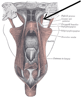
The styloglossus muscle is a bilaterally paired muscle of the tongue. It originates at the styloid process of the temporal bone. It inserts onto the side of the tongue. It acts to elevate and retract the tongue. It is innervated by the hypoglossal nerve.
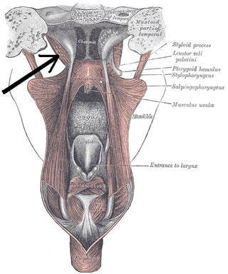
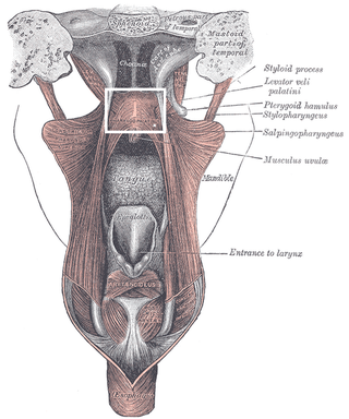
The levator veli palatini is a muscle of the soft palate and pharynx. It is innervated by the vagus nerve via its pharyngeal plexus. During swallowing, it contracts, elevating the soft palate to help prevent food from entering the nasopharynx.
The palatopharyngeusmuscle is a small muscle in the roof of the mouth.
The salpingopharyngeus muscle is a muscle of the pharynx. It arises from the lower part of the cartilage of the Eustachian tube, and inserts into the palatopharyngeus muscle by blending with its posterior fasciculus. It is innervated by vagus nerve via the pharyngeal plexus. It raises the pharynx and larynx during deglutition (swallowing) and laterally draws the pharyngeal walls up. It opens the pharyngeal orifice of the Eustachian tube during swallowing to allow for the equalization of pressure between it and the pharynx.

The middle pharyngeal constrictor is a fan-shaped muscle located in the neck. It is one of three pharyngeal constrictor muscles. It is smaller than the inferior pharyngeal constrictor muscle.

The superior pharyngeal constrictor muscle is a quadrilateral muscle of the pharynx. It is the uppermost and thinnest of the three pharyngeal constrictors.

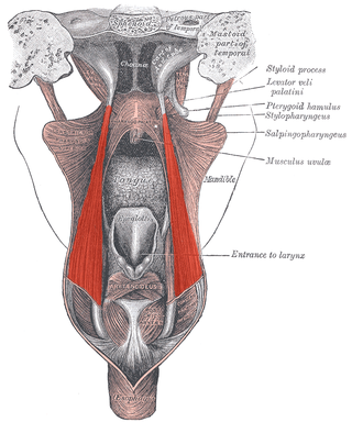
The stylopharyngeus muscle is a muscle in the head. It originates from the temporal styloid process. Some of its fibres insert onto the thyroid cartilage, while others end by intermingling with proximal structures. It is innervated by the glossopharyngeal nerve. It acts to elevate the larynx and pharynx, and dilate the pharynx, thus facilitating swallowing.
The tensor veli palatini muscle is a thin, triangular muscle of the head that tenses the soft palate and opens the Eustachian tube to equalise pressure in the middle ear.

The musculus uvulae is a bilaterally muscle of the soft palate that acts to shorten the uvula when both muscles contract. It forms most of the mass of the uvula. It is innervated by the pharyngeal plexus of vagus nerve.
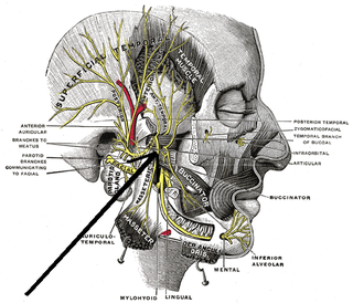
The medial pterygoid nerve (nerve to medial pterygoid, or internal pterygoid nerve) is a nerve of the head. It is a branch of the mandibular nerve (CN V3). It supplies the medial pterygoid muscle, the tensor veli palatini muscle, and the tensor tympani muscle.
The pharyngeal branch of the vagus nerve is the principal motor nerve of the pharynx. It represents the motor component of the pharyngeal plexus of vagus nerve and ultimately provides motor innervation to most of the muscles of the soft palate, and of the pharynx.
The pharyngeal plexus is a nerve plexus located upon the outer surface of the pharynx. It contains a motor component, a sensory component, and sympathetic component.

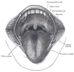
The fauces, isthmus of fauces, or the oropharyngeal isthmus, is the opening at the back of the mouth into the throat. It is a narrow passage between the velum and the base of the tongue.

The pharynx is the part of the throat behind the mouth and nasal cavity, and above the esophagus and trachea. It is found in vertebrates and invertebrates, though its structure varies across species. The pharynx carries food to the esophagus and air to the larynx. The flap of cartilage called the epiglottis stops food from entering the larynx.

In human anatomy, the mouth is the first portion of the alimentary canal that receives food and produces saliva. The oral mucosa is the mucous membrane epithelium lining the inside of the mouth.