
The internal obturator muscle or obturator internus muscle originates on the medial surface of the obturator membrane, the ischium near the membrane, and the rim of the pubis.

The stylohyoid muscle is one of the suprahyoid muscles. Its originates from the styloid process of the temporal bone; it inserts onto hyoid bone. It is innervated by a branch of the facial nerve. It acts draw the hyoid bone upwards and backwards.

The epicranial aponeurosis is an aponeurosis. It covers the upper part of the skull in humans and many other animals.

The teres major muscle is a muscle of the upper limb. It attaches to the scapula and the humerus and is one of the seven scapulohumeral muscles. It is a thick but somewhat flattened muscle.
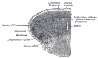
The inferior longitudinal muscle of tongue is an intrinsic muscle of the tongue. It is situated on the under surface of the tongue between the genioglossus and hyoglossus. It is innervated by the hypoglossal nerve. Its contraction shortens and thickens the tongue.
The vertical muscle of the tongue is an intrinsic muscle of the tongue. Its fibers extend from the upper to the under surface of the tongue. It is innervated by the hypoglossal nerve. Its contraction flattens, widens and elongates the tongue.

The transverse muscle of tongue is an intrinsic muscle of the tongue. It consists of fibers which arise from the median fibrous septum. It passes laterally to insert into the submucous fibrous tissue at the sides of the tongue. It is innervated by the hypoglossal nerve. Its contraction elongates and narrows the tongue.

The superior cerebellar artery (SCA) is an artery of the head. It arises near the end of the basilar artery. It is a branch of the basilar artery. It supplies parts of the cerebellum, the midbrain, and other nearby structures. It is the cause of trigeminal neuralgia in some patients.

The superior gluteal nerve is a mixed nerve of the sacral plexus that originates in the pelvis. It provides motor innervation to the gluteus medius, gluteus minimus, tensor fasciae latae, and piriformis muscles; it also has a cutaneous branch.

The lingual artery arises from the external carotid artery between the superior thyroid artery and facial artery. It can be located easily in the tongue.

The thyrocervical trunk is a short artery of the neck. It arises from the subclavian artery, then promptly divides into its branches: the inferior thyroid artery, suprascapular artery, and (sometimes) the transverse cervical artery.

The perineal nerve is a nerve of the pelvis. It arises from the pudendal nerve in the pudendal canal. It gives superficial branches to the skin, and a deep branch to muscles. It supplies the skin and muscles of the perineum. Its latency is tested with electrodes.

The short ciliary nerves are nerves of the orbit around the eye. They are branches of the ciliary ganglion. They supply parasympathetic and sympathetic nerve fibers to the ciliary muscle, iris, and cornea. Damage to the short ciliary nerve may result in loss of the pupillary light reflex, or mydriasis.

The middle cerebellar peduncle is a paired structure of the brain. It connects the pons to the cerebellum, with fibres originating from the pontine nucleus and travelling to the opposite hemisphere of the cerebellar cortex. It is supplied by the anterior inferior cerebellar artery (AICA) and branches from the basilar artery. It conveys information from the cerebrum and the pons to the cerebellum.

The anterior perforated substance is a part of the brain. It is bilateral. It is irregular and quadrilateral. It lies in front of the optic tract and behind the olfactory trigone.

The posterior auricular nerve is a nerve of the head. It is a branch of the facial nerve. It communicates with branches from the vagus nerve, the great auricular nerve, and the lesser occipital nerve. Its auricular branch supplies the posterior auricular muscle, the intrinsic muscles of the auricle, and gives sensation to the auricle. Its occipital branch supplies the occipitalis muscle.

The dorsal artery of the penis is an artery on the top surface of the penis. It is a branch of the internal pudendal artery. It runs forward on the dorsum of the penis to the glans, where it divides into two branches to the glans penis and the foreskin (prepuce).
The zygomatic branches of the facial nerve (malar branches) are nerves of the face. They run across the zygomatic bone to the lateral angle of the orbit. Here, they supply the orbicularis oculi muscle, and join with filaments from the lacrimal nerve and the zygomaticofacial branch of the maxillary nerve (CN V2).

The corpus cavernosum of clitoris is one of a pair of sponge-like regions of erectile tissue of the clitoris. It is made of a sponge-like tissue that fills with blood during erection. This is homologous to the corpus cavernosum penis. The term corpora cavernosa literally means "cave-like bodies".
The quadrigeminal cistern is a subarachnoid cistern situated between splenium of corpus callosum, and the superior surface of the cerebellum. It contains a part of the great cerebral vein, the posterior cerebral artery, quadrigeminal artery, glossopharyngeal nerve, and the pineal gland.
