avoiding foods that irritate the mouth, including acidic, hot or spicy foods; avoid irritation from gum chewing; avoiding oral hygiene products containing sodium lauryl sulfate, brushing with a soft-bristled brush after meals and flossing daily.
Aphthous stomatitis,[2] or recurrent aphthous stomatitis (RAS), commonly referred to as a Canker Sore, is a common condition characterized by the repeated formation of benign and non-contagiousmouth ulcers (aphthae) in otherwise healthy individuals. The cause is not completely understood but involves a T cell-mediated immune response triggered by a variety of factors which may include nutritional deficiencies, local trauma, stress, hormonal influences, allergies, genetic predisposition, certain foods, dehydration, some food additives, or some hygienic chemical additives like SDS (common in toothpaste).
These ulcers occur periodically and heal completely between attacks. In the majority of cases, the individual ulcers last about 7–10 days, and ulceration episodes occur 3–6 times per year. Most appear on the non-keratinizing epithelial surfaces in the mouth – i.e. anywhere except the attached gingiva, the hard palate and the dorsum of the tongue– although the more severe forms, which are less common, may also involve keratinizing epithelial surfaces. Symptoms range from a minor nuisance to interfering with eating and drinking. The severe forms may be debilitating, even causing weight loss due to malnutrition.
The condition is very common, affecting about 20% of the general population to some degree.[1] The onset is often during childhood or adolescence, and the condition usually lasts for several years before gradually disappearing. There is no cure, but treatments such as corticosteroids aim to manage pain, reduce healing time and reduce the frequency of episodes of ulceration.
Signs and symptoms
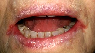
The lower lip is retracted, revealing aphthous ulcers on the labial mucosa (note erythematous "halo" surrounding ulcers)Ulcers can take many shapes and sizes. This one is long and narrow
Persons with aphthous stomatitis have no detectable systemic symptoms or signs (i.e., outside the mouth).[3] Generally, symptoms may include prodromal sensations such as burning, itching, or stinging, which may precede the appearance of any lesion by some hours; and pain, which is often out of proportion to the extent of the ulceration and is worsened by physical contact, especially with certain foods and drinks (e.g., if they are acidic or abrasive). Pain is worst in the days immediately following the initial formation of the ulcer, and then recedes as healing progresses.[4] If there are lesions on the tongue, speaking and chewing can be uncomfortable, and ulcers on the soft palate, back of the throat, or esophagus can cause painful swallowing.[4] Signs are limited to the lesions themselves.
Ulceration episodes usually occur about 3–6 times per year.[5] However, severe disease is characterized by virtually constant ulceration (new lesions developing before old ones have healed) and may cause debilitating chronic pain and interfere with comfortable eating. In severe cases, this prevents adequate nutrient intake leading to malnutrition and weight loss.[4]
Aphthous ulcers typically begin as erythematousmacules (reddened, flat area of mucosa) which develop into ulcers that are covered with a yellow-grey fibrinousmembrane that can be scraped away. A reddish "halo" surrounds the ulcer.[6] The size, number, location, healing time, and periodicity between episodes of ulcer formation are all dependent upon the subtype of aphthous stomatitis.
Causes
The cause is not entirely clear,[3] but is thought to be multifactorial.[7] It has been suggested that aphthous stomatitis is not a single entity, but rather a group of conditions with different causes.[3] Multiple research studies have attempted to identify a causative organism, but aphthous stomatitis appears to be non-contagious, non-infectious, and not sexually transmissible.[3] The mucosal destruction is thought to be the result of a T cell (T lymphocyte) mediated immune response which involves the generation of interleukins and tumor necrosis factor alpha (TNF-α).[7]Mast cells and macrophages are also involved, secreting TNF-α along with the T cells. When early aphthous ulcers are biopsied, the histologic appearance shows a dense inflammatory infiltrate, 80% of which is made up of T cells.[6] Persons with aphthous stomatitis also have circulating lymphocytes which react with peptides 91–105 of heat shock protein 65–60,[3] and the ratio of CD4+ T cells to CD8+ T cells in the peripheral blood of individuals with aphthous stomatitis is decreased.[6]
Aphthous stomatitis has been associated with other autoimmune diseases, namely systemic lupus erythematosus, Behçet's disease and inflammatory bowel diseases. However, common autoantibodies are not detected in most patients, and the condition tends to resolve spontaneously with advancing age rather than worsen.
Evidence for the T cell-mediated mechanism of mucosal destruction is strong, but the exact triggers for this process are unknown and are thought to be multiple and varied from one person to the next. This suggests that there are a number of possible triggers, each of which is capable of producing the disease in different subgroups. In other words, different subgroups appear to have different causes for the condition. These can be considered in three general groups, namely primary immuno-dysregulation, decrease of the mucosal barrier and states of heightened antigenic sensitivity (see below).[6] Risk factors in aphthous stomatitis are also sometimes considered as either host-related or environmental.[8]
Immunity
At least 40% of people with aphthous stomatitis have a positive family history, suggesting that some people are genetically predisposed to developing oral ulceration.[7]HLA-B12, HLA-B51, HLA-Cw7, HLA-A2, HLA-A11, and HLA-DR2 are examples of human leukocyte antigen types associated with aphthous stomatitis.[3][6] However, these HLA types are inconsistently associated with the condition, and also vary according to ethnicity.[9] People who have a positive family history of aphthous stomatitis tend to develop a more severe form of the condition, and at an earlier age than is typical.[9]
Stress has effects on the immune system, which may explain why some cases directly correlate with stress. It is often stated that in studies of students with the condition, ulceration is exacerbated during examination periods and lessened during periods of vacation.[3][6] Alternatively, it has been suggested that oral parafunctional activities such as lip or cheek chewing become more pronounced during periods of stress, and hence the mucosa is subjected to more minor trauma.[9]
Aphthous-like ulceration also occurs in conditions involving systemic immuno-dysregulation, e.g. cyclic neutropenia and human immunodeficiency virus infection. In cyclic neutropenia, more severe oral ulceration occurs during periods of severe immuno-dysregulation, and resolution of the underlying neutropenia is associated with healing of the ulcers. The relative increase in percentage of CD8+ T cells, caused by a reduction in numbers of CD4+ T cells may be implicated in RAS-type ulceration in HIV infection.[6]
Mucosal barrier
The thickness of the mucosa may be an important factor in aphthous stomatitis. Usually, ulcers form on the thinner, non-keratinizing mucosal surfaces in the mouth. Factors which decrease the thickness of the mucosa increase the frequency of occurrence, and factors which increase the thickness of the mucosa correlate with decreased ulceration.[6]
The nutritional deficiencies associated with aphthous stomatitis (vitamin B12, folate, and iron) can all cause a decrease in the thickness of the oral mucosa (atrophy).[6]
Local trauma is also associated with aphthous stomatitis, and it is known that trauma can decrease the mucosal barrier. Trauma could occur during injections of local anesthetic in the mouth, or otherwise during dental treatments, frictional trauma from a sharp surface in the mouth such as broken tooth, or from tooth brushing.[9]
Hormonal factors are capable of altering the mucosal barrier. In one study, a small group of females with aphthous stomatitis had fewer occurrences of aphthous ulcers during the luteal phase of the menstrual cycle or with use of the contraceptive pill.[3][6] This phase is associated with a fall in progestogen levels, mucosal proliferation and keratinization. This subgroup often experiences remission during pregnancy. However, other studies report no correlation between aphthous stomatitis and menstrual period, pregnancy or menopause.[9]
Aphthous stomatitis is more common in people who smoke,[7][10][unreliable medical source] and there is also a correlation between habit duration and severity of the condition.[11] Tobacco use is associated with an increase in keratinization of the oral mucosa.[6] In extreme forms, this may manifest as leukoplakia or stomatitis nicotina (smoker's keratosis). This increased keratinization may mechanically reinforce the mucosa and reduce the tendency of ulcers to form after minor trauma, or present a more substantial barrier to microbes and antigens, but this is unclear. Nicotine is also known to stimulate production of adrenal steroids and reduce production of TNF-α, interleukin-1 and interleukin-6.[9]Smokeless tobacco products also seem to protect against aphthous stomatitis.[11] Cessation of smoking is known to sometimes precede the onset of aphthous stomatitis in people previously unaffected, or exacerbate the condition in those who were already experiencing aphthous ulceration.[3] Despite this correlation, starting smoking again does not usually lessen the condition.[12]
In some instances, recurrent mouth ulcers may be a manifestation of an allergic reaction.[13] Possible allergens include certain foods (e.g., chocolate, coffee, strawberries, eggs, nuts, tomatoes, cheese, citrus fruits,benzoates,cinnamaldehyde, and highly acidic foods), toothpastes, and mouthwashes.[8][13] Where dietary allergens are responsible, mouth ulcers usually develop within about 12–24 hours of exposure.[8]
Sodium lauryl sulphate (SLS), a detergent present in some brands of toothpaste and other oral healthcare products, may produce oral ulceration in some individuals.[3] It has been shown that aphthous stomatitis is more common in people using toothpastes containing SLS, and that some reduction in ulceration occurs when a SLS-free toothpaste is used.[14]
Systemic disease
Systemic disorders associated with aphthous-like ulceration[6]
Aphthous-like ulceration may occur in association with several systemic disorders (see table). These ulcers are clinically and histopathologically identical to the lesions of aphthous stomatitis, but this type of oral ulceration is not considered to be true aphthous stomatitis by some sources.[7][15] Some of these conditions may cause ulceration on other mucosal surfaces in addition to the mouth such as the conjunctiva or the genital mucous membranes. Resolution of the systemic condition often leads to decreased frequency and severity of the oral ulceration.[6]
Behçet's disease is a triad of mouth ulcers, genital ulcers and anterior uveitis.[8] The main feature of Behçet's disease is aphthous-like ulceration, but this is usually more severe than seen in aphthous stomatitis without a systemic cause, and typically resembles major or herpetiforme ulceration or both.[7][16] Aphthous-like ulceration is the first sign of the disease in 25–75% of cases.[6] Behçet's is more common in individuals whose ethnic origin is from regions along the Silk Road (between the Mediterranean and the Far East).[17] It tends to be rare in other countries such as the United States and the United Kingdom.[8]MAGIC syndrome is a possible variant of Behçet's disease, and is associated with aphthous-like ulceration. The name stands for "mouth and genital ulcers with inflamed cartilage" (relapsing polychondritis).[9]
PFAPA syndrome is a rare condition that tends to occur in children.[9] The name stands for "periodic fever, aphthae, pharyngitis (sore throat) and cervical adenitis" (inflammation of the lymph nodes in the neck). The fevers occur periodically about every 3–5 weeks. The condition appears to improve with tonsillectomy or immunosuppression, suggesting an immunologic cause.[16]
In cyclic neutropenia, there is a reduction in the level of circulating neutrophils in the blood that occurs about every 21 days. Opportunistic infections commonly occur and aphthous-like ulceration is worst during this time.[16]
Hematinic deficiencies (vitamin B12, folic acid and iron), occurring singly or in combination,[8] and with or without any underlying gastrointestinal disease, may be twice as common in people with RAS. However, iron and vitamin supplements only infrequently improve the ulceration.[16] The relationship to vitamin B12 deficiency has been the subject of many studies. Although these studies found that 0–42% of those with recurrent ulcers have a vitamin B12 deficiency, an association with deficiency is rare. Even in the absence of deficiency, vitamin B12 supplementation may be helpful due to unclear mechanisms.[18] Hematinic deficiencies can cause anemia, which is also associated with aphthous-like ulceration.[7]
Gastrointestinal disorders are sometimes associated with aphthous-like stomatitis, e.g. most commonly celiac disease, but also inflammatory bowel disease such as Crohn's disease or ulcerative colitis.[7] The link between gastrointestinal disorders and aphthous stomatitis is probably related to nutritional deficiencies caused by malabsorption.[16] Less than 5% of people with RAS have celiac disease, which usually presents with severe malnutrition, anemia, abdominal pain, diarrhea and glossitis (inflammation of the tongue).[9] Sometimes aphthous-like ulcerations can be the only sign of celiac disease.[9] Despite this association, a gluten-free diet does not usually improve the oral ulceration.[16]
Blood is often taken to assess the hemoglobin, iron, folate and vitamin B12 levelsA patch test is sometimes carried out. Areas of the skin on the back are stimulated with various common allergens. The ones which cause an inflammatory reaction may also be involved in recurrent oral ulceration
Diagnosis is mostly based on the clinical appearance and the medical history.[3] The most important diagnostic feature is a history of recurrent, self healing ulcers at fairly regular intervals.[20] Although there are many causes of oral ulceration, recurrent oral ulceration has relatively few causes, most commonly aphthous stomatitis, but rarely Behçet's disease, erythema multiforme, ulceration associated with gastrointestinal disease,[12][20] and recurrent intra-oral herpes simplex infection. A systemic cause is more likely in adults who suddenly develop recurrent oral ulceration with no prior history.[16]
Special investigations may be indicated to rule out other causes of oral ulceration. These include blood tests to exclude anemia, deficiencies of iron, folate or vitamin B12, or celiac disease.[8] However, the nutritional deficiencies may be latent and the peripheral blood picture may appear relatively normal.[8] Some suggest that screening for celiac disease should form part of the routine work up for individuals complaining of recurrent oral ulceration.[9] Many of the systemic diseases cause other symptoms apart from oral ulceration, which is in contrast to aphthous stomatitis where there is isolated oral ulceration. Patch testing may be indicated if allergies are suspected (e.g. a strong relationship between certain foods and episodes of ulceration). Several drugs can cause oral ulceration (e.g. nicorandil), and a trial substitution to an alternative drug may highlight a causal relationship.[3]
Tissue biopsy is not usually required, unless to rule out other suspected conditions such as oral squamous cell carcinoma.[20] The histopathologic appearance is not pathognomonic (the microscopic appearance is not specific to the condition). Early lesions have a central zone of ulceration covered by a fibrinous membrane. In the connective tissue deep to the ulcer there is increased vascularity and a mixed inflammatory infiltrate composed of lymphocytes, histiocytes and polymorphonuclear leukocytes. The epithelium on the margins of the ulcer shows spongiosis and there are many mononuclear cells in the basal third. There are also lymphocytes and histiocytes in the connective tissue surrounding deeper blood vessels near to the ulcer, described histologically as "perivascular cuffing".[6][20]
Classification
Aphthous stomatitis has been classified as a type of non-infectious stomatitis (inflammation of the mouth).[20] One classification distinguishes "common simple aphthae", accounting for 95% of cases, with 3–6 attacks per year, rapid healing, minimal pain and restriction of ulceration to the mouth; and "complex aphthae", accounting for 5% of cases, where ulcers may be present on the genital mucosa in addition to mouth, healing is slower and pain is more severe.[5] A more common method of classifying aphthous stomatitis is into three variants, distinguished by the size, number and location of the lesions, the healing time of individual ulcers and whether a scar is left after healing (see below).
Minor aphthous ulceration
This is the most common type of aphthous stomatitis, accounting for about 80–85% of all cases.[8] This subtype is termed minor aphthous ulceration (MiAU),[3] or minor recurrent aphthous stomatitis (MiRAS). The lesions themselves may be referred to as minor aphthae or minor aphthous ulcers. These lesions are generally less than 10mm in diameter (usually about 2–3mm),[8] and affect non-keratinized mucosal surfaces (i.e. the labial and buccal mucosa, lateral borders of the tongue and the floor of the mouth). Usually several ulcers appear at the same time, but single ulcers are possible. Healing usually takes seven to ten days and leaves no scar. Between episodes of ulceration, there is usually an ulcer-free period of variable length.[7]
Major aphthous ulceration
This subtype makes up about 10% of all cases of aphthous stomatitis.[6] It is termed major aphthous ulceration (MaAU) or major recurrent aphthous stomatitis (MaRAS). Major aphthous ulcers (major aphthae) are similar to minor aphthous ulcers, but are more than 10mm in diameter and the ulceration is deeper.[6][7] Because the lesions are larger, healing takes longer (about twenty to thirty days), and may leave scars. Each episode of ulceration usually produces a greater number of ulcers, and the time between attacks is less than seen in minor aphthous stomatitis.[6] Major aphthous ulceration usually affects non-keratinized mucosal surfaces, but less commonly keratinized mucosa may also be involved, such as the dorsum (top surface) of the tongue or the gingiva (gums).[9] The soft palate or the fauces (back of the throat) may also be involved,[9] the latter being part of the oropharynx rather than the oral cavity. Compared to minor aphthous ulceration, major aphthae tend to have an irregular outline.[8]
Herpetiform ulceration
Herpetiform ulcers,[7] (also termed stomatitis herpetiformis,[21] or herpes-like ulcerations) is a subtype of aphthous stomatitis so named because the lesions resemble a primary infection with herpes simplex virus (primary herpetic gingivostomatitis).[6] However, herpetiform ulceration is not caused by herpes viruses. As with all types of aphthous stomatitis, it is not contagious. Unlike true herpetic ulcers, herpetiforme ulcers are not preceded by vesicles (small, fluid-filled blisters).[9] Herpetiforme ulcers are less than 1mm in diameter and occur in variably sized crops up to one hundred at a time. Adjacent ulcers may merge to form larger, continuous areas of ulceration. Healing occurs within fifteen days without scarring.[8] The ulceration may affect keratinized mucosal surfaces in addition to non keratinized. Herpetiform ulceration is often extremely painful, and the lesions recur more frequently than minor or major aphthous ulcers. Recurrence may be so frequent that ulceration is virtually continuous. It generally occurs in a slightly older age group than the other subtypes,[9] and females are affected slightly more frequently than males.[3]
RAS type ulceration
Recurrent oral ulceration associated with systemic conditions is termed "RAS-type ulceration", "RAS-like ulceration", or "aphthous-like ulcers".[3] Aphthous stomatitis occurs in individuals with no associated systemic disease.[7] Persons with certain systemic diseases may be prone to oral ulceration, but this is secondary to the underlying medical condition (see the systemic disease section).[7] This kind of ulceration is considered by some to be separate from true aphthous stomatitis.[7][15] However, this definition is not strictly applied. For example, many sources refer to oral ulceration caused by anemia and/or nutritional deficiencies as aphthous stomatitis, and some also consider Behçet's disease to be a variant.[6][8]
Treatment
The vast majority of people with aphthous stomatitis have minor symptoms and do not require any specific therapy. The pain is often tolerable with simple dietary modification during an episode of ulceration such as avoiding spicy and acidic foods and beverages.[4] Many different topical and systemic medications have been proposed (see table), sometimes showing little or no evidence of usefulness when formally investigated.[7] Some of the results of interventions for RAS may in truth represent a placebo effect.[16] No therapy is curative, with treatment aiming to relieve pain, promote healing and reduce the frequency of episodes of ulceration.[7]
Medication
The first line of therapy for aphthous stomatitis are topical agents rather than systemic medication,[7] with topical corticosteroids being the mainstay treatment.[3][16] Systemic treatment is usually reserved for severe disease due to the risk of adverse side effects associated with many of these agents. A systematic review found that no single systemic intervention was found to be effective.[7] Good oral hygiene is important to prevent secondary infection of the ulcers.[3]
Occasionally, in females where ulceration is correlated to the menstrual cycle or to birth control pills, progestogen or a change in birth control may be beneficial.[3] Use of nicotine replacement therapy for people who have developed oral ulceration after stopping smoking has also been reported.[9] Starting smoking again does not usually lessen the condition.[12] Trauma can be reduced by avoiding rough or sharp foodstuffs and by brushing teeth with care. If sodium lauryl sulfate is suspected to be the cause, avoidance of products containing this chemical may be useful and prevent recurrence in some individuals.[22] Similarly patch testing may indicate that food allergy is responsible, and the diet modified accordingly.[3] If investigations reveal deficiency states, correction of the deficiency may result in resolution of the ulceration. For example, there is some evidence that vitamin B12 supplementation may prevent recurrence in some individuals.[22]
Surgical excision of aphthous ulcers has been described, but it is an ineffective and inappropriate treatment.[6]Silver nitrate has also been used as a chemical cauterant.[16] Apart from the mainstream approaches detailed above, there are numerous treatments of unproven effectiveness, ranging from herbal remedies to otherwise alternative treatments, including Aloe vera, Myrtus communis, Rosa damascena, potassium alum, nicotine, polio virusvaccine and prostaglandin E2.[3] A 2023 systematic review found that supplementation with vitamin B12, zinc sulfate and omega-3 seem to be beneficial in the management of RAS.[24]
Anecdotal evidence suggests that vitamin C may have a beneficial preventative impact on ulceration.[citation needed] However, the benefits of vitamin C are limited to the first 8 to 12 hours after the first tingling is sensed. Thereafter, vitamin C has far less effect. Vitamin C may cure the condition in 24 - 48 hours only if it is consumed within the first 8 to 12 hours.
Prognosis
By definition, there is no serious underlying medical condition, and most importantly, the ulcers do not represent oral cancer nor are they infectious. However, aphthae are capable of causing significant discomfort. There is a spectrum of severity, with symptoms ranging from a minor nuisance to disabling.[4] Due to pain during eating, weight loss may develop as a result of not eating in severe cases of aphthous stomatitis. Usually, the condition lasts for several years before spontaneously disappearing in later life.[3]
Epidemiology
Aphthous stomatitis affects between 5% and 66% of people, with about 20% of individuals in most populations having the condition to some degree.[6][8] This makes it the most common disease of the oral mucosa.[20] Aphthous stomatitis occurs worldwide, but is more common in developed countries.[3]
Within nations, it is more common in higher socioeconomic groups.[3] Males and females are affected in an equal ratio, and the peak age of onset between 10 and 19 years.[7] About 80% of people with aphthous stomatitis first developed the condition before the age of 30.[6] There have been reports of ethnic variation. For example, in the United States, aphthous stomatitis may be three times more common in white-skinned people than black-skinned people.[16]
History, society and culture
"Aphthous affectations" and "aphthous ulcerations" of the mouth are mentioned several times in the treatise "Of the Epidemics" (part of the Hippocratic corpus, in the 4th century BCE),[25] although it seems likely that this was oral ulceration as a manifestation of some infectious disease, since they are described as occurring in epidemic-like patterns, with concurrent symptoms such as fever.
Aphthous stomatitis was once thought to be a form of recurrent herpes simplex virus infection, and some clinicians still refer to the condition as "herpes" despite this cause having been disproven.[26]
The informal term "canker sore" is sometimes used, mainly in North America,[27] either to describe this condition generally, or to refer to the individual ulcers of this condition,[28] or mouth ulcers of any cause unrelated to this condition. The origin of the word "canker" is thought to have been influenced by Latin, Old English, Middle English and Old North French.[29] In Latin, cancer translates to "malignant tumor" or literally "crab" (related to the likening of sectioned tumors to the limbs of a crab). The closely related word in Middle English and Old North French, chancre, now more usually applied to syphilis, is also thought to be involved.[29] Despite this etymology, aphthous stomatitis is not a form of cancer but rather entirely benign.
An aphtha (plural aphthae) is a non specific term that refers to an ulcer of the mouth. The word is derived from the Greek word aphtha meaning "eruption" or "ulcer".[9] The lesions of several other oral conditions are sometimes described as aphthae, including Bednar's aphthae (infected, traumatic ulcers on the hard palate in infants),[30]oral candidiasis, and foot-and-mouth disease. When used without qualification, aphthae commonly refers to lesions of recurrent aphthous stomatitis. Since the word aphtha is often taken to be synonymous with ulcer, it has been suggested that the term "aphthous ulcer" is redundant, but it remains in common use.[31]Stomatitis is also a non-specific term referring to any inflammatory process in the mouth, with or without oral ulceration.[32] It may describe many different conditions apart from aphthous stomatitis such as angular stomatitis.
The current most widely used medical term is "recurrent aphthous stomatitis" or simply "aphthous stomatitis".[4] Historically, many different terms have been used to refer to recurrent aphthous stomatitis or its sub-types, and some are still in use. Mikulicz's aphthae is a synonym of minor RAS,[9] named after Jan Mikulicz-Radecki. Synonyms for major RAS include Sutton's ulcers (named after Richard Lightburn Sutton), Sutton's disease,[33] Sutton's syndrome and periadenitis mucosa necrotica recurrens.[3][9] Synonyms for aphthous stomatitis as a whole include (recurrent) oral aphthae, (recurrent) aphthous ulceration and (oral) aphthosis.[6][15]
Rembrandt Gentle White toothpaste did not contain sodium lauryl sulfate, and was specifically marketed as being for the benefit of "canker sore sufferers". When the manufacturer Johnson & Johnson discontinued the product in 2014, it caused a backlash of anger from long-term customers, and the toothpaste began to sell for many times the original price on the auction website eBay.[35][36]
A mouth ulcer (aphtha) is an ulcer that occurs on the mucous membrane of the oral cavity. Mouth ulcers are very common, occurring in association with many diseases and by many different mechanisms, but usually there is no serious underlying cause. Rarely, a mouth ulcer that does not heal may be a sign of oral cancer. These ulcers may form individually or multiple ulcers may appear at once. Once formed, an ulcer may be maintained by inflammation and/or secondary infection.
Necrotizing gingivitis (NG) is a common, non-contagious infection of the gums with sudden onset. The main features are painful, bleeding gums, and ulceration of inter-dental papillae. This disease, along with necrotizing periodontitis (NP) and necrotizing stomatitis, is classified as a necrotizing periodontal disease, one of the three general types of gum disease caused by inflammation of the gums (periodontitis).
Oral candidiasis (Acute pseudomembranous candidiasis), also known as oral thrush among other names, is candidiasis that occurs in the mouth. That is, oral candidiasis is a mycosis (yeast/fungal infection) of Candida species on the mucous membranes of the mouth.
Oral leukoplakia is a potentially malignant disorder affecting the oral mucosa. It is defined as "essentially an oral mucosal white lesion that cannot be considered as any other definable lesion." Oral leukoplakia is a white patch or plaque that develops in the oral cavity and is strongly associated with smoking. Leukoplakia is a firmly attached white patch on a mucous membrane which is associated with increased risk of cancer. The edges of the lesion are typically abrupt and the lesion changes with time. Advanced forms may develop red patches. There are generally no other symptoms. It usually occurs within the mouth, although sometimes mucosa in other parts of the gastrointestinal tract, urinary tract, or genitals may be affected.
Stomatitis is inflammation of the mouth and lips. It refers to any inflammatory process affecting the mucous membranes of the mouth and lips, with or without oral ulceration.
Glossitis can mean soreness of the tongue, or more usually inflammation with depapillation of the dorsal surface of the tongue, leaving a smooth and erythematous (reddened) surface,. In a wider sense, glossitis can mean inflammation of the tongue generally. Glossitis is often caused by nutritional deficiencies and may be painless or cause discomfort. Glossitis usually responds well to treatment if the cause is identified and corrected. Tongue soreness caused by glossitis is differentiated from burning mouth syndrome, where there is no identifiable change in the appearance of the tongue, and there are no identifiable causes.
The oral mucosa is the mucous membrane lining the inside of the mouth. It comprises stratified squamous epithelium, termed "oral epithelium", and an underlying connective tissue termed lamina propria. The oral cavity has sometimes been described as a mirror that reflects the health of the individual. Changes indicative of disease are seen as alterations in the oral mucosa lining the mouth, which can reveal systemic conditions, such as diabetes or vitamin deficiency, or the local effects of chronic tobacco or alcohol use. The oral mucosa tends to heal faster and with less scar formation compared to the skin. The underlying mechanism remains unknown, but research suggests that extracellular vesicles might be involved.
Geographic tongue, also known by several other terms, is a condition of the mucous membrane of the tongue, usually on the dorsal surface. It is a common condition, affecting approximately 2–3% of the general population. It is characterized by areas of smooth, red depapillation which migrate over time. The name comes from the map-like appearance of the tongue, with the patches resembling the islands of an archipelago. The cause is unknown, but the condition is entirely benign, and there is no curative treatment. Uncommonly, geographic tongue may cause a burning sensation on the tongue, for which various treatments have been described with little formal evidence of efficacy.
Gingivostomatitis is a combination of gingivitis and stomatitis, or an inflammation of the oral mucosa and gingiva. Herpetic gingivostomatitis is often the initial presentation during the first ("primary") herpes simplex infection. It is of greater severity than herpes labialis which is often the subsequent presentations. Primary herpetic gingivostomatitis is the most common viral infection of the mouth.
Angular cheilitis (AC) is inflammation of one or both corners of the mouth. Often the corners are red with skin breakdown and crusting. It can also be itchy or painful. The condition can last for days to years. Angular cheilitis is a type of cheilitis.
An oral medicine or stomatology doctor/dentist has received additional specialized training and experience in the diagnosis and management of oral mucosal abnormalities including oral cancer, salivary gland disorders, temporomandibular disorders and facial pain, taste and smell disorders; and recognition of the oral manifestations of systemic and infectious diseases. It lies at the interface between medicine and dentistry. An oral medicine doctor is trained to diagnose and manage patients with disorders of the orofacial region.
Stomatitis nicotina is a diffuse white patch on the hard palate, usually caused by tobacco smoking, usually pipe or cigar smoking. It is painless, and it is caused by a response of the palatal oral mucosa to chronic heat. A more pronounced appearance can occur with reverse smoking, sometimes distinguished from stomatitis nicotina by the term reverse smoker's stomatitis. While stomatitis nicotina that is caused by heat is not a premalignant condition, the condition that is caused by reverse smoking is premalignant.
Mucositis is the painful inflammation and ulceration of the mucous membranes lining the digestive tract, usually as an adverse effect of chemotherapy and radiotherapy treatment for cancer. Mucositis can occur anywhere along the gastrointestinal (GI) tract, but oral mucositis refers to the particular inflammation and ulceration that occurs in the mouth. Oral mucositis is a common and often debilitating complication of cancer treatment.
Orofacial granulomatosis (OFG) is a condition characterized by persistent enlargement of the soft tissues of the mouth, lips and the area around the mouth on the face, causing in most cases extreme pain. The mechanism of the enlargement is granulomatous inflammation. The underlying cause of the condition is not completely understood, and there is disagreement as to how it relates to Crohn's disease and sarcoidosis.
Corneal ulcer, also called keratitis, is an inflammatory or, more seriously, infective condition of the cornea involving disruption of its epithelial layer with involvement of the corneal stroma. It is a common condition in humans particularly in the tropics and in farming. In developing countries, children afflicted by vitamin A deficiency are at high risk for corneal ulcer and may become blind in both eyes persisting throughout life. In ophthalmology, a corneal ulcer usually refers to having an infection, while the term corneal abrasion refers more to a scratch injury.
Amlexanox is an anti-inflammatory antiallergic immunomodulator used to treat recurrent aphthous ulcers, and several inflammatory conditions. This drug has been discontinued in the U.S.
Sulfonated phenolics/sulfuric acid liquid topical agent that is used in the treatment of ulcerating oral mucosal lesions and minor oral abrasions.
An ulcer is a discontinuity or break in a bodily membrane that impedes normal function of the affected organ. According to Robbins's pathology, "ulcer is the breach of the continuity of skin, epithelium or mucous membrane caused by sloughing out of inflamed necrotic tissue." Common forms of ulcers recognized in medicine include:
Oral and maxillofacial pathology refers to the diseases of the mouth, jaws and related structures such as salivary glands, temporomandibular joints, facial muscles and perioral skin. The mouth is an important organ with many different functions. It is also prone to a variety of medical and dental disorders.
Oral manifestations of systematic disease are signs and symptoms of disease occurring elsewhere in the body detected in the oral cavity and oral secretions. High blood sugar can be detected by sampling saliva. Saliva sampling may be a non-invasive way to detect changes in the gut microbiome and changes in systemic disease. Another example is tertiary syphilis, where changes to teeth can occur. Syphilis infection can be associated with longitudinal furrows of the tongue.
1 2 3 Bailey J, McCarthy C, Smith RF (October 2011). "Clinical inquiry. What is the most effective way to treat recurrent canker sores?". The Journal of Family Practice. 60 (10): 621–32. PMID21977491.
↑ Tricarico A, Molteni G, Mattioli F, Guerra A, Mordini B, Presutti L, Iughetti L (November–December 2012). "Nipple trauma in infants? Bednar aphthae". American Journal of Otolaryngology. 33 (6): 756–7. doi:10.1016/j.amjoto.2012.06.009. PMID22884485.
This page is based on this Wikipedia article Text is available under the CC BY-SA 4.0 license; additional terms may apply. Images, videos and audio are available under their respective licenses.