
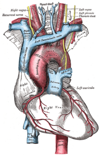
The vagus nerve, historically cited as the pneumogastric nerve, is the tenth cranial nerve or CN X, and interfaces with the parasympathetic control of the heart, lungs, and digestive tract. It actually comprises two nerves—the left and right vagus nerves—but they are typically referred to collectively in the singular. The vagus is the longest nerve of the autonomic nervous system in the human body and comprises sensory and motor fibers. The sensory fibers originate from neurons of the nodose ganglion, whereas the motor fibers come from neurons of the dorsal motor nucleus of the vagus and the nucleus ambiguus.

The autonomic nervous system (ANS), formerly the vegetative nervous system, is a division of the peripheral nervous system that supplies smooth muscle and glands, and thus influences the function of internal organs. The autonomic nervous system is a control system that acts largely unconsciously and regulates bodily functions, such as the heart rate, digestion, respiratory rate, pupillary response, urination, and sexual arousal. This system is the primary mechanism in control of the fight-or-flight response.
A chemoreceptor, also known as chemosensor, is a specialized sensory receptor cell which transduces a chemical substance to generate a biological signal. This signal may be in the form of an action potential, if the chemoreceptor is a neuron, or in the form of a neurotransmitter that can activate a nerve fiber if the chemoreceptor is a specialized cell, such as taste receptors, or an internal peripheral chemoreceptor, such as the carotid bodies. In physiology, a chemoreceptor detects changes in the normal environment, such as an increase in blood levels of carbon dioxide (hypercapnia) or a decrease in blood levels of oxygen (hypoxia), and transmits that information to the central nervous system which engages body responses to restore homeostasis.
The control of ventilation refers to the physiological mechanisms involved in the control of breathing, which is the movement of air into and out of the lungs. Ventilation facilitates respiration. Respiration refers to the utilization of oxygen and balancing of carbon dioxide by the body as a whole, or by individual cells in cellular respiration.

The carotid body is a small cluster of chemoreceptor cells, and supporting sustentacular cells. The carotid body is located in the adventitia, in the bifurcation (fork) of the common carotid artery, which runs along both sides of the neck.

Chromaffin cells, also pheochromocytes, are neuroendocrine cells found mostly in the medulla of the adrenal glands in mammals. These cells serve a variety of functions such as serving as a response to stress, monitoring carbon dioxide and oxygen concentrations in the body, maintenance of respiration and the regulation of blood pressure. They are in close proximity to pre-synaptic sympathetic ganglia of the sympathetic nervous system, with which they communicate, and structurally they are similar to post-synaptic sympathetic neurons. In order to activate chromaffin cells, the splanchnic nerve of the sympathetic nervous system releases acetylcholine, which then binds to nicotinic acetylcholine receptors on the adrenal medulla. This causes the release of catecholamines. The chromaffin cells release catecholamines: ~80% of adrenaline (epinephrine) and ~20% of noradrenaline (norepinephrine) into systemic circulation for systemic effects on multiple organs, and can also send paracrine signals. Hence they are called neuroendocrine cells.

In the human brainstem, the solitary nucleus(SN) is a series of purely sensory nuclei forming a vertical column of grey matter embedded in the medulla oblongata. Through the center of the SN runs the solitary tract, a white bundle of nerve fibers, including fibers from the facial, glossopharyngeal and vagus nerves, that innervate the SN. The SN projects to, among other regions, the reticular formation, parasympathetic preganglionic neurons, hypothalamus and thalamus, forming circuits that contribute to autonomic regulation. Cells along the length of the SN are arranged roughly in accordance with function; for instance, cells involved in taste are located in the rostral part, while those receiving information from cardio-respiratory and gastrointestinal processes are found in the caudal part.

A paraganglioma is a rare neuroendocrine neoplasm that may develop at various body sites. When the same type of tumor is found in the adrenal gland, they are referred to as a pheochromocytoma. They are rare tumors, with an overall estimated incidence of 1/300,000. Unlike other types of cancer, there is no test that determines benign from malignant tumors; long-term follow-up is therefore recommended for all individuals with paraganglioma.
The aortic bodies are one of several small clusters of peripheral chemoreceptors located along the aortic arch. They are important in measuring partial pressures of oxygen and carbon dioxide in the blood, and blood pH.
Neuroendocrine cells are cells that receive neuronal input and, as a consequence of this input, release message molecules (hormones) into the blood. In this way they bring about an integration between the nervous system and the endocrine system, a process known as neuroendocrine integration. An example of a neuroendocrine cell is a cell of the adrenal medulla, which releases adrenaline to the blood. The adrenal medullary cells are controlled by the sympathetic division of the autonomic nervous system. These cells are modified postganglionic neurons. Autonomic nerve fibers lead directly to them from the central nervous system. The adrenal medullary hormones are kept in vesicles much in the same way neurotransmitters are kept in neuronal vesicles. Hormonal effects can last up to ten times longer than those of neurotransmitters. Sympathetic nerve fiber impulses stimulate the release of adrenal medullary hormones. In this way the sympathetic division of the autonomic nervous system and the medullary secretions function together.
The inferior ganglion of the glossopharyngeal nerve is a sensory ganglion. It is larger than and below the superior ganglion of the glossopharyngeal nerve. It is located within the jugular foramen.

A paraganglion is a group of non-neuronal cells derived of the neural crest. They are named for being generally in close proximity to sympathetic ganglia. They are essentially of two types: (1) chromaffin or sympathetic paraganglia made of chromaffin cells and (2) nonchromaffin or parasympathetic ganglia made of glomus cells. They are neuroendocrine cells, the former with primary endocrine functions and the latter with primary chemoreceptor functions.
Peripheral chemoreceptors are so named because they are sensory extensions of the peripheral nervous system into blood vessels where they detect changes in chemical concentrations. As transducers of patterns of variability in the surrounding environment, carotid and aortic bodies count as chemosensors in a similar way as taste buds and photoreceptors. However, because carotid and aortic bodies detect variation within the body’s internal organs, they are considered interoceptors. Taste buds, olfactory bulbs, photoreceptors, and other receptors associated with the five traditional sensory modalities, by contrast, are exteroceptors in that they respond to stimuli outside the body. The body also contains proprioceptors, which respond to the amount of stretch within the organ, usually muscle, that they occupy.
Central chemoreceptors of the central nervous system, located on the ventrolateral medullary surface in the vicinity of the exit of the 9th and 10th cranial nerves, are sensitive to the pH of their environment.

The pre-Bötzinger complex (preBötC) is a cluster of interneurons in the ventral respiratory group of the medulla of the brainstem. This complex has been proven to be essential for the generation of the respiratory rhythm in mammals. The exact mechanism of the rhythm generation and transmission to motor nuclei remains controversial and the topic of much research.

The respiratory center is located in the medulla oblongata and pons, in the brainstem. The respiratory center is made up of three major respiratory groups of neurons, two in the medulla and one in the pons. In the medulla they are the dorsal respiratory group, and the ventral respiratory group. In the pons, the pontine respiratory group includes two areas known as the pneumotaxic centre and the apneustic centre.
Hypoxic ventilatory response (HVR) is the increase in ventilation induced by hypoxia that allows the body to intake and process oxygen at higher rates. It is initially elevated in lowlanders who travel to high altitude, but reduces significantly over time as people acclimatize. In biological anthropology, HVR also refers to human adaptation to environmental stresses resulting from high altitude.
Fish are exposed to large oxygen fluctuations in their aquatic environment since the inherent properties of water can result in marked spatial and temporal differences in the concentration of oxygen (see oxygenation and underwater). Fish respond to hypoxia with varied behavioral, physiological, and cellular responses in order to maintain homeostasis and organism function in an oxygen-depleted environment. The biggest challenge fish face when exposed to low oxygen conditions is maintaining metabolic energy balance, as 95% of the oxygen consumed by fish is used for ATP production releasing the chemical energy of O2 through the mitochondrial electron transport chain. Therefore, hypoxia survival requires a coordinated response to secure more oxygen from the depleted environment and counteract the metabolic consequences of decreased ATP production at the mitochondria. This article is a review of the effects of hypoxia on all aspects of fish, ranging from behavior down to genes.
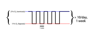
Intermittent hypoxia (also known as episodic hypoxia) is an intervention in which a person or animal undergoes alternating periods of normoxia and hypoxia. Normoxia is defined as exposure to oxygen levels normally found in earth's atmosphere (~21% O2) and hypoxia as any oxygen levels lower than those of normoxia. Normally, exposure to hypoxia is negatively associated to physiological changes to the body, such as altitude sickness. However, when used in moderation, intermittent hypoxia may be used clinically as a means to alleviate various pathological conditions.

Cell-based therapies for Parkinson's disease include various investigational procedures which transplant specific populations of cells into the brains of people with Parkinson's disease. The investigation of cell transplantation therapies followed the discovery that the death of dopaminergic neurons in the substantia nigra pars compacta resulted in the motor symptoms of the disease. Thus, cell transplantation has focused on various dopamine producing cells throughout the body.

