Glossary of developmental biology

Last updated

This glossary of developmental biology is a list of definitions of terms and concepts commonly used in the study of developmental biology and related disciplines in biology, including embryology and reproductive biology, primarily as they pertain to vertebrate animals and particularly to humans and other mammals. The developmental biology of invertebrates, plants, fungi, and other organisms is treated in other articles; e.g terms relating to the reproduction and development of insects are listed in Glossary of entomology, and those relating to plants are listed in Glossary of botany.

Contents

This glossary is intended as introductory material for novices; for more specific and technical detail, see the article corresponding to each term. Additional terms relevant to vertebrate reproduction and development may also be found in Glossary of biology, Glossary of cell biology, Glossary of genetics, and Glossary of evolutionary biology.

A

acrosomal vesicle
acrosome
allantois
One of four extraembryonic membranes formed during the development of amniotes, arising as a sac-like extension of the hindgut and having the dual function of excretion and respiration. [1] In mammals, the allantois arises as a diverticulum made of splanchnopleure almost as soon as the hindgut is established, and quickly builds a dense network of circulatory vessels which eventually assumes responsibility for all metabolic interchange between the fetus and mother. [2]
amniocentesis
amnion
One of four extraembryonic membranes formed during the development of animals belonging to the clade Amniota, all of which are terrestrial vertebrates. Arising from somatopleure very early in development, the amnion grows to surround and define the amniotic cavity, which contains amniotic fluid that cushions and protects the developing embryo from injury. [2]
amniotic fluid
amniotic sac
archenteron

Also gastrocoel.

The central internal cavity of the gastrula in most animal embryos, fated to develop into the lumen of the digestive tube; the primitive gut. The archenteron initially has only one open end, known as the blastopore.

B

birth
blastocoel

Also blastocoele, blastocele, cleavage cavity, and segmentation cavity.

The fluid-filled or yolk-filled cavity that forms in the developing blastula or blastocyst in virtually all animal species. The blastocoel appears to serve two functions: it permits cell migration during gastrulation, and it prevents cells beneath it from interacting prematurely with cells above it by physically separating them. [2]
blastocyst
blastodisc
blastomere
blastopore
blastula
An early form of the animal embryo generally consisting of a hollow sphere of cells (blastomeres) in a single layer (the blastoderm) surrounding a fluid-filled cavity (the blastocoel). [2] Mammalian embryos develop into a specialized blastula known as a blastocyst, containing a differentiated inner cell mass and trophectoderm.
blastulation
A stage in the embryonic development of all animals defined by the formation of the blastula, following morulation and preceding gastrulation. During blastulation, the early embryo develops from a solid ball of cells called a morula into a hollow sphere consisting of an internal cavity surrounded by a single layer of blastomeres; a significant amount of embryonic activity is also dedicated to establishing cell polarity and the basic axes of the body plan, determining the fates of specific cells, activating the embryonic genome, and ultimately transferring full control of gene expression from the mother to the embryo. In mammals, blastulation results in a blastocyst, a specialized blastula marked by very early differentiation of cell populations.

C

cavitation
The hollowing out of a fluid-filled space within a solid mass of cells, e.g. during blastulation or secondary neurulation. [2]
chorion

Also serosa and false amnion.

1.  One of four extraembryonic membranes formed during the development of amniotes, arising from trophoblast and somatopleure as a corollary of the amnion and enclosing both the amnion and the allantois. The chorion provides the fetal contribution to the formation of the placenta. [1] [2]
2.  A tough coat surrounding the eggs of some insects and fish. [2]
cleavage
concealed ovulation
conception

D

delamination
deuterostome
developmental biology
diakinesis
dioestrus

Also diestrus.

In the mammalian estrous cycle, the long period of quiescence following ovulation, during which the uterus prepares to receive a fertilized ovum. [2]
differentiation
diplonema
diplont
An organism having diploid somatic cells and haploid gametes. [2]
diverticulum

E

ectoderm
embryo
embryo transfer
embryoblast
Another name for the inner cell mass, i.e. that portion of the blastocyst that actually gives rise to embryonic tissues, as opposed to extraembryonic tissues. [2]
embryogenesis

Also embryogeny.

The growth and formation of the embryo; the course of development that occurs during the time period beginning with the fertilization of the ovum and ending when the developing animal can no longer be considered an embryo, the criteria for which may vary widely and arbitrarily depending on species. In humans, the embryonic period ends nine weeks after conception, after which time the term fetus is used instead of embryo. In many other animals, embryogenesis is considered complete only after hatching or birth.
embryology
embryonate
Containing a developing embryo; e.g. an embryonated hen's egg, as opposed to an unfertilized egg. [2]
endoderm
endometrium
epiblast
epiboly
estrous cycle

Also oestrous cycle.

F

Fallopian tubes
false amnion
See chorion .
fate map
A diagram that shows what will become of each region of the embryo during the course of normal development. Fate maps are created by selectively marking populations of cells in distinct regions of the early embryo with distinct visual reporters (by any of a variety of methods designed to permit easy visualization of the marked cells, e.g. vital stains, fluorescent compounds, or retroviral transfection) and then allowing the embryo to proceed normally through the subsequent stages of development, after which each specific reporter can again be visualized, thereby revealing the new positions and morphologies of the marked cells and/or their daughter cells. Visualizing the reporters at two or more different developmental stages shows how the different parts of the embryo have moved and changed over time. [2]
fertilization
follicle-stimulating hormone (FSH)

G

gastrocoel
See archenteron .
gastrula
gastrulation
A stage in the embryonic development of most animals defined by the formation of the gastrula, following blastulation and preceding neurulation. During gastrulation, the blastula or blastocyst undergoes a major reorganization from a single, continuous layer of cells surrounding a single cavity into the complex, multilayered, multicavity gastrula, in which all of the primordial germ layers are present. Though the precise pattern of morphogenetic changes constituting gastrulation varies considerably between species, all types of gastrulation are unified by five basic classes of cell movements: the invagination of one side of the blastula into the blastocoel; the involution of the inner layer of cells over the basal surface of the outer layer; the ingression of individual cells into the embryo; the delamination of one layer into two layers via splitting or migration; and the epiboly or expansion of one layer over other cells or layers. [3] By the end of gastrulation, the cells of the embryo have begun differentiation into distinct lineages, the basic axes of the body plan have been established (e.g. dorsal-ventral, anterior-posterior, etc.), and one or more layers of cells have been internalized, including the prospective gut.
germ cell
germ layer
gonad

H

histogenesis
The process by which the definite cells and tissues that make up the body of an organism arise from embryonic cells; or, more generally, the generation of new tissues at any stage of life. [2]
hypoblast

I

in vitro fertilization (IVF)
ingression
inner cell mass
invagination
involution

J

K

L

leptonema
Leydig cell

M

meiosis
meiotic arrest
mesoderm
mid-blastula transition (MBT)
morula
A very early form of the animal embryo consisting of a solid ball of 16 to 32 blastomeres. By the morula stage, these cells have become flattened and have begun to develop stronger cell-to-cell adhesion, as well as to pump fluid into an internal cavity that will eventually become the blastocoel.

N

neonatal
neurula
neurulation
notochord

O

oestrous cycle
See estrous cycle .
oocyte
oogenesis
organogenesis
ovary
oviduct
ovulation
ovum

P

pachynema
parthenogenesis
parturition
placenta
polar body
postimplantation
postnatal
postpartum
preimplantation
prenatal
primary spermatocyte
primitive streak
primordium
pronucleus
Either of the haploid gametic nuclei, i.e. that of the ovum or sperm, as they exist prior to syngamy. The female pronucleus is formed during oogenesis at the time of the second meiotic division, which occurs before fertilization; in contrast, the nucleus of a spermatozoon is generally only considered a pronucleus after fertilization, once it is inside the cytoplasm of the egg and has begun to decondense. [2]
protoblast
protostome

Q

R

reproductive biology

S

secondary spermatocyte
semen
serosa
See chorion .
Sertoli cell
somatic cell nuclear transfer (SCNT)
somatopleure
somite
sperm
spermatid
spermatocyte
spermatogonium
spermiogenesis
splanchnopleure
superovulation

T

teratogen
testis
trophectoderm
trophoblast

U

uterus

V

vas deferens

W

X

Y

Z

zona pellucida
zygonema
zygote
zygotic genome activation (ZGA)

See also

Related Research Articles

<span class="mw-page-title-main">Ontogeny</span> Origination and development of an organism

Ontogeny is the origination and development of an organism, usually from the time of fertilization of the egg to adult. The term can also be used to refer to the study of the entirety of an organism's lifespan.

<span class="mw-page-title-main">Embryo</span> Multicellular diploid eukaryote in its earliest stage of development

An embryo is an initial stage of development of a multicellular organism. In organisms that reproduce sexually, embryonic development is the part of the life cycle that begins just after fertilization of the female egg cell by the male sperm cell. The resulting fusion of these two cells produces a single-celled zygote that undergoes many cell divisions that produce cells known as blastomeres. The blastomeres are arranged as a solid ball that when reaching a certain size, called a morula, takes in fluid to create a cavity called a blastocoel. The structure is then termed a blastula, or a blastocyst in mammals.

The amniotic sac, also called the bag of waters or the membranes, is the sac in which the embryo and later fetus develops in amniotes. It is a thin but tough transparent pair of membranes that hold a developing embryo until shortly before birth. The inner of these membranes, the amnion, encloses the amniotic cavity, containing the amniotic fluid and the embryo. The outer membrane, the chorion, contains the amnion and is part of the placenta. On the outer side, the amniotic sac is connected to the yolk sac, the allantois, and via the umbilical cord, the placenta.

<span class="mw-page-title-main">Amnion</span> Innermost membranous sac that surrounds and protects the developing embryo

The amnion is a membrane that closely covers the human and various other embryos when first formed. It fills with amniotic fluid, which causes the amnion to expand and become the amniotic sac that provides a protective environment for the developing embryo. The amnion, along with the chorion, the yolk sac and the allantois protect the embryo. In birds, reptiles and monotremes, the protective sac is enclosed in a shell. In marsupials and placental mammals, it is enclosed in a uterus.

<span class="mw-page-title-main">Blastulation</span> Sphere of cells formed during early embryonic development in animals

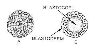
Blastulation is the stage in early animal embryonic development that produces the blastula. In mammalian development the blastula develops into the blastocyst with a differentiated inner cell mass and an outer trophectoderm. The blastula is a hollow sphere of cells known as blastomeres surrounding an inner fluid-filled cavity called the blastocoel. Embryonic development begins with a sperm fertilizing an egg cell to become a zygote, which undergoes many cleavages to develop into a ball of cells called a morula. Only when the blastocoel is formed does the early embryo become a blastula. The blastula precedes the formation of the gastrula in which the germ layers of the embryo form.

<span class="mw-page-title-main">Gastrulation</span> Stage in embryonic development in which germ layers form

Gastrulation is the stage in the early embryonic development of most animals, during which the blastula, or in mammals the blastocyst is reorganized into a two-layered or three-layered embryo known as the gastrula. Before gastrulation, the embryo is a continuous epithelial sheet of cells; by the end of gastrulation, the embryo has begun differentiation to establish distinct cell lineages, set up the basic axes of the body, and internalized one or more cell types including the prospective gut.

<span class="mw-page-title-main">Blastocyst</span> Structure formed around day 5 of mammalian embryonic development

The blastocyst is a structure formed in the early embryonic development of mammals. It possesses an inner cell mass (ICM) also known as the embryoblast which subsequently forms the embryo, and an outer layer of trophoblast cells called the trophectoderm. This layer surrounds the inner cell mass and a fluid-filled cavity known as the blastocoel. In the late blastocyst the trophectoderm is known as the trophoblast. The trophoblast gives rise to the chorion and amnion, the two fetal membranes that surround the embryo. The placenta derives from the embryonic chorion and the underlying uterine tissue of the mother.

<span class="mw-page-title-main">Invagination</span> Process in embryonic development

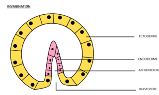
Invagination is the process of a surface folding in on itself to form a cavity, pouch or tube. In developmental biology, invagination is a mechanism that takes place during gastrulation. This mechanism or cell movement happens mostly in the vegetal pole. Invagination consists of the folding of an area of the exterior sheet of cells towards the inside of the blastula. In each organism, the complexity will be different depending on the number of cells. Invagination can be referenced as one of the steps of the establishment of the body plan. The term, originally used in embryology, has been adopted in other disciplines as well.

<span class="mw-page-title-main">Blastocoel</span> Fluid-filled or yolk-filled cavity that forms in the blastula

The blastocoel, also spelled blastocoele and blastocele, and also called cleavage cavity, or segmentation cavity is a fluid-filled or yolk-filled cavity that forms in the blastula during very early embryonic development. At this stage in mammals the blastula develops into the blastocyst containing an inner cell mass, and outer trophectoderm.

A germ layer is a primary layer of cells that forms during embryonic development. The three germ layers in vertebrates are particularly pronounced; however, all eumetazoans produce two or three primary germ layers. Some animals, like cnidarians, produce two germ layers making them diploblastic. Other animals such as bilaterians produce a third layer between these two layers, making them triploblastic. Germ layers eventually give rise to all of an animal's tissues and organs through the process of organogenesis.

<span class="mw-page-title-main">Yolk sac</span> Membranous sac attached to an embryo

The yolk sac is a membranous sac attached to an embryo, formed by cells of the hypoblast layer of the bilaminar embryonic disc. This is alternatively called the umbilical vesicle by the Terminologia Embryologica (TE), though yolk sac is far more widely used. In humans, the yolk sac is important in early embryonic blood supply, and much of it is incorporated into the primordial gut during the fourth week of embryonic development.

<span class="mw-page-title-main">Animal embryonic development</span> Process by which the embryo forms and develops

In developmental biology, animal embryonic development, also known as animal embryogenesis, is the developmental stage of an animal embryo. Embryonic development starts with the fertilization of an egg cell (ovum) by a sperm cell, (spermatozoon). Once fertilized, the ovum becomes a single diploid cell known as a zygote. The zygote undergoes mitotic divisions with no significant growth and cellular differentiation, leading to development of a multicellular embryo after passing through an organizational checkpoint during mid-embryogenesis. In mammals, the term refers chiefly to the early stages of prenatal development, whereas the terms fetus and fetal development describe later stages.

<span class="mw-page-title-main">Inner cell mass</span> Early embryonic mass that gives rise to the fetus

The inner cell mass (ICM) or embryoblast is a structure in the early development of an embryo. It is the mass of cells inside the blastocyst that will eventually give rise to the definitive structures of the fetus. The inner cell mass forms in the earliest stages of embryonic development, before implantation into the endometrium of the uterus. The ICM is entirely surrounded by the single layer of trophoblast cells of the trophectoderm.

<span class="mw-page-title-main">Somatopleuric mesenchyme</span>

In the anatomy of an embryo, the somatopleure is a structure created during embryogenesis when the lateral plate mesoderm splits into two layers. The outer layer becomes applied to the inner surface of the ectoderm, and with it (partially) forms the somatopleure.

<span class="mw-page-title-main">Epiblast</span> Embryonic inner cell mass tissue that forms the embryo itself, through the three germ layers

In amniote embryonic development, the epiblast is one of two distinct cell layers arising from the inner cell mass in the mammalian blastocyst, or from the blastula in reptiles and birds, the other layer is the hypoblast. It drives the embryo proper through its differentiation into the three primary germ layers, ectoderm, mesoderm and endoderm, during gastrulation. The amniotic ectoderm and extraembryonic mesoderm also originate from the epiblast.

<span class="mw-page-title-main">Bilaminar embryonic disc</span>

The bilaminar embryonic disc, bilaminar blastoderm or embryonic disc is the distinct two-layered structure of cells formed in an embryo. In the development of the human embryo this takes place by day eight. It is formed when the inner cell mass, also known as the embryoblast, forms a bilaminar disc of two layers, an upper layer called the epiblast and a lower layer called the hypoblast, which will eventually form into fetus. These two layers of cells are stretched between two fluid-filled cavities at either end: the primitive yolk sac and the amniotic sac.

<span class="mw-page-title-main">Human embryonic development</span> Development and formation of the human embryo

Human embryonic development, or human embryogenesis, is the development and formation of the human embryo. It is characterised by the processes of cell division and cellular differentiation of the embryo that occurs during the early stages of development. In biological terms, the development of the human body entails growth from a one-celled zygote to an adult human being. Fertilization occurs when the sperm cell successfully enters and fuses with an egg cell (ovum). The genetic material of the sperm and egg then combine to form the single cell zygote and the germinal stage of development commences. Embryonic development in the human, covers the first eight weeks of development; at the beginning of the ninth week the embryo is termed a fetus. The eight weeks has 23 stages.

<span class="mw-page-title-main">Cavitation (embryology)</span>

Cavitation is a process in early embryonic development that follows cleavage. Cavitation is the formation of the blastocoel, a fluid-filled cavity that defines the blastula, or in mammals the blastocyst. After fertilization, cell division of the zygote occurs which results in the formation of a solid ball of cells (blastomeres) called the morula. Further division of cells increases their number in the morula, and the morula differentiates them into two groups. The internal cells become the inner cell mass, and the outer cells become the trophoblast. Before cell differentiation takes place there are two transcription factors, Oct-4 and nanog that are uniformly expressed on all of the cells, but both of these transcription factors are turned off in the trophoblast once it has formed.

References

  1. 1 2 Rugh, Roberts (1968). The Mouse: Its Reproduction and Development. Minneapolis, Minn.: Burgess Publishing Company.
  2. 1 2 3 4 5 6 7 8 9 10 11 12 13 14 Dye, Frank J. (2002). Dictionary of Developmental Biology and Embryology. New York: Wiley-Liss. ISBN   9780471443575.
  3. Gilbert, Scott F. (2000). Developmental Biology (6th ed.). Sunderland, Massachusetts: Sinauer Associates. ISBN   0-87893-243-7.