The endometrium is the inner epithelial layer, along with its mucous membrane, of the mammalian uterus. It has a basal layer and a functional layer: the basal layer contains stem cells which regenerate the functional layer. The functional layer thickens and then is shed during menstruation in humans and some other mammals, including apes, Old World monkeys, some species of bat, the elephant shrew and the Cairo spiny mouse. In most other mammals, the endometrium is reabsorbed in the estrous cycle. During pregnancy, the glands and blood vessels in the endometrium further increase in size and number. Vascular spaces fuse and become interconnected, forming the placenta, which supplies oxygen and nutrition to the embryo and fetus. The speculated presence of an endometrial microbiota has been argued against.

The placenta is a temporary embryonic and later fetal organ that begins developing from the blastocyst shortly after implantation. It plays critical roles in facilitating nutrient, gas and waste exchange between the physically separate maternal and fetal circulations, and is an important endocrine organ, producing hormones that regulate both maternal and fetal physiology during pregnancy. The placenta connects to the fetus via the umbilical cord, and on the opposite aspect to the maternal uterus in a species-dependent manner. In humans, a thin layer of maternal decidual (endometrial) tissue comes away with the placenta when it is expelled from the uterus following birth. Placentas are a defining characteristic of placental mammals, but are also found in marsupials and some non-mammals with varying levels of development.

The menstrual cycle is a series of natural changes in hormone production and the structures of the uterus and ovaries of the female reproductive system that makes pregnancy possible. The ovarian cycle controls the production and release of eggs and the cyclic release of estrogen and progesterone. The uterine cycle governs the preparation and maintenance of the lining of the uterus (womb) to receive an embryo. These cycles are concurrent and coordinated, normally last between 21 and 35 days, with a median length of 28 days, and continue for about 30–45 years.

The blastocyst is a structure formed in the early embryonic development of mammals. It possesses an inner cell mass (ICM) also known as the embryoblast which subsequently forms the embryo, and an outer layer of trophoblast cells called the trophectoderm. This layer surrounds the inner cell mass and a fluid-filled cavity known as the blastocoel. In the late blastocyst, the trophectoderm is known as the trophoblast. The trophoblast gives rise to the chorion and amnion, the two fetal membranes that surround the embryo. The placenta derives from the embryonic chorion and the underlying uterine tissue of the mother.
The trophoblast is the outer layer of cells of the blastocyst. Trophoblasts are present four days after fertilization in humans. They provide nutrients to the embryo and develop into a large part of the placenta. They form during the first stage of pregnancy and are the first cells to differentiate from the fertilized egg to become extraembryonic structures that do not directly contribute to the embryo. After blastulation, the trophoblast is contiguous with the ectoderm of the embryo and is referred to as the trophectoderm. After the first differentiation, the cells in the human embryo lose their totipotency because they can no longer form a trophoblast. They become pluripotent stem cells.

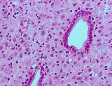
The decidua is the modified mucosal lining of the uterus that forms every month, in preparation for pregnancy. It is shed off each month when there is no fertilised egg to support. The decidua is under the influence of progesterone. Endometrial cells become highly characteristic. The decidua forms the maternal part of the placenta and remains for the duration of the pregnancy. After birth the decidua is shed together with the placenta.

"Cytotrophoblast" is the name given to both the inner layer of the trophoblast or the cells that live there. It is interior to the syncytiotrophoblast and external to the wall of the blastocyst in a developing embryo.

Implantation, also known as nidation, is the stage in the embryonic development of mammals in which the blastocyst hatches, attaches, adheres, and invades into the wall of the female's uterus. Implantation is the first stage of gestation, and, when successful, the female is considered to be pregnant. An implanted embryo is detected by the presence of increased levels of human chorionic gonadotropin (hCG) in a pregnancy test. The implanted embryo will receive oxygen and nutrients in order to grow.
Before the fertilized ovum reaches the uterus, the mucous membrane of the body of the uterus undergoes important changes and is then known as the decidua. The thickness and vascularity of the mucous membrane are greatly increased; its glands are elongated and open on its free surface by funnel-shaped orifices, while their deeper portions are tortuous and dilated into irregular spaces. The interglandular tissue is also increased in quantity, and is crowded with large round, oval, or polygonal cells, termed decidual cells. Their enlargement is due to glycogen and lipid accumulation in the cytoplasm allowing these cells to provide a rich source of nutrition for the developing embryo. Decidual cells are also thought to control the invasion of the endometrium by trophoblast cells.
Human embryonic development or human embryogenesis is the development and formation of the human embryo. It is characterised by the processes of cell division and cellular differentiation of the embryo that occurs during the early stages of development. In biological terms, the development of the human body entails growth from a one-celled zygote to an adult human being. Fertilization occurs when the sperm cell successfully enters and fuses with an egg cell (ovum). The genetic material of the sperm and egg then combine to form the single cell zygote and the germinal stage of development commences. Embryonic development in the human, covers the first eight weeks of development; at the beginning of the ninth week the embryo is termed a fetus. The eight weeks has 23 stages.
Arcuate arteries located in the uterus branch out and supply blood to different layers of the uterus. These arteries meet the myometrial-endometrial junction and lead to straight and endometrial arteries. The endometrium receives blood from endometrial arteries which are also called spiral arteries. Endometrial arteries proliferate rapidly and react to different hormones released. These hormones are progesterone and estrogen released by the ovaries and produced by the endocrine system. The endometrial arteries not only supply blood to the endometrium but are also important during pregnancy. They are the initial site of transportation of blood from the mother to the baby.

Glycodelin(GD) also known as human placental protein-14 (PP-14)progestogen-associated endometrial protein (PAEP) or pregnancy-associated endometrial alpha-2 globulin is a glycoprotein that inhibits cell immune function and plays an essential role in the pregnancy process. In humans is encoded by the PAEP gene.

Homeobox protein Hox-A10 is a protein that in humans is encoded by the HOXA10 gene.
Reproductive immunology refers to a field of medicine that studies interactions between the immune system and components related to the reproductive system, such as maternal immune tolerance towards the fetus, or immunological interactions across the blood-testis barrier. The concept has been used by fertility clinics to explain fertility problems, recurrent miscarriages and pregnancy complications observed when this state of immunological tolerance is not successfully achieved. Immunological therapy is a method for treating many cases of previously "unexplained infertility" or recurrent miscarriage.
Hormonal regulation occurs at every stage of development. A milieu of hormones simultaneously affects development of the fetus during embryogenesis and the mother, including human chorionic gonadotropin (hCG) and progesterone (P4).

Choriogonadotropin subunit beta (CG-beta) also known as chorionic gonadotrophin chain beta is a protein that in humans is encoded by the CGB gene.
Menstruation is the shedding of the uterine lining (endometrium). It occurs on a regular basis in uninseminated sexually reproductive-age females of certain mammal species.

Preimplantation factor(PIF) is a peptide secreted by trophoblast cells prior to placenta formation in early embryonic development. Human embryos begin to express PIF at the 4-cell stage, with expression increasing by the morula stage and continuing to do so throughout the first trimester. Expression of preimplantation factor in the blastocyst was discovered as an early correlate of the viability of the eventual pregnancy. Preimplantation factor was identified in 1994 by a lymphocyte platelet-binding assay, where it was thought to be an early biomarker of pregnancy. It has a simple primary structure with a short sequence of fifteen amino acids without any known quaternary structure. A synthetic analogue of preimplantation factor (commonly abbreviated in studies as sPIF or PIF*) that has an identical amino acid sequence and mimics the normal biological activity of PIF has been developed and is commonly used in research studies, particularly those that aim to study potential adult therapeutics.

Maternal recognition of pregnancy is a crucial aspect of carrying a pregnancy to full term. Without maternal recognition to maintain pregnancy, the initial messengers which stop luteolysis and promote foetal implantation, growth and uterine development finish with nothing to replace them and the pregnancy is lost.
Extravillous trophoblasts(EVTs), are one form of differentiated trophoblast cells of the placenta. They are invasive mesenchymal cells which function to establish critical tissue connection in the developing placental-uterine interface. EVTs derive from progenitor cytotrophoblasts (CYTs), as does the other main trophoblast subtype, syncytiotrophoblast (SYN). They are sometimes called intermediate trophoblast.