Elevated alkaline phosphatase

Last updated
Elevated alkaline phosphatase
Feathery degeneration high mag.jpg
Micrograph showing changes that may be associated with an elevated alkaline phosphatase (cholestasis and feathery degeneration). Liver biopsy. H&E stain.
Specialty Pathology

Elevated alkaline phosphatase occurs when levels of alkaline phosphatase (ALP) exceed the reference range. This group of enzymes has a low substrate specificity and catalyzes the hydrolysis of phosphate esters in a basic environment. The major function of alkaline phosphatase is transporting chemicals across cell membranes. [1] Alkaline phosphatases are present in many human tissues, including bone, intestine, kidney, liver, placenta and white blood cells. [2] Damage to these tissues causes the release of ALP into the bloodstream. Elevated levels can be detected through a blood test. Elevated alkaline phosphate is associated with certain medical conditions [3] or syndromes (e.g., hyperphosphatasia with mental retardation syndrome, HPMRS). It serves as a significant indicator for certain medical conditions, diseases and syndromes.[ citation needed ]

Contents

If the cause for alkaline phosphatase elevation is unknown, isoenzyme studies using electrophoresis can confirm the source of the ALP. Heat stability also distinguishes bone and liver isoenzymes ("bone burns, liver lasts").[ clarification needed ]

It is essential to use age-specific normal reference values, as healthy infants and children usually have levels that would be considered elevated in adults. [4] Additionally, ALP levels are "not well defined" as of 2020, and can vary by sex and by racial group. [5] Latinos tend to have higher normal levels, followed by those of African heritage, Europeans, and Asians or Pacific Islanders tend to have the lowest baseline ALP levels (all groups assuming body mass index within normal weight range). [5] Men tend to have slightly higher normal ALP levels than women, [5] except during pregnancy.

Causes

Liver

Bones

Other unlisted musculoskeletal conditions may also cause elevated alkaline phosphatase.

Obesity

Elevated levels of the alkaline phosphatase enzyme are reported among those who have obesity. A study reported there were higher serum levels of alkaline phosphatase in obese than in the non-obese. With elevated alkaline phosphatase levels, there is an increase in disproportionate intracellular fat depots and thereby releasing itself into the bloodstream. The nature of the relationship between alkaline phosphatase and obesity is still being investigated. [8]

Kidney

Elevated serum levels of alkaline phosphatase has been associated with chronic kidney disease (CKD). [9] Studies have shown that elevated levels may predict mortality independent of bone metabolism factors and liver function tests in CKD. Elevated levels are also associated with diabetes, hypertension, and cardiovascular disease; it was found that elevated levels are associated with elevated serum C-reactive protein (CRP), which could reflect an inflammatory and atherogenic milieu, possibly an alternative cause for elevated serum alkaline phosphatase. [10]

Cancer

Elevated alkaline phosphatase in patients with cancer normally spans[ clarification needed ] throughout the bones or liver. Metastases that exist in the lung, breast, prostate, colon, thyroid, and further organs can also enter the liver or bone. [12] Cancers that are already present in certain organs and tissues can produce alkaline phosphatase elevations if metastasis is not present. In experiential studies, isoenzymes, which are distinct forms of alkaline phosphatase generated by these tumors, can raise the total volume of alkaline phosphatase in the blood. The Regan isoenzyme [ clarification needed ] [13] is one of the best studies[ clarification needed ] of these isoenzymes that is linked to several human cancers. Basically, the Regan isozenzyme is an alkaline phosphatase that is located in the placenta and associated with gonadal and urologic cancers.[ citation needed ]

Other causes

Diagnosis

An alkaline phosphatase isoenzyme test can be done to check for elevated ALP levels. Tissues that contain high levels of ALP include the liver, bile ducts, and bones. Normal levels of ALP range from (44 to 147) U/L (units per liter) and significantly elevated levels may be an indication of conditions such as various types of cancer, bone diseases such as Paget disease, liver diseases such as hepatitis, blood disorders, or other conditions. [20]

Elevated alkaline phosphatase is most commonly caused by liver disease or bone disorders. Testing for ALP primarily consists of obtaining a blood sample from a patient along with several other tests for the disorder in question that may be associated with the increase in ALP in the blood serum. [21] It is possible to distinguish between the different forms (isoenzymes) of ALP produced by different types of body tissues, in order to identify the cause of elevated ALP; this can facilitate choosing a treatment course.

Normally, children and adolescents have higher alkaline phosphatase levels than adults, due to accelerated bone growth. ALP is especially high during the pubertal growth spurt. [22]

Treatments

The following are the most common treatments of elevated alkaline phosphatase. [23]

Related Research Articles

<span class="mw-page-title-main">Jaundice</span> Abnormal pigmentation symptom for disease of the liver

Jaundice, also known as icterus, is a yellowish or greenish pigmentation of the skin and sclera due to high bilirubin levels. Jaundice in adults is typically a sign indicating the presence of underlying diseases involving abnormal heme metabolism, liver dysfunction, or biliary-tract obstruction. The prevalence of jaundice in adults is rare, while jaundice in babies is common, with an estimated 80% affected during their first week of life. The most commonly associated symptoms of jaundice are itchiness, pale feces, and dark urine.

Liver function tests, also referred to as a hepatic panel, are groups of blood tests that provide information about the state of a patient's liver. These tests include prothrombin time (PT/INR), activated partial thromboplastin time (aPTT), albumin, bilirubin, and others. The liver transaminases aspartate transaminase and alanine transaminase are useful biomarkers of liver injury in a patient with some degree of intact liver function.

<span class="mw-page-title-main">Eosinophilia</span> Blood condition

Eosinophilia is a condition in which the eosinophil count in the peripheral blood exceeds 5×10⁸/L (500/μL). Hypereosinophilia is an elevation in an individual's circulating blood eosinophil count above 1.5 × 109/L (i.e. 1,500/μL). The hypereosinophilic syndrome is a sustained elevation in this count above 1.5 × 109/L (i.e. 1,500/μL) that is also associated with evidence of eosinophil-based tissue injury.

<span class="mw-page-title-main">Gamma-glutamyltransferase</span> Class of enzymes

Gamma-glutamyltransferase is a transferase that catalyzes the transfer of gamma-glutamyl functional groups from molecules such as glutathione to an acceptor that may be an amino acid, a peptide or water. GGT plays a key role in the gamma-glutamyl cycle, a pathway for the synthesis and degradation of glutathione as well as drug and xenobiotic detoxification. Other lines of evidence indicate that GGT can also exert a pro-oxidant role, with regulatory effects at various levels in cellular signal transduction and cellular pathophysiology. This transferase is found in many tissues, the most notable one being the liver, and has significance in medicine as a diagnostic marker.

Hypercalcemia, also spelled hypercalcaemia, is a high calcium (Ca2+) level in the blood serum. The normal range is 2.1–2.6 mmol/L (8.8–10.7 mg/dL, 4.3–5.2 mEq/L), with levels greater than 2.6 mmol/L defined as hypercalcemia. Those with a mild increase that has developed slowly typically have no symptoms. In those with greater levels or rapid onset, symptoms may include abdominal pain, bone pain, confusion, depression, weakness, kidney stones or an abnormal heart rhythm including cardiac arrest.

<span class="mw-page-title-main">Paget's disease of bone</span> Disease affecting bone remodeling

Paget's disease of bone is a condition involving cellular remodeling and deformity of one or more bones. The affected bones show signs of dysregulated bone remodeling at the microscopic level, specifically excessive bone breakdown and subsequent disorganized new bone formation. These structural changes cause the bone to weaken, which may result in deformity, pain, fracture or arthritis of associated joints.

<span class="mw-page-title-main">Hyperparathyroidism</span> Increase in parathyroid hormone levels in the blood

Hyperparathyroidism is an increase in parathyroid hormone (PTH) levels in the blood. This occurs from a disorder either within the parathyroid glands or as response to external stimuli.

<span class="mw-page-title-main">Primary sclerosing cholangitis</span> Medical condition

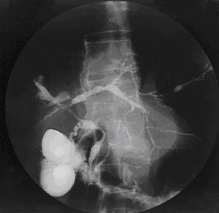
Primary sclerosing cholangitis (PSC) is a long-term progressive disease of the liver and gallbladder characterized by inflammation and scarring of the bile ducts, which normally allow bile to drain from the gallbladder. Affected individuals may have no symptoms or may experience signs and symptoms of liver disease, such as yellow discoloration of the skin and eyes, itching, and abdominal pain.

<span class="mw-page-title-main">Liver disease</span> Medical condition

Liver disease, or hepatic disease, is any of many diseases of the liver. If long-lasting it is termed chronic liver disease. Although the diseases differ in detail, liver diseases often have features in common.

<span class="mw-page-title-main">Alkaline phosphatase</span> Homodimeric protein enzyme

The enzyme alkaline phosphatase has the physiological role of dephosphorylating compounds. The enzyme is found across a multitude of organisms, prokaryotes and eukaryotes alike, with the same general function, but in different structural forms suitable to the environment they function in. Alkaline phosphatase is found in the periplasmic space of E. coli bacteria. This enzyme is heat stable and has its maximum activity at high pH. In humans, it is found in many forms depending on its origin within the body – it plays an integral role in metabolism within the liver and development within the skeleton. Due to its widespread prevalence in these areas, its concentration in the bloodstream is used by diagnosticians as a biomarker in helping determine diagnoses such as hepatitis or osteomalacia.

<span class="mw-page-title-main">Glycogen storage disease type I</span> Medical condition

Glycogen storage disease type I is an inherited disease that prevents the liver from properly breaking down stored glycogen, which is necessary to maintain adequate blood sugar levels. GSD I is divided into two main types, GSD Ia and GSD Ib, which differ in cause, presentation, and treatment. There are also possibly rarer subtypes, the translocases for inorganic phosphate or glucose ; however, a recent study suggests that the biochemical assays used to differentiate GSD Ic and GSD Id from GSD Ib are not reliable, and are therefore GSD Ib.

<span class="mw-page-title-main">Cholestasis</span> Medical condition

Cholestasis is a condition where the flow of bile from the liver to the duodenum is impaired. The two basic distinctions are:

<span class="mw-page-title-main">Osteitis fibrosa cystica</span> Medical condition

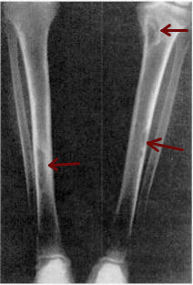
Osteitis fibrosa cystica is a skeletal disorder resulting in a loss of bone mass, a weakening of the bones as their calcified supporting structures are replaced with fibrous tissue, and the formation of cyst-like brown tumors in and around the bone. Osteitis fibrosis cystica (OFC), also known as osteitis fibrosa, osteodystrophia fibrosa, and von Recklinghausen's disease of bone, is caused by hyperparathyroidism, which is a surplus of parathyroid hormone from over-active parathyroid glands. This surplus stimulates the activity of osteoclasts, cells that break down bone, in a process known as osteoclastic bone resorption. The hyperparathyroidism can be triggered by a parathyroid adenoma, hereditary factors, parathyroid carcinoma, or renal osteodystrophy. Osteoclastic bone resorption releases minerals, including calcium, from the bone into the bloodstream, causing both elevated blood calcium levels, and the structural changes which weaken the bone. The symptoms of the disease are the consequences of both the general softening of the bones and the excess calcium in the blood, and include bone fractures, kidney stones, nausea, moth-eaten appearance in the bones, appetite loss, and weight loss.

<span class="mw-page-title-main">Metastatic calcification</span>

Metastatic calcification is deposition of calcium salts in otherwise normal tissue, because of elevated serum levels of calcium, which can occur because of deranged metabolism as well as increased absorption or decreased excretion of calcium and related minerals, as seen in hyperparathyroidism.

<span class="mw-page-title-main">Comprehensive metabolic panel</span> Blood test assessing vital organ health

The comprehensive metabolic panel, or chemical screen, is a panel of 14 blood tests that serves as an initial broad medical screening tool. The CMP provides a rough check of kidney function, liver function, diabetic and parathyroid status, and electrolyte and fluid balance, but this type of screening has its limitations. Abnormal values from a CMP are often the result of false positives and thus the CMP may need to be repeated, requiring a second blood drawing procedure and possibly additional expense for the patient, even though no disease is present. This test is also known as SMA12+2 test.

<span class="mw-page-title-main">ALPL</span> Protein-coding gene in the species Homo sapiens

Alkaline phosphatase, tissue-nonspecific isozyme is an enzyme that in humans is encoded by the ALPL gene.

<span class="mw-page-title-main">Metastatic liver disease</span> Medical condition

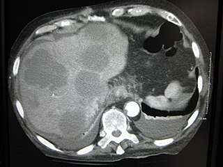
A liver metastasis is a malignant tumor in the liver that has spread from another organ affected by cancer. The liver is a common site for metastatic disease because of its rich, dual blood supply. Metastatic tumors in the liver are 20 times more common than primary tumors. In 50% of all cases the primary tumor is of the gastrointestinal tract; other common sites include the breast, ovaries, bronchus and kidney. Patients with Colorectal cancer will develop liver metastases during the disease

CA 15-3, for Carcinoma Antigen 15-3, is a tumor marker for many types of cancer, most notably breast cancer.

<span class="mw-page-title-main">Cirrhosis</span> Chronic disease of the liver, characterized by fibrosis

Cirrhosis, also known as liver cirrhosis or hepatic cirrhosis, and end-stage liver disease, is the impaired liver function caused by the formation of scar tissue known as fibrosis due to damage caused by liver disease. Damage to the liver leads to repair of liver tissue and subsequent formation of scar tissue. Over time, scar tissue can replace normal functioning tissue, leading to the impaired liver function of cirrhosis. The disease typically develops slowly over months or years. Early symptoms may include tiredness, weakness, loss of appetite, unexplained weight loss, nausea and vomiting, and discomfort in the right upper quadrant of the abdomen. As the disease worsens, symptoms may include itchiness, swelling in the lower legs, fluid build-up in the abdomen, jaundice, bruising easily, and the development of spider-like blood vessels in the skin. The fluid build-up in the abdomen may develop into spontaneous infections. More serious complications include hepatic encephalopathy, bleeding from dilated veins in the esophagus, stomach, or intestines, and liver cancer.

<span class="mw-page-title-main">Feathery degeneration</span>

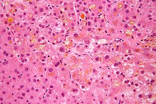
In histopathology, feathery degeneration, formally feathery degeneration of hepatocytes, is a form of liver parenchymal cell death associated with cholestasis.

References

  1. Celik, Handan; Tosun, Midraci; Cetinkaya, Mehmet Bilge; Bektab, Ahmet; Malatyalýodlu, Erdal (2009-08-01). "Markedly elevated serum alkaline phosphatase level in an uncomplicated pregnancy". The Journal of Maternal-Fetal & Neonatal Medicine. 22 (8): 705–707. doi:10.1080/14767050802702323. ISSN   1476-4954. PMID   19544151. S2CID   37866377.
  2. Kaplan, Marshall M. (1972). "Alkaline Phosphatase". New England Journal of Medicine. 286 (4): 200–202. doi:10.1056/nejm197201272860407. PMID   4550137.
  3. Li-Fern H, Rajasoorya C (February 1999). "The elevated serum alkaline phosphatase—the chase that led to two endocrinopathies and one possible unifying diagnosis". Eur. J. Endocrinol. 140 (2): 143–7. doi: 10.1530/eje.0.1400143 . PMID   10069658.
  4. Gholami Bahnemiri M, Mirabedini S, Mohammadi P, Barmaki H, Qaffaripour Z, Rezapour M, Alijanpour M. Determination of serum alkaline phosphatase reference in healthy children aged 1-18 years. Caspian J Intern Med. 2022 Fall;13(4):749-756. doi: 10.22088/cjim.13.4.749. PMID: 36420337; PMCID: PMC9659844.
  5. 1 2 3 Gonzalez H, Imam Z, Wong R, Li J, Lu M, Trudeau S, Gordon S, Imam M, Gish R. Normal alkaline phosphatase levels are dependent on race/ethnicity: NationalGEP Health and Nutrition Examination Survey data. BMJ Open Gastroenterol. 2020 Oct;7(1):e000502. doi: 10.1136/bmjgast-2020-000502. PMID: 33055108; PMCID: PMC7559036.
  6. "Paget's Disease of Bone". The Lecturio Medical Concept Library. Retrieved 9 July 2021.
  7. L Tibi; A W Patrick; P Leslie; A D Toft; A F Smith (1989-07-01). "Alkaline phosphatase isoenzymes in plasma in hyperthyroidism". Clinical Chemistry. Clinchem.org. pp. 1427–1430. doi:10.1093/clinchem/35.7.1427. PMID   2758588 . Retrieved 2014-07-03.
  8. Khan, Abdul Rehman; Awan, Fazli Rabbi; Najam, SyedaSadia; Islam, Mehboob; Siddique, Tehmina; Zain, Maryam (2015). "Elevated serum level of human alkaline phosphatase in obesity". Journal of Pakistan Medical Association . 65 (11): 1182–1185. PMID   26564289 . Retrieved 2016-05-19.
  9. "Chronic Kidney Disease". The Lecturio Medical Concept Library. Retrieved 8 July 2021.
  10. Damera, Sriharsha; Raphael, Kalani L.; Baird, Bradley C.; Cheung, Alfred K.; Greene, Tom; Beddhu, Srinivasan (2011-01-02). "Serum alkaline phosphatase levels associate with elevated serum C-reactive protein in chronic kidney disease". Kidney International. 79 (2): 228–233. doi:10.1038/ki.2010.356. PMC   5260661 . PMID   20881941.
  11. Damera, Sriharsha (September 2010). "Serum alkaline phosphatase levels associate with elevated serum C-reactive protein in chronic kidney disease". International Society of Nephrology. 79 (2): 228–233. doi:10.1038/ki.2010.356. PMC   5260661 . PMID   20881941.
  12. Christensen, Stephen. "Elevated Alkaline Phosphatase & Cancer |". LIVESTRONG.COM . Retrieved 2016-05-10.
  13. Fishman, W. H. (1980-01-01). "Immunology and biochemistry of the Regan isoenzyme". The Prostate. 1 (4): 399–410. doi:10.1002/pros.2990010403. ISSN   0270-4137. PMID   7024957. S2CID   10948252.
  14. Cappello, Maria; Randazzo, Claudia; Bravatà, Ivana; Licata, Anna; Peralta, Sergio; Craxì, Antonio; Almasio, Piero Luigi (2014). "Liver Function Test Abnormalities in Patients with Inflammatory Bowel Diseases: A Hospital-based Survey". Clinical Medicine Insights: Gastroenterology. 7: 25–31. doi:10.4137/CGast.S13125. ISSN   1179-5522. PMC   4069044 . PMID   24966712.
  15. Lange PH, Millan JL, Stigbrand T, Vessella RL, Ruoslahti E, Fishman WH (August 1982). "Placental alkaline phosphatase as a tumor marker for seminoma". Cancer Res. 42 (8): 3244–7. PMID   7093962.
  16. Pruessner, Harold T, “Detecting Celiac Disease in your Patients” American Family Physician: 57 (5), March 1, 1998 pp1023-1034.
  17. Eur J Gastroenterol Hepatol. 2012 Jan;24(1):17–24. doi: 10.1097/MEG.0b013e32834c7b71. Liver-test abnormalities in sarcoidosis. Cremers J, Drent M, Driessen A, Nieman F, Wijnen P, Baughman R, Koek G.
  18. Pareek, S. S., “Liver involvement in secondary syphilis” Digestive Diseases and Sciences: 24 (1), January 1979 pp 41–43.
  19. Matsushita M, Harajiri S, Tabata S, Yukimasa N, Muramoto Y, Komoda T. [Alkaline phosphatase activity in blood group B or O secretors is fluctuated by the dinner intake of previous night]. Rinsho Byori. 2013 Apr;61(4):307-12. Japanese. PMID: 23855186.
  20. "ALP – blood test". MedlinePlus . Retrieved 2016-05-10.
  21. Majeska, Robert (April 10, 1982). "The Effect of 1,25(OH)zD3 on Alkaline Phosphatase in Osteoblastic Osteosarcoma Cell". The Journal of Biological Chemistry. 257 (7): 3362–3365. doi: 10.1016/S0021-9258(18)34781-1 . PMID   6949902.
  22. "Transient hyperphosphatasemia of infancy and early childhood". www.uptodate.com. Retrieved 2017-05-08.
  23. "Elevated Alkaline Phosphatase – Levels, Causes and Treatment". healthh.com. Retrieved 2016-05-19.