General anaesthetics are often defined as compounds that induce a loss of consciousness in humans or loss of righting reflex in animals. Clinical definitions are also extended to include an induced coma that causes lack of awareness to painful stimuli, sufficient to facilitate surgical applications in clinical and veterinary practice. General anaesthetics do not act as analgesics and should also not be confused with sedatives. General anaesthetics are a structurally diverse group of compounds whose mechanisms encompass multiple biological targets involved in the control of neuronal pathways. The precise workings are the subject of some debate and ongoing research.
An evoked potential or evoked response is an electrical potential in a specific pattern recorded from a specific part of the nervous system, especially the brain, of a human or other animals following presentation of a stimulus such as a light flash or a pure tone. Different types of potentials result from stimuli of different modalities and types. Evoked potential is distinct from spontaneous potentials as detected by electroencephalography (EEG), electromyography (EMG), or other electrophysiologic recording method. Such potentials are useful for electrodiagnosis and monitoring that include detections of disease and drug-related sensory dysfunction and intraoperative monitoring of sensory pathway integrity.

General anaesthesia (UK) or general anesthesia (US) is a method of medically inducing loss of consciousness that renders a patient unarousable even with painful stimuli. This effect is achieved by administering either intravenous or inhalational general anaesthetic medications, which often act in combination with an analgesic and neuromuscular blocking agent. Spontaneous ventilation is often inadequate during the procedure and intervention is often necessary to protect the airway. General anaesthesia is generally performed in an operating theater to allow surgical procedures that would otherwise be intolerably painful for a patient, or in an intensive care unit or emergency department to facilitate endotracheal intubation and mechanical ventilation in critically ill patients. Depending on the procedure, general anaesthesia may be optional or required. Regardless of whether a patient may prefer to be unconscious or not, certain pain stimuli could result in involuntary responses from the patient that may make an operation extremely difficult. Thus, for many procedures, general anaesthesia is required from a practical perspective.

Delta waves are high amplitude neural oscillations with a frequency between 0.5 and 4 hertz. Delta waves, like other brain waves, can be recorded with electroencephalography (EEG) and are usually associated with the deep stage 3 of NREM sleep, also known as slow-wave sleep (SWS), and aid in characterizing the depth of sleep. Suppression of delta waves leads to inability of body rejuvenation, brain revitalization and poor sleep.
Awareness under anesthesia, also referred to as intraoperative awareness or accidental awareness during general anesthesia (AAGA), is a rare complication of general anesthesia where patients regain varying levels of consciousness during their surgical procedures. While anesthesia awareness is possible without resulting in any long-term memory of the experience, it is also possible for victims to have awareness with explicit recall, where they can remember the events related to their surgery.
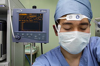
Bispectral index (BIS) is one of several technologies used to monitor depth of anesthesia. BIS monitors are used to supplement Guedel's classification system for determining depth of anesthesia. Titrating anesthetic agents to a specific bispectral index during general anesthesia in adults allows the anesthetist to adjust the amount of anesthetic agent to the needs of the patient, possibly resulting in a more rapid emergence from anesthesia. Use of the BIS monitor could reduce the incidence of intraoperative awareness during anaesthesia. The exact details of the algorithm used to create the BIS index have not been disclosed by the company that developed it.

Heart rate variability (HRV) is the physiological phenomenon of variation in the time interval between heartbeats. It is measured by the variation in the beat-to-beat interval.
The sensorimotor rhythm (SMR) is a brain wave. It is an oscillatory idle rhythm of synchronized electric brain activity. It appears in spindles in recordings of EEG, MEG, and ECoG over the sensorimotor cortex. For most individuals, the frequency of the SMR is in the range of 7 to 11 Hz.
In mathematics, in the area of statistical analysis, the bispectrum is a statistic used to search for nonlinear interactions.
Theta waves generate the theta rhythm, a neural oscillation in the brain that underlies various aspects of cognition and behavior, including learning, memory, and spatial navigation in many animals. It can be recorded using various electrophysiological methods, such as electroencephalogram (EEG), recorded either from inside the brain or from electrodes attached to the scalp.

Transcranial Doppler (TCD) and transcranial color Doppler (TCCD) are types of Doppler ultrasonography that measure the velocity of blood flow through the brain's blood vessels by measuring the echoes of ultrasound waves moving transcranially. These modes of medical imaging conduct a spectral analysis of the acoustic signals they receive and can therefore be classified as methods of active acoustocerebrography. They are used as tests to help diagnose emboli, stenosis, vasospasm from a subarachnoid hemorrhage, and other problems. These relatively quick and inexpensive tests are growing in popularity. The tests are effective for detecting sickle cell disease, ischemic cerebrovascular disease, subarachnoid hemorrhage, arteriovenous malformations, and cerebral circulatory arrest. The tests are possibly useful for perioperative monitoring and meningeal infection. The equipment used for these tests is becoming increasingly portable, making it possible for a clinician to travel to a hospital, to a doctor's office, or to a nursing home for both inpatient and outpatient studies. The tests are often used in conjunction with other tests such as MRI, MRA, carotid duplex ultrasound and CT scans. The tests are also used for research in cognitive neuroscience.
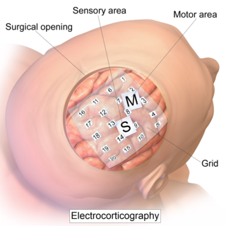
Electrocorticography (ECoG), a type of intracranial electroencephalography (iEEG), is a type of electrophysiological monitoring that uses electrodes placed directly on the exposed surface of the brain to record electrical activity from the cerebral cortex. In contrast, conventional electroencephalography (EEG) electrodes monitor this activity from outside the skull. ECoG may be performed either in the operating room during surgery or outside of surgery. Because a craniotomy is required to implant the electrode grid, ECoG is an invasive procedure.

Electroencephalography (EEG) is a method to record an electrogram of the spontaneous electrical activity of the brain. The biosignals detected by EEG have been shown to represent the postsynaptic potentials of pyramidal neurons in the neocortex and allocortex. It is typically non-invasive, with the EEG electrodes placed along the scalp using the International 10–20 system, or variations of it. Electrocorticography, involving surgical placement of electrodes, is sometimes called "intracranial EEG". Clinical interpretation of EEG recordings is most often performed by visual inspection of the tracing or quantitative EEG analysis.

In medicine, monitoring is the observation of a disease, condition or one or several medical parameters over time.
Quantitative electroencephalography is a field concerned with the numerical analysis of electroencephalography (EEG) data and associated behavioral correlates.

Burst suppression is an electroencephalography (EEG) pattern that is characterized by periods of high-voltage electrical activity alternating with periods of no activity in the brain. The pattern is found in patients with inactivated brain states, such as from general anesthesia, coma, or hypothermia. This pattern can be physiological, as during early development, or pathological, as in diseases such as Ohtahara syndrome.
Electroencephalography (EEG) is the science of recording the spontaneous rhythmic electrical activity of a living brain through electrodes on the scalp. Brain rhythms have origins similar to the electrical activity of the heart. The rhythmic activity varies in frequency and amplitude with age, attention, sleep, and chemical concentrations of oxygen, carbon dioxide, glucose, ammonia, and hormones. Chemicals that affect brain functions change brain rhythms in systematic and identifiable ways. As new psychoactive drugs were discovered that changed behavior, the basis for the science of psychopharmacology, the accompanying changes in the rhythms were found to be drug class specific. The measurement of the changes in rhythms became the basis for the science of pharmaco-EEG.
EEG analysis is exploiting mathematical signal analysis methods and computer technology to extract information from electroencephalography (EEG) signals. The targets of EEG analysis are to help researchers gain a better understanding of the brain; assist physicians in diagnosis and treatment choices; and to boost brain-computer interface (BCI) technology. There are many ways to roughly categorize EEG analysis methods. If a mathematical model is exploited to fit the sampled EEG signals, the method can be categorized as parametric, otherwise, it is a non-parametric method. Traditionally, most EEG analysis methods fall into four categories: time domain, frequency domain, time-frequency domain, and nonlinear methods. There are also later methods including deep neural networks (DNNs).

High-frequency oscillations (HFO) are brain waves of the frequency faster than ~80 Hz, generated by neuronal cell population. High-frequency oscillations can be recorded during an electroencephalagram (EEG), local field potential (LFP) or electrocorticogram (ECoG) electrophysiology recordings. They are present in physiological state during sharp waves and ripples - oscillatory patterns involved in memory consolidation processes. HFOs are associated with pathophysiology of the brain like epileptic seizure and are often recorded during seizure onset. It makes a promising biomarker for the identification of the epileptogenic zone. Other studies points to the HFO role in psychiatric disorders and possible implications to psychotic episodes in schizophrenia.
Total intravenous anesthesia (TIVA) refers to the intravenous administration of anesthetic agents to induce a temporary loss of sensation or awareness. The first study of TIVA was done in 1872 using chloral hydrate, and the common anesthetic agent propofol was licensed in 1986. TIVA is currently employed in various procedures as an alternative technique of general anesthesia in order to improve post-operative recovery.
