
Macrocephaly is a condition in which circumference of the human head is abnormally large. It may be pathological or harmless, and can be a familial genetic characteristic. People diagnosed with macrocephaly will receive further medical tests to determine whether the syndrome is accompanied by particular disorders. Those with benign or familial macrocephaly are considered to have megalencephaly.

Bone healing, or fracture healing, is a proliferative physiological process in which the body facilitates the repair of a bone fracture.

An osteosarcoma (OS) or osteogenic sarcoma (OGS) is a cancerous tumor in a bone. Specifically, it is an aggressive malignant neoplasm that arises from primitive transformed cells of mesenchymal origin and that exhibits osteoblastic differentiation and produces malignant osteoid.

Osteomyelitis (OM) is an infection of bone. Symptoms may include pain in a specific bone with overlying redness, fever, and weakness. The long bones of the arms and legs are most commonly involved in children e.g. the femur and humerus, while the feet, spine, and hips are most commonly involved in adults.
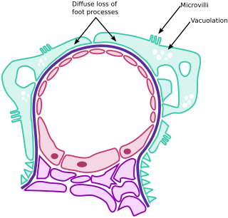
Minimal change disease is a disease affecting the kidneys which causes a nephrotic syndrome. Nephrotic syndrome leads to the loss of significant amounts of protein in the urine, which causes the widespread edema and impaired kidney function commonly experienced by those affected by the disease. It is most common in children and has a peak incidence at 2 to 6 years of age. MCD is responsible for 10–25% of nephrotic syndrome cases in adults. It is also the most common cause of nephrotic syndrome of unclear cause (idiopathic) in children.

A periosteal reaction is the formation of new bone in response to injury or other stimuli of the periosteum surrounding the bone. It is most often identified on X-ray films of the bones.

Hypophosphatasia (; also called deficiency of alkaline phosphatase, phosphoethanolaminuria, or Rathbun's syndrome; sometimes abbreviated HPP) is a rare, and sometimes fatal, metabolic bone disease. Clinical symptoms are heterogeneous, ranging from the rapidly fatal, perinatal variant, with profound skeletal hypomineralization, respiratory compromise or vitamin B6 dependent seizures to a milder, progressive osteomalacia later in life. Tissue non-specific alkaline phosphatase (TNSALP) deficiency in osteoblasts and chondrocytes impairs bone mineralization, leading to rickets or osteomalacia. The pathognomonic finding is subnormal serum activity of the TNSALP enzyme, which is caused by one of 388 genetic mutations identified to date, in the gene encoding TNSALP. Genetic inheritance is autosomal recessive for the perinatal and infantile forms but either autosomal recessive or autosomal dominant in the milder forms.
Pachydermoperiostosis (PDP) is a rare genetic disorder that affects both bones and skin. Other names are idiopathic hypertrophic osteoarthropathy or Touraine-Solente-Golé syndrome. It is mainly characterized by pachyderma, periostosis and finger clubbing.
Craniomandibular osteopathy, also known as lion's jaw, is a developmental disease in dogs causing extensive bony changes in the mandible and skull. In this disease, a cyclical resorption of normal bone and replacement by immature bone occurs along the inner and outer surfaces of the affected bones. It usually occurs between the ages of 3 and 8 months. Breeds most commonly affected include the West Highland White Terrier, Scottish Terrier, Cairn Terrier, and Boston Terrier. It is rare in large-breed dogs, but it has been reported. Symptoms include firm swelling of the jaw, drooling, pain, and difficulty eating.
A Brodie abscess is a subacute osteomyelitis, which may persist for years before progressing to a chronic, frank osteomyelitis. Classically, this may present after progression to a draining abscess extending from the tibia out through the skin. Occasionally acute osteomyelitis may be contained to a localized area and walled off by fibrous and granulation tissue. This is termed Brodie's abscess.

Commonly known as a dental cyst, the periapical cyst is the most common odontogenic cyst. It may develop rapidly from a periapical granuloma, as a consequence of untreated chronic periapical periodontitis.

The endosteum is a thin vascular membrane of connective tissue that lines the inner surface of the bony tissue that forms the medullary cavity of long bones.

Osteoblastoma is an uncommon osteoid tissue-forming primary neoplasm of the bone.

Ewing sarcoma is a type of cancer that may be a bone sarcoma or a soft-tissue sarcoma. Symptoms may include swelling and pain at the site of the tumor, fever, and a bone fracture. The most common areas where it begins are the legs, pelvis, and chest wall. In about 25% of cases, the cancer has already spread to other parts of the body at the time of diagnosis. Complications may include a pleural effusion or paraplegia.
Orthopedic pathology, also known as bone pathology is a subspecialty of surgical pathology which deals with the diagnosis and feature of many bone diseases, specifically studying the cause and effects of disorders of the musculoskeletal system. It uses gross and microscopic findings along with the findings of in vivo radiological studies, and occasionally, specimen radiographs to diagnose diseases of the bones.

Schindler disease, also known as Kanzaki disease and alpha-N-acetylgalactosaminidase deficiency is a rare disease found in humans. This lysosomal storage disorder is caused by a deficiency in the enzyme alpha-NAGA (alpha-N-acetylgalactosaminidase), attributable to mutations in the NAGA gene on chromosome 22, which leads to excessive lysosomal accumulation of glycoproteins. A deficiency of the alpha-NAGA enzyme leads to an accumulation of glycosphingolipids throughout the body. This accumulation of sugars gives rise to the clinical features associated with this disorder. Schindler disease is an autosomal recessive disorder, meaning that one must inherit an abnormal allele from both parents in order to have the disease.
A child bone fracture or a pediatric fracture is a medical condition in which a bone of a child is cracked or broken. About 15% of all injuries in children are fracture injuries. Bone fractures in children are different from adult bone fractures because a child's bones are still growing. Also, more consideration needs to be taken when a child fractures a bone since it will affect the child in his or her growth.
Osteomyelitis of the jaws is osteomyelitis which occurs in the bones of the jaws. Historically, osteomyelitis of the jaws was a common complication of odontogenic infection. Before the antibiotic era, it was frequently a fatal condition.
Glomerulocystic kidney disease (GCKD) is a cystic disorder of the kidneys. GCKD involves cystic dilation of Bowman's capsule. It can occur with or without congenital abnormality.
An occult fracture is a fracture that is not readily visible, generally in regard to projectional radiography ("X-ray"). Radiographically, occult and subtle fractures are a diagnostic challenge. They may be divided into 1) high energy trauma fracture, 2) fatigue fracture from cyclical and sustained mechanical stress, and 3) insufficiency fracture occurring in weakened bone. Independently of the cause, the initial radiographic examination can be negative either because the findings seem normal or are too subtle. Advanced imaging tools such as computed tomography, magnetic resonance imaging (MRI), and scintigraphy are highly valuable in the early detection of these fractures.
