Monoclonal B-cell lymphocytosis

Last updated
Monoclonal B-cell lymphocytosis
Other namesmonoclonal lymphocytosis of undetermined significance
Specialty Hematology, oncology
Symptoms None
Complications May progress to chronic lymphocytic leukemia or certain lymphoma types; increased risk of developing non-hematologic cancers, serious infections, and kidney disease
Durationchronic
TypesCLL/SLL, atypical CLL/SLL, non-CLL/SLL, and MBL-MZ

Monoclonal B-cell lymphocytosis (MBL) is an asymptomatic condition in which individuals have increased blood levels of particular subtypes of monoclonal lymphocytes (i.e. an aberrant and potentially malignant group of lymphocytes produced by a single ancestral cell). This increase must persist for at least 3 months. [1] The lymphocyte subtypes are B-cells that share certain features with the abnormal clones of lymphocytes that circulate in chronic lymphocytic leukemia/small lymphocyte lymphoma (CLL/SLL) or, less frequently, other types of B-cell malignancies. Some individuals with these circulating B-cells develop CLL/SLL or the lymphoma types indicated by their circulating monoclonal B-cells. Hence, MBL is a premalignant disorder [2]

Contents

In 2017, the World Health Organization (WHO) reclassified MBL as a distinct entity in which individuals have: 1) an excessive number of circulating monoclonal B-cells; 2) lack evidence of lymphadenopathy, organomegaly, or other tissue involvements caused by these cells; 3) no features of any other B cell lymphoproliferative disease such as one of the B-cell lymphomas; and 4) evidence that these cells have either a CLL/SLL, atypical CLL/SLL, or non-CLL/SLL phenotype based on these cells' expression of certain marker proteins. [3] [4] A fourth MBL phenotype, monoclonal B-cell lymphocytosis-marginal zone (i.e. MBL-MZ) appears to be emerging as a distinct form of non-CLL/SLL MBL. [2]

MBL consist of two groups: low-count MBL has blood B-cell counts <0.5x9 cells/liter (i.e. 0.5x9/L) whereas high-count MBL has blood B-cell counts ≥0.5x9/L but <5x10⁹/L. [5] While low-count MBL does not progress to a malignant disease, high-count MBL does so at a rate of 1-2% per year. [3] MLP-MZ is an exception to this rule in that it s usually associated with B-cell counts >3x10⁹/L and all cases, regardless of B cell counts, have a somewhat higher risk of progressing to a malignant stage. [6]

The incidence of all MBL phenotypes increases with age and is strikingly high in the elderly. Below age 40, MBL's incidence is <1% of the general population in most countries but above this age it is found in ~10% of all individuals. The disorder's incidence in individuals >90 may be as high as 75%. Age along with B-cell blood counts, MBL phenotype, and certain genomic abnormalities in the monoclonal B cells are critical considerations in evaluating the clinical implications of MBL and its need for management. [2]

MBL phenotypes

MBL falls into three phenotypes that are distinguished based on the cell surface marker proteins which they express viz., the CLL/SLL, atypical CLL/SLL, and non-CLL/SLL phenotypes. These markers are: CD5, CD19, CD20, CD23, and immunoglobulins (Ig) (either Ig light chains or complete Ig, i.e. light chains bound to Ig heavy chains. [2] [3] Distinguishing between these phenotypes is important because they progress to different lymphocyte malignancies. The following table gives the markers for the three MBL phenotypes with (+) indicating the expression (either dim, moderate, or bright depending or the intensity of their expression), (−) indicating the absence of expression, and na indicating not applicable as determined using fluorescent probes that bind the marker proteins. Detection of fluorescent probe binding by the cells requires the use of flow cytometry preferably employing 6 to 8 different fluorescent probes that bind to different markers on 5 million cells from the patient's blood. The table also includes the percentage of MLB cases with the phenotype and the malignancies to which they progress. [7]

MBL phenotypeCD5 [3] CD19 [3] CD20 [3] CD23 [3] Light chains or immunolboulins [3] percent with phenotype [2] potential malignancy complication [2] [8] [9]
CLL/SLL MLB+++ (dim)+light chain Ig, either +, + (dim), or −68-75%CLL/SLL
Atypical CLL/SLL MLB+++ (bright)− or +complete Ig, either + (moderate) or + (bright)~15% mantle cell lymphoma, follicular lymphoma
Non-CLL/SLL MLBeither − or + (dim)++nalight chain Ig, either + (moderate) or + (bright)~14% splenic marginal zone lymphoma, splenic lymphoma/leukemia unclassifiable

Monoclonal B-cell lymphocytosis of the marginal zone

Cases of non-CLL/SLL MBL in which the monoclonal B cells do not express CD5, CD23, CD10, or CD103 but strongly express CD79B and light chain Ig have been tentatively designated as having monoclonal B-cell lymphocytosis of the marginal zone (i.e. CBL-MZ). This term is used because normal marginal zone B-cell lymphocytes express these markers. Individuals with CBL-MZ commonly present with: B-cell blood counts that are extremely high (>4.0x10⁹; range 3.0x10⁹/L to 37.1x10⁹/L);, [6] represent a large percentage of cases that would otherwise be designated as non-CLL/SLL MLB; [2] often have an IgM monoclonal gammopathy, i.e. high blood levels of a monoclonal IgM antibody; and in addition to the IgM gammopathy, other features that are seen in Waldenström's macroglobulinemia and IgM monoclonal gammopathy of undetermined significance. These individuals are more likely than those with other types of MBL to have their disorder progress to a malignancy. These malignancies appear to have been primarily marginal zone B-cell lymphomas of the splenic marginal zone B-cell, splenic lymphoma/leukemia unclassifiable, hairy cell leukemia, and possibly Waldenström's macroglobulinemia. MBL-MZ requires further studies to evaluate its frequencies, rate of progression to malignancy, and treatment. [6]

Pathophysiology

Genome abnormalities

Most studies on the genomic abnormalities in MBL did not distinguish between the disorder's phenotypes. However, familial studies have found that hereditary factors can contributor to the development of specifically CLL/SLL MLB. [10] Of all the hematologic malignancies, CLL/SLL is the most likely to afflict multiple family members with estimates of familial CLL/SLL ranging from 6 to 10% of all CLL/SLL cases. About 18% of first-degree relatives of individuals with familial CLL/SLL and ~16% of the close relatives of patients with non-hereditary CLL/SLL have CLL/SLL MBL. [9] These associations strongly suggest that inheritable genomic abnormalities contribute to the development of CLL/SLL MLB and, possibly, the progression of this disorder to CLL/SLL. [9] [11]

Chromosome abnormalities, single nucleotide polymorphisms (SNPs, i.e. substitutions of a single nucleotide in a DNA sequence at a specific position in the genome) and gene mutations, while each occurs in <15% of cases, [12] are present in CLL/SLL MBL and to some extent are similar to those found in CLL/SLL. For example, position 21.33 to 22.2 on the long (i.e. "q" ) arm of chromosome 13 is a potential susceptibility locus for familial CLL/SLL. This locus has been identified not only in individuals with familial CLL/SLL but also in their blood relatives who have CLL/SLL MBL. [11] More than 20 SNPs are confirmed risk factors for the development of CLL/SLL; at least 6 of these are also risk factors for CLL/SLL MBL. [9] Finally, the following studies were done on individuals defined as having MBL but din not give its phenotype. Here, these patients are presumed to have the CLL/SLL MBL phenotype. Individuals with low-count and high-count MBL shared with CLL/SLL patients many genomic abnormalities including: deletions of the long arm (i.e. "q" arm) of chromosomes 11 and 13; deletion of the short arm (i.e. "p" arm) of chromosome 17; trisomy of chromosome 12; and mutations in NOTCH1, BIRC3, SF3B1, MYD88, ATM , and TP53 genes. In general, the presence and frequency of the mutations in high count MBL were closer than the low count MLB in resembling those in CLL/SLL. [2] All three groups had mutations in the IGH@ region of chromosome 14. This region contains the complex gene that encodes the VDJ region of the heavy chain component of antibodies. Among these mutations, IVGH4-59/61 is most often mutated in low-count MBL while IGHV1-69, IGH2-5, IGHV3-23, IGH23-33, IGHV3-48, and IGHV4-34 are most often mutated in high-count MBL and CLL/SLL. [2] [9] Finally, genetic abnormalities such as the deletion of the q arm in chromosome 13 found in low count MBL are more commonly associated with a favorable prognosis in CLL/SLL while those found in high count MBL, e.g. deletions in the q arm of chromosome 11 or p arm of chromosome 17 [13] are commonly associated with unfavorable prognoses in CLL/SLL. [9]

Individuals with MBL-MZ have monoclonal B cell cells that bear complex and distinctive genomic abnormalities, such as deletions and translocations involving chromosome 7, presence of an isochromosome 17, and, rarely, mutations in the NOTCH2 and KLF2 genes. [6] Some of these genomic abnormalities are similar to those found in splenic marginal zone lymphomas and some of the MBL-MZ patients that bore these abnormalities developed this lymphoma. [2] The genetic abnormalities in atypical and non-CLL/SLL MBL have not been well-defined.[ citation needed ]

The cited studies suggest that there is a step-wise accumulation of genomic abnormalities that lead to CLL/SLL MBL and MBL-MZ and then to overt malignancy. [9] It presumed that similar accumulations led to the development of atypical and non-Cll/SLL MLB and than to their respective malignancies. [6] However, given the number and diversity of these abnormalities, it is unclear which are critical determinants of these disorders. [2] A recent model based on laboratory studies of normal CD19+ B cell, monoclonal CLL/SLL MBL cells, and CLL/SLL malignant cells found that their accumulation of genomic abnormalities may be caused by progressively increasing: 1) double strand breaks in DNA, 2) activation of non-homologous end joining error-prone DNA repair mechanisms, and 3) consequential accumulation of genomic abnormalities which promote the clonal development, survival, proliferation, and ultimately malignancy of the involved B cells. [14]

Infectious diseases

Studies have identified MBL in ~30% of patients infected with the hepatitis C virus, found increased risks of CLL/SLL-MLB in patients with pneumonia, and decreased risk of CLL/CSS MBL in patients who have been vaccinated for influenza or pneumonia. Herpes Zoster and various upper respiratory tract infections are also considered to be risk factors for developing CLL/SLL MBL. It seems possible that the pathogens involved in these diseases provide antigens that stimulate the development of MBL although further studies are required to explore this hypothesis further. [9]

Blood transfusion

One study diagnosed MBL in 0.14% of blood donors and suggested a possibility that MBL is transmitted through blood transfusions. [15] This concern as well as the concern of transmitting CLL/SLL in transfusions of blood from patients with CLL/SLL has been voiced elsewhere. [16] However, a study conducted in Sweden and Denmark on 7,413 recipients of blood from 796 donors diagnosed with CLL/SLL found no evidence of CLL/SLL clustering among recipients of blood from these donors. It therefore appears that CLL/SLL and, by implication, CLL/SLL MBL have little or no ability to be transmitted by blood transfusions, at least when the donors and recipients are unrelated. [17]

Bone marrow transplantation

Rare cases of MBL have been reported to develop in individuals who receive a, autologous stem cell bone marrow transplant from donors who have MBL. Currently, the risk of this development is unclear and requires further study. In those special cases where related donors are used for transplantation, it may be useful to screen these donors for MBL. [9]

Diagnosis

Blood B-cell counts

Individuals with MBL usually present with unexplained increases in blood lymphocyte counts (i.e. lymphocytosis). The most common causes for lymphocytosis are viral infections, autoimmune diseases (particularly connective tissue diseases), hypersensitivity reactions, acute stress reactions, and prior splenectomy. [9] Unlike many individuals with lymphocytosis due to the latter disorders, individuals with MBL are asymptomatic, may have a family history of CLL/SLL, are usually >40 years old, and may have a history of serious infections (high-count MBL is ~3-fold more likely than age-matched healthy controls to have a history of serious infections and infection-related hospitalizations [9] ). [2] The diagnosis of MBL in these patients depends on finding 0.5-5x10⁹ monoclonal B cells that express the makers characteristic of CLL/SLL MLB, atypical CLL/SLL MLB, non-CLL/SLL MLB, or MLB-MZ. [3] However, individuals with CBL-MZ commonly present with B-cell blood counts that are extremely high (>4.0x10⁹; range 3.0x10⁹/L to 37.1x10⁹/L); [2] and may have an IgM monoclonal gammopathy. [6]

Bone marrow involvement

Most individuals with MBL have at presentation an abnormal infiltrate of monoclonal B-cells in their bone marrow as determined by biopsy. These B cells represent a median value of ~20% of all nucleated cells in the marrow. Regardless of the percentage of these cells, the presence of monoclonal B cells in bone marrow does not appear to influence the malignant progression of MBL [9] and is not part of the criteria used to diagnose the disorder. [3]

Nodal MBL

MBL patients may present with asymptomatic lymphadenopathy (i.e. lymph nodes that are enlarged or abnormal in consistency). In one study, ~42% of MBL patients had enlarged lymph nodes as detected by CT scans. Nonetheless, these patients' rate of progression to malignant disease does not differ from that for MBL patients that had normal CT scans. However, patients who have grossly enlarged (i.e. >1.5 centimeters) (cm) lymph nodes on physical examination do have a greater risk of progression. It has been recommended that patients with ≥1 lymph node larger than 1.5 cm be diagnosed as having Cll/SLL, that patients with lymph nodes ≤1.5 cm in size be diagnoses as having normal MBL, and that CT scans should not be used in diagnosing or staging MBL. [9]

MBL with autoimmune cytopenia

Rare patients with MBL may present with the autoimmune disease-induced cytopenias of hemolytic anemia (reduced circulating red blood cell numbers) or thrombocytopenic purpura (reduced circulating platelet numbers). [2] In the past, cases of CLL/SLL MBL associated with an autoimmune disease were diagnosed as CLL/SLL. However, patients with these autoimmune disorders who have very small B cell clones either never develop a lymphocyte malignancy or, rarely, do so and only after many years. Consequently, it is now widely recognized that such cases, when associated with very small numbers of monoclonal B-cells, are best diagnosed as CLL/SLL MBL with autoimmune cytopenia rather than CLL/SLL. [9]

Tissue-based MBL

Monoclonal CLL/SLL phenotype B cells have been found using sensitive flow cytometry methods in various tissues. [3] They have been identified as infiltrates in 1.9% of liver biopsies and 0.4% of prostate tissues obtained at prostatectomy. While the significance of these lesions is unknown, the presence of extensive infiltrations that replace normal tissue is more consistent with a diagnose of CLL/SLL than CLL/SLL MBL. [9]

Differential diagnosis

The key factor that distinguishes low-count CLL/SLL-MLB, high-count CLL/SLL-MLB, and CLL/SLL is the number of circulating monoclonal B cells, as described above. However, the other MLB phenotypes may progress to and/or be mimicked by various monoclonal B-cell lymphocyte malignancies. The key cell markers and other points that help distinguish the following MBL phenotypes from these malignancies include the following (refer to Table for comparisons to non-malignant predecessor cells):

In situ lymphoid neoplasia

In situ lymphoid neoplasia (ISLN), a lymphocyte disorder newly categorized by the World Health Organization (2016,) has several features in common with MLB. Like MBL, it is an asymptomatic, premalignant disorder of B-cells that is associated with the circulation of these cells and may progress to follicular lymphoma, mantle cell lymphoma, or CLL/SLL. ISLN differs from MBL in that its neoplastic B-cells accumulate in the follicles of lymphoid tissue, usually circulate in very low numbers, and bear distinctive genetic abnormalities that differ from those in MBL. ISNL is diagnosed based on, and requiring, the finding of these neoplastic B-cells in lymphoid follicles. [20]

Treatment

Low-count MBL is an indolent disorder that in virtually all individuals does not progress to a malignant phase. Overall survival in low count MBL does not differ from that found in age-matched healthy individuals. [2] MBL-MZ is an exception to this rule: this disorder generally presents with high monoclonal B-cell counts and regardless of the level of these counts may progress to a malignant phase at a greater than that found in other forms of MBL. [6]

Depending on its phenotype (see above Table), high-count MBL progresses to CLL/SLL, mantel cell lymphoma, follicular lymphoma, splenic marginal zone lymphoma, or splenic lymphoma/leukemia unclassifiable at a rate of 1-2% per year [21] whereas MBL-MZ progresses to a marginal zone B-cell lymphoma, Waldenström's macroglobulinemia, or Hairy cell leukemia at a ~3% per year. [6] Factors predisposing to this progression in CLL/SLL MBL include the expression of CD38 cell-surface glycoprotein on the monoclonal B-cells, deletion of the short arm of chromosome 17 in these cells, [4] high serum levels of beta-2 macroglobulin, [2] and circulating B cell levels >10x109/L. [9] There is relatively little information on the features promoting the progression of atypical CLL/SLL MBL, non-CLL/SLL MBL, and MBL-MZ to their respective lymphomas.[ citation needed ]

Individuals with high-count MBL (studies based primarily on the CLL/SLL phenotype) are at an increased risk for developing: 1) cancers of the breast, lung, and gastrointestinal tract in up to 13% of all cases; 2) autoimmune hemolytic anemia and immune thrombocytopenic purpura; 3) unexplained kidney disease as manifested by chronic kidney disease and/or the nephrotic syndrome; and 4) serious infections. [2] While earlier studies suggested that only very high-count MBL (i.e. >10x109 B-cells/L) was associated with a decrease in survival, [2] more recent studies indicate that high-count MBL (i.e. (i.e. >0.5x10⁹ B-cells/L) do show a shorter overall survival. [2] In addition to having very high numbers of B-cells, high-count CLL/SLL MLB patients whose monoclonal B cell clone lacks mutions in IGVH genes (see above section on genome abnormalities) or whose |β2 macroglobulin is elevated have a shorten survival. However, the shortened survival times in high-count CLL/SLL MBL does not appear due to its progression to CLL/SLL. Rather, this shortened survival appears due to the disorders' susceptibility to serious infections, other types of cancers, immune cytopenias, and renal disease. [2]

Recommended treatments for patients with high-count MBL [9] and MBL-MZ [6] include yearly follow-up evaluations to test for the malignant progression of their disorder and for the development of other forms of cancer, infections, immune cytopenias, and renal disease. It may also be beneficial to ensure that high-count MLB patients are up to date on vaccinations including those for influenza, pneumococcal pneumonia, and tetanus before they become more severely immunocompromised by the progression of their disorder. In all cases live vaccines should be avoided in these individuals. [2]

See also

Related Research Articles

<span class="mw-page-title-main">Eosinophilia</span> Blood condition

Eosinophilia is a condition in which the eosinophil count in the peripheral blood exceeds 5×10⁸/L (500/μL). Hypereosinophilia is an elevation in an individual's circulating blood eosinophil count above 1.5 × 109/L (i.e. 1,500/μL). The hypereosinophilic syndrome is a sustained elevation in this count above 1.5 × 109/L (i.e. 1,500/μL) that is also associated with evidence of eosinophil-based tissue injury.

<span class="mw-page-title-main">Lymphocytosis</span> Medical condition

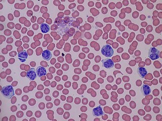
Lymphocytosis is an increase in the number or proportion of lymphocytes in the blood. Absolute lymphocytosis is the condition where there is an increase in the lymphocyte count beyond the normal range while relative lymphocytosis refers to the condition where the proportion of lymphocytes relative to white blood cell count is above the normal range. In adults, absolute lymphocytosis is present when the lymphocyte count is greater than 5000 per microliter (5.0 x 10⁹/L), in older children greater than 7000 per microliter and in infants greater than 9000 per microliter. Lymphocytes normally represent 20% to 40% of circulating white blood cells. When the percentage of lymphocytes exceeds 40%, it is recognized as relative lymphocytosis.

<span class="mw-page-title-main">Chronic lymphocytic leukemia</span> Medical condition

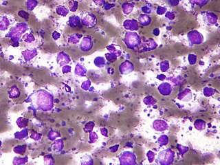
Chronic lymphocytic leukemia (CLL) is a type of cancer in which the bone marrow makes too many lymphocytes. Early on, there are typically no symptoms. Later, non-painful lymph node swelling, feeling tired, fever, night sweats, or weight loss for no clear reason may occur. Enlargement of the spleen and low red blood cells (anemia) may also occur. It typically worsens gradually over years.

<span class="mw-page-title-main">Tumors of the hematopoietic and lymphoid tissues</span> Medical condition

Tumors of the hematopoietic and lymphoid tissues or tumours of the haematopoietic and lymphoid tissues are tumors that affect the blood, bone marrow, lymph, and lymphatic system. Because these tissues are all intimately connected through both the circulatory system and the immune system, a disease affecting one will often affect the others as well, making aplasia, myeloproliferation and lymphoproliferation closely related and often overlapping problems. While uncommon in solid tumors, chromosomal translocations are a common cause of these diseases. This commonly leads to a different approach in diagnosis and treatment of hematological malignancies. Hematological malignancies are malignant neoplasms ("cancer"), and they are generally treated by specialists in hematology and/or oncology. In some centers "hematology/oncology" is a single subspecialty of internal medicine while in others they are considered separate divisions. Not all hematological disorders are malignant ("cancerous"); these other blood conditions may also be managed by a hematologist.

<span class="mw-page-title-main">Monoclonal gammopathy of undetermined significance</span> Medical condition

Monoclonal gammopathy of undetermined significance (MGUS) is a plasma cell dyscrasia in which plasma cells or other types of antibody-producing cells secrete a myeloma protein, i.e. an abnormal antibody, into the blood; this abnormal protein is usually found during standard laboratory blood or urine tests. MGUS resembles multiple myeloma and similar diseases, but the levels of antibodies are lower, the number of plasma cells in the bone marrow is lower, and it rarely has symptoms or major problems. However, since MGUS can lead to multiple myeloma, which develops at the rate of about 1.5% a year, or other symptomatic conditions, yearly monitoring is recommended.

Lymphoproliferative disorders (LPDs) refer to a specific class of diagnoses, comprising a group of several conditions, in which lymphocytes are produced in excessive quantities. These disorders primarily present in patients who have a compromised immune system. Due to this factor, there are instances of these conditions being equated with "immunoproliferative disorders"; although, in terms of nomenclature, lymphoproliferative disorders are a subclass of immunoproliferative disorders—along with hypergammaglobulinemia and paraproteinemias.

<span class="mw-page-title-main">B-cell lymphoma</span> Blood cancer that affects B-type white blood cells

The B-cell lymphomas are types of lymphoma affecting B cells. Lymphomas are "blood cancers" in the lymph nodes. They develop more frequently in older adults and in immunocompromised individuals.

Splenic marginal zone lymphoma (SMZL) is a type of cancer made up of B-cells that replace the normal architecture of the white pulp of the spleen. The neoplastic cells are both small lymphocytes and larger, transformed lymphoblasts, and they invade the mantle zone of splenic follicles and erode the marginal zone, ultimately invading the red pulp of the spleen. Frequently, the bone marrow and splenic hilar lymph nodes are involved along with the peripheral blood. The neoplastic cells circulating in the peripheral blood are termed villous lymphocytes due to their characteristic appearance.

Richter's transformation (RT), also known as Richter's syndrome, is the conversion of chronic lymphocytic leukemia (CLL) or its variant, small lymphocytic lymphoma (SLL), into a new and more aggressively malignant disease. CLL is the circulation of malignant B lymphocytes with or without the infiltration of these cells into lymphatic or other tissues while SLL is the infiltration of these malignant B lymphocytes into lymphatic and/or other tissues with little or no circulation of these cells in the blood. CLL along with its SLL variant are grouped together in the term CLL/SLL.

<span class="mw-page-title-main">B-cell prolymphocytic leukemia</span> Medical condition

B-cell prolymphocytic leukemia, referred to as B-PLL, is a rare blood cancer. It is a more aggressive, but still treatable, form of leukemia.

Plasma cell dyscrasias are a spectrum of progressively more severe monoclonal gammopathies in which a clone or multiple clones of pre-malignant or malignant plasma cells over-produce and secrete into the blood stream a myeloma protein, i.e. an abnormal monoclonal antibody or portion thereof. The exception to this rule is the disorder termed non-secretory multiple myeloma; this disorder is a form of plasma cell dyscrasia in which no myeloma protein is detected in serum or urine of individuals who have clear evidence of an increase in clonal bone marrow plasma cells and/or evidence of clonal plasma cell-mediated tissue injury. Here, a clone of plasma cells refers to group of plasma cells that are abnormal in that they have an identical genetic identity and therefore are descendants of a single genetically distinct ancestor cell.

Hematologic diseases are disorders which primarily affect the blood & blood-forming organs. Hematologic diseases include rare genetic disorders, anemia, HIV, sickle cell disease & complications from chemotherapy or transfusions.

<span class="mw-page-title-main">Marginal zone B-cell lymphoma</span> Group of lymphomas

Marginal zone B-cell lymphomas, also known as marginal zone lymphomas (MZLs), are a heterogeneous group of lymphomas that derive from the malignant transformation of marginal zone B-cells. Marginal zone B cells are innate lymphoid cells that normally function by rapidly mounting IgM antibody immune responses to antigens such as those presented by infectious agents and damaged tissues. They are lymphocytes of the B-cell line that originate and mature in secondary lymphoid follicles and then move to the marginal zones of mucosa-associated lymphoid tissue, the spleen, or lymph nodes. Mucosa-associated lymphoid tissue is a diffuse system of small concentrations of lymphoid tissue found in various submucosal membrane sites of the body such as the gastrointestinal tract, mouth, nasal cavity, pharynx, thyroid gland, breast, lung, salivary glands, eye, skin and the human spleen.

<span class="mw-page-title-main">Ibrutinib</span> Medication used in cancer treatment

Ibrutinib, sold under the brand name Imbruvica among others, is a small molecule drug that inhibits B-cell proliferation and survival by irreversibly binding the protein Bruton's tyrosine kinase (BTK). Blocking BTK inhibits the B-cell receptor pathway, which is often aberrantly active in B cell cancers. Ibrutinib is therefore used to treat such cancers, including mantle cell lymphoma, chronic lymphocytic leukemia, and Waldenström's macroglobulinemia. Ibrutinib also binds to C-terminal Src Kinases. These are off-target receptors for the BTK inhibitor. Ibrutinib binds to these receptors and inhibits the kinase from promoting cell differentiation and growth. This leads to many different side effects like left atrial enlargement and atrial fibrillation during the treatment of Chronic Lymphocytic Leukemia.

<span class="mw-page-title-main">BENTA disease</span> Medical condition

BENTA disease is a rare genetic disorder of the immune system. BENTA stands for "B cell expansion with NF-κB and T cell anergy" and is caused by germline heterozygous gain-of-function mutations in the gene CARD11. This disorder is characterized by polyclonal B cell lymphocytosis with onset in infancy, splenomegaly, lymphadenopathy, mild immunodeficiency, and increased risk of lymphoma. Investigators Andrew L. Snow and Michael J. Lenardo at the National Institute of Allergy and Infectious Diseases at the U.S. National Institutes of Health first characterized BENTA disease in 2012. Dr. Snow's current laboratory at the Uniformed Services University of the Health Sciences is now actively studying this disorder.

<span class="mw-page-title-main">Venetoclax</span> Medication

Venetoclax, sold under the brand names Venclexta and Venclyxto, is a medication used to treat adults with chronic lymphocytic leukemia (CLL), small lymphocytic lymphoma (SLL), or acute myeloid leukemia (AML).

Lymphocyte-variant hypereosinophilia is a rare disorder in which eosinophilia or hypereosinophilia is caused by an aberrant population of lymphocytes. These aberrant lymphocytes function abnormally by stimulating the proliferation and maturation of bone marrow eosinophil-precursor cells termed colony forming unit-Eosinophils or CFU-Eos.

Epstein–Barr virus–associated lymphoproliferative diseases are a group of disorders in which one or more types of lymphoid cells, i.e. B cells, T cells, NK cells, and histiocytic-dendritic cells, are infected with the Epstein–Barr virus (EBV). This causes the infected cells to divide excessively, and is associated with the development of various non-cancerous, pre-cancerous, and cancerous lymphoproliferative disorders (LPDs). These LPDs include the well-known disorder occurring during the initial infection with the EBV, infectious mononucleosis, and the large number of subsequent disorders that may occur thereafter. The virus is usually involved in the development and/or progression of these LPDs although in some cases it may be an "innocent" bystander, i.e. present in, but not contributing to, the disease.

In situ lymphoid neoplasia is a precancerous condition newly classified by the World Health Organization in 2016. The Organization recognized two subtypes of ISLN: in situ follicular neoplasia (ISFN) and in situ mantle cell neoplasia (ISMCL). ISFN and ISMCL are pathological accumulations of lymphocytes in the germinal centers and mantle zones, respectively, of the follicles that populate lymphoid organs such as lymph nodes. These lymphocytes are monoclonal B-cells that may develop into follicular (FL) and mantle cell (MCL) lymphomas, respectively.

<span class="mw-page-title-main">Indolent lymphoma</span> Medical condition

Indolent lymphoma, also known as low-grade lymphoma, is a group of slow-growing non-Hodgkin lymphomas (NHLs). Because they spread slowly, they tend to have fewer signs and symptoms when first diagnosed and may not require immediate treatment. Symptoms can include swollen but painless lymph nodes, unexplained fever, and unintended weight loss.

References

  1. Jaffe ES (January 2019). "Diagnosis and classification of lymphoma: Impact of technical advances". Seminars in Hematology. 56 (1): 30–36. doi: 10.1053/j.seminhematol.2018.05.007 . PMC   7394061 . PMID   30573042.
  2. 1 2 3 4 5 6 7 8 9 10 11 12 13 14 15 16 17 18 19 20 21 22 23 24 25 Angelillo P, Capasso A, Ghia P, Scarfò L (December 2018). "Monoclonal B-cell lymphocytosis: Does the elderly patient need a specialistic approach?". European Journal of Internal Medicine. 58: 2–6. doi:10.1016/j.ejim.2018.09.006. PMID   30268574. S2CID   52892403.
  3. 1 2 3 4 5 6 7 8 9 10 11 12 Choi SM, O'Malley DP (December 2018). "Diagnostically relevant updates to the 2017 WHO classification of lymphoid neoplasms". Annals of Diagnostic Pathology. 37: 67–74. doi:10.1016/j.anndiagpath.2018.09.011. PMID   30308438. S2CID   52963674.
  4. 1 2 Hallek M (September 2017). "Chronic lymphocytic leukemia: 2017 update on diagnosis, risk stratification, and treatment". American Journal of Hematology. 92 (9): 946–965. doi: 10.1002/ajh.24826 . PMID   28782884.
  5. Tresckow JV, Eichhorst B, Bahlo J, Hallek M (January 2019). "The Treatment of Chronic Lymphatic Leukemia". Deutsches Ärzteblatt International. 116 (4): 41–46. doi:10.3238/arztebl.2019.0041. PMC   6415618 . PMID   30855005.
  6. 1 2 3 4 5 6 7 8 9 10 11 Xochelli A, Oscier D, Stamatopoulos K (2017). "Clonal B-cell lymphocytosis of marginal zone origin". Best Practice & Research. Clinical Haematology. 30 (1–2): 77–83. doi:10.1016/j.beha.2016.08.028. PMID   28288720.
  7. Hallek M, Cheson BD, Catovsky D, et al. (June 2008). "Guidelines for the diagnosis and treatment of chronic lymphocytic leukemia: a report from the International Workshop on Chronic Lymphocytic Leukemia updating the National Cancer Institute-Working Group 1996 guidelines". Blood. 111 (12): 5446–56. doi:10.1182/blood-2007-06-093906. PMC   2972576 . PMID   18216293.
  8. Swerdlow SH, Campo E, Pileri SA, Harris NL, Stein H, Siebert R, Advani R, Ghielmini M, Salles GA, Zelenetz AD, Jaffe ES (May 2016). "The 2016 revision of the World Health Organization classification of lymphoid neoplasms". Blood. 127 (20): 2375–90. doi:10.1182/blood-2016-01-643569. PMC   4874220 . PMID   26980727.
  9. 1 2 3 4 5 6 7 8 9 10 11 12 13 14 15 16 17 18 19 20 21 Strati P, Shanafelt TD (July 2015). "Monoclonal B-cell lymphocytosis and early-stage chronic lymphocytic leukemia: diagnosis, natural history, and risk stratification". Blood. 126 (4): 454–62. doi:10.1182/blood-2015-02-585059. PMC   4624440 . PMID   26065657.
  10. Marti G, Abbasi F, Raveche E, Rawstron AC, Ghia P, Aurran T, Caporaso N, Shim YK, Vogt RF (December 2007). "Overview of monoclonal B-cell lymphocytosis". Br. J. Haematol. 139 (5): 701–8. doi:10.1111/j.1365-2141.2007.06865.x. PMID   18021084.
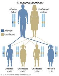
  11. 1 2 Wiernik PH (February 2015). "Familial leukemias". Current Treatment Options in Oncology. 16 (2): 8. doi:10.1007/s11864-014-0323-3. PMID   25762123. S2CID   9926252.
  12. Milpied P, Nadel B, Roulland S (July 2015). "Premalignant cell dynamics in indolent B-cell malignancies". Current Opinion in Hematology. 22 (4): 388–96. doi:10.1097/MOH.0000000000000159. PMID   26049761. S2CID   26080460.
  13. Rodríguez D, Bretones G, Arango JR, Valdespino V, Campo E, Quesada V, López-Otín C (March 2015). "Molecular pathogenesis of CLL/SLL and its evolution". International Journal of Hematology. 101 (3): 219–28. doi: 10.1007/s12185-015-1733-0 . PMID   25630433.
  14. Popp HD, Flach J, Brendel S, Ruppenthal S, Kleiner H, Seifarth W, Schneider S, Schulze TJ, Weiss C, Wenz F, Hofmann WK, Fabarius A (March 2019). "Accumulation of DNA damage and alteration of the DNA damage response in monoclonal B-cell lymphocytosis and chronic lymphocytic leukemia". Leukemia & Lymphoma. 60 (3): 795–804. doi:10.1080/10428194.2018.1498494. PMID   30376743. S2CID   53110926.
  15. Rachel JM, Zucker ML, Fox CM, Plapp FV, Menitove JE, Abbasi F, Marti GE (December 2007). "Monoclonal B-cell lymphocytosis in blood donors". British Journal of Haematology. 139 (5): 832–6. doi:10.1111/j.1365-2141.2007.06870.x. PMID   17961190. S2CID   32218085.
  16. Stetler-Stevenson M (February 2014). "Monoclonal B-cell lymphocytosis in donors". Blood. 123 (9): 1281–2. doi:10.1182/blood-2014-01-546739. PMC   3938144 . PMID   24578489.
  17. Hjalgrim H, Rostgaard K, Vasan SK, Ullum H, Erikstrup C, Pedersen OB, Nielsen KR, Titlestad KE, Melbye M, Nyrén O, Edgren G (October 2015). "No evidence of transmission of chronic lymphocytic leukemia through blood transfusion". Blood. 126 (17): 2059–61. doi: 10.1182/blood-2015-03-632844 . PMID   26302757.
  18. 1 2 Raess PW, Mintzer D, Husson M, Nakashima MO, Morrissette JJ, Daber R, Bagg A (October 2013). "BRAF V600E is also seen in unclassifiable splenic B-cell lymphoma/leukemia, a potential mimic of hairy cell leukemia". Blood. 122 (17): 3084–5. doi: 10.1182/blood-2013-07-513523 . PMID   24159168.
  19. Wotherspoon A, Attygalle A, Mendes LS (December 2015). "Bone marrow and splenic histology in hairy cell leukaemia". Best Practice & Research. Clinical Haematology. 28 (4): 200–7. doi:10.1016/j.beha.2015.10.019. PMID   26614898.
  20. Oishi N, Montes-Moreno S, Feldman AL (January 2018). "In situ neoplasia in lymph node pathology". Seminars in Diagnostic Pathology. 35 (1): 76–83. doi:10.1053/j.semdp.2017.11.001. PMID   29129357.
  21. Marti GE, Rawstron AC, Ghia P, Hillmen P, Houlston RS, Kay N, Schleinitz TA, Caporaso N (August 2005). "Diagnostic criteria for monoclonal B-cell lymphocytosis". British Journal of Haematology. 130 (3): 325–32. doi: 10.1111/j.1365-2141.2005.05550.x . PMID   16042682.